
Abstract
We investigated immunohistochemically the expression of 1,25 dihydroxy vitamin D3 receptors (VDRs) in normal human breast tissue and in breast carcinomas. For the first time, a VDR immunoreactivity score (VDR-IRS) in breast tissue is presented. Mean VDRIRS in breast carcinomas was 7.28 compared to 1.55 in normal breast tissue. Comparing staining patterns for VDR and Ki-67, no visual correlation was found, indicating that VDR upregulation in breast carcinomas is not exclusively controlled by the proliferative activity of these tumor cells. Our study adds to the body of evidence that breast tissue may be a sensitive target organ for therapeutically applied new vitamin D analogues that exert few calcemic side effects.
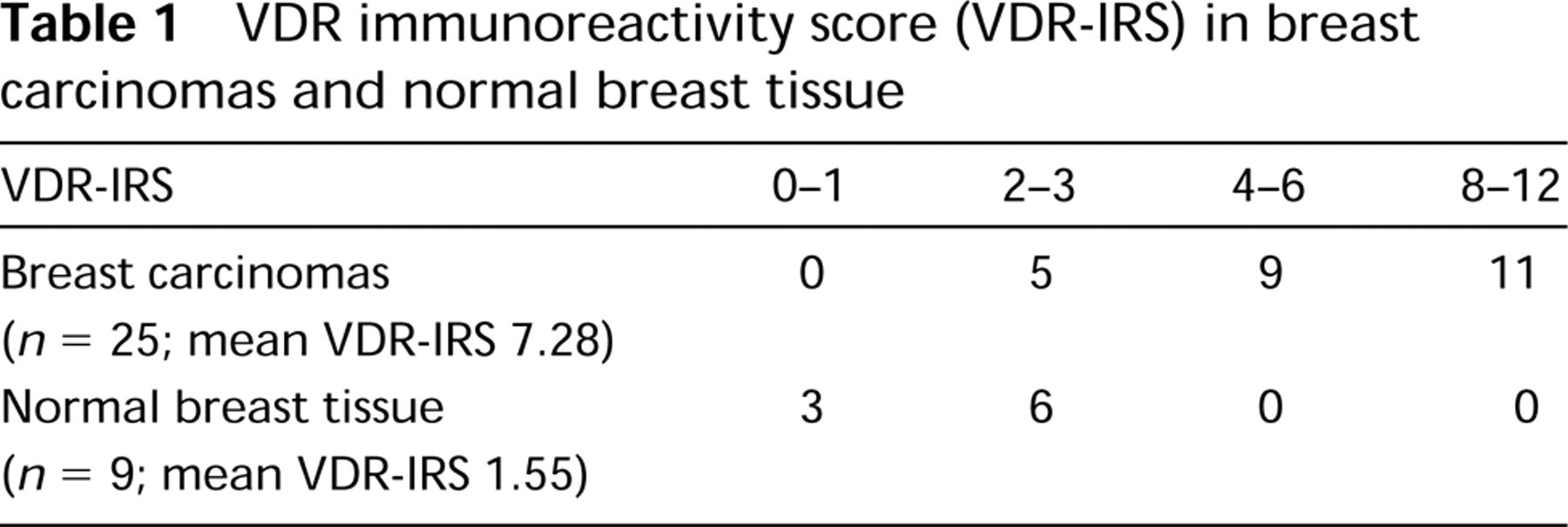
VDRs were detected on frozen sections of breast tissue (lobular carcinomas, n = 7; duct carcinomas, n = 18; normal breast tissue, n = 9) using a previously described and highly sensitive immunohistochemical streptavidin-peroxidase technique (Milde et al. 1991) employing the mAb 9A7γ (Dianova; Hamburg, Germany) (Pike et al. 1982). Microscopic analysis of all specimens was performed by two independent observers (MF and JR). VDR staining intensity (VDR-SI), percentage of VDR-positive tumor cells (VDR-PP), and an immunoreactive VDR score (VDR-IRS) were assessed as a modification of the method described previously for estrogen and progesterone receptors (Remmele et al. 1986): no (VDR-IRS 0–1), weak (VDR-IRS 2–3), moderate (VDR-IRS 4–6), and strong (VDR-IRS 8–12) VDR immunoreactivity. VDR-IRS was increased in breast carcinomas (mean VDR-IRS 7.28) compared to normal breast tissue (mean VDR-IRS 1.55), indicating increased VDR protein levels in these tumors (Table 1).
The steroid hormone responsiveness is directly proportional to the number of corresponding receptors (Costa and Feldman 1987). Because VDR mediates the biological effects of calcitriol and analogues on proliferation and differentiation in target cells (Haussler et al. 1988), VDR upregulation may indicate an increased sensitivity of breast carcinomas to endogeneously or therapeutically applied calcitriol.
VDR immunoreactivity score (VDR-IRS) in breast carcinomas and normal breast tissue
It has recently been shown that 1,25(OH)2D3 potentiates TNF-induced cytotoxicity in human cancer cells (Rocker et al. 1994). This biological activity is most likely the result of a VDR-mediated genomic effect and therefore may be potentiated in breast carcinomas by VDR upregulation.
It has been demonstrated that breast carcinomas contain a high percentage of apoptotic cells (Brinkmann et al. 1996). Recently it has been shown that calcitriol induces apoptosis in various tumor cells (Welsh et al. 1994). Increased expression of VDRs may be a mechanism involved in the induction of apoptosis in breast carcinomas.
Analyzing coexpression of VDR with markers of proliferation, no visual correlation was found. To assess proliferation of breast tissue, we immunostained specimens using a mouse monoclonal antibody (MAb) directed against Ki-67 antigen (clone Ki-67; Dakopatts, Copenhagen, Denmark) (Gerdes et al. 1984). Sections stained for Ki-67 were assessed by counting the number of Ki-67-positive and -negative cells in the most strongly stained tumor area (magnification × 400; at least 200 tumor cells were counted), resulting in an immunoreactivity score for Ki-67 (Ki-67-IRS): 0, no cells labeled/tumor; 1, 1–9% of cells labeled/tumor; 2, 10–25% of cells labeled/tumor; 3, more than 25% of cells labeled/tumor. Seventeen of 25 breast carcinomas revealed 0–9% of Ki-67-positive tumor cells, six specimens showed 10–25%, and the remaining two specimens contained more than 25% Ki-67-immunoreactive tumor cells.
Statistical analysis was performed by the Pearson Chi-Square test. No significant correlation was found between VDR-IRS and the histological type of the breast carcinomas analyzed (p = 0.731). In addition, VDR-IRS was not statistically significantly correlated with the histological grading of breast carcinomas (p = 0.930). No statistically significant correlation was observed between VDR-IRS and Ki-67-IRS (p = 0.407). These findings indicate that VDR expression is not exclusively a function of cell proliferation in these tumor cells but is most probably determined additionally by different, currently unknown mechanisms.
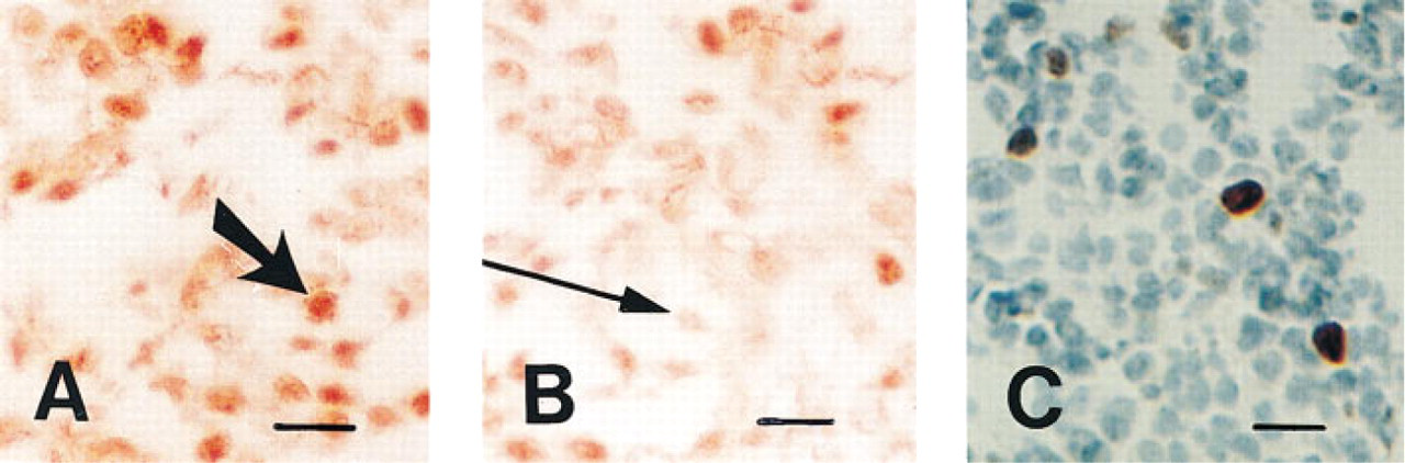
Expression of VDR (
To our knowledge, this is the first report presenting a VDR-immunoreactivity score in breast carcinomas. Because VDR mediates the biological effects of 1,25-dihydroxyvitamin D3, VDR upregulation indicates that breast carcinomas may be sensitive target tissues for therapeutically applied new vitamin D analogues that exert few calcemic side effects.