
Abstract
In primary or cultured cells, in situ hybridization (ISH) or immunocytochemistry (ICC) is often performed on tissue that has been fixed by paraformaldehyde or Carnoy's. Recently we reported an optimized HOPE (HEPES–glutamic acid buffer-mediated organic solvent protection effect) fixation protocol for ISH targeting mRNA in lung tissues. We have now examined whether HOPE fixation could also be used on in vitro cultured cells for targeting mRNA by ISH or proteins by ICC on cytospin preparations. Using the myeloid stem cell line KG-1a as a model system, we showed that HOPE fixation can be applied for ISH and ICC on cultured cells. HOPE can be used with cells and tissues and with a broad spectrum of immunohistocytochemical and molecular techniques.
HOPE (HEPES–glutamic acid buffer-mediated organic solvent protection effect) fixation with subsequent paraffin embedding has recently been described to be a useful new tool for assessment of RNA by in situ hybridization (ISH) in tissues and expanded possibilities for immunohistochemistry (IHC) due to low denaturation of proteins and nucleic acids together with a well-preserved histomorphology (Olert et al. 2001; Goldmann et al. 2002; Pechkovsky et al. 2002a,b; Uhlig et al. 2002). Here we describe the application of HOPE fixation for cultured cells on cytospin preparations.
We first investigated the expression of IL-1β, GMCSF, and INF–γ in KG-1a cells by RT-PCR. Whereas expression of GM-CSF and IL-1β mRNA was detectable in untreated KG-1a cells (Figure 1A), INF–γ expression (Figure 1B) was observed in KG-1a cells incubated with a supernatant of LPS (500 ng/ml, highly purified from Salmonella enterica; Serva, Friedenau, Germany)-stimulated mononuclear cells (SUPLPS) but not in untreated KG-1a cells. Comparison of IL-1β, GM-CSF, and INF–γ expression with GAPDH mRNA expression by quantitative RT-PCR showed 10-fold lower expression of IL-1β, 100-fold lower expression of GM-CSF (in untreated KG-1a cells), and 20-fold lower expression of INF–γ (in SUPLPS-stimulated KG-1a cells) with regard to GAPDH mRNA expression (data not shown).
We next examined whether expression of IL-1β, GM-CSF, and INF–γ mRNA could also be detected by ISH on cytospin preparations of HOPE-fixed KG-1a cells. To provide at least some comparability to established techniques, we also applied ISH on KG-1a cells fixed with Carnoy's solution or paraformaldehyde (PFA) by targeting INF–γ mRNA under the same hybridization conditions used for HOPE fixation, and finally evaluated the results in comparison to HOPE fixation.
For ISH or ICC, KG-1a cells (ATTC No. CCL-246.1; American Type Culture Collection, Rockville, MD) were maintained in Iscove's modified DMEM supplemented with 1% penicillin/streptomycin solution (Gibco/Invitrogen; Karlsruhe, Germany) and 20% heat-inactivated FCS (Linaris; Wertheim–Bettingen, Germany) and were washed several times with PBS. For targeting INF–γ mRNA by ISH, KG-1a cells were incubated in SUPLPS or medium alone, harvested after 16 hr, and washed several times in PBS. Finally KG-1a cells were resuspended at a concentration of 2 × 10⁶/ml in PBS. KG-1a cells (50,000) were attached to SuperFrost Plus microscope slides (Menzel–Gläser; Braunschweig, Germany) by centrifugation for 5 min at 450 rpm at high acceleration in a Cytospin 2 centrifuge (Shandon; Frankfurt, Germany) and dried for 10 min at 37C in a sterilizer. After overnight fixation at 4C in HOPE solution (DCS Innovative Diagnostik Systeme; Hamburg, Germany), cells were incubated with acetone/glyoxal for 1 hr at 4C, and dehydrated six times with acetone for 30 min at 4C, followed by two incubations in isopropanol (10 min at 60C, 2 min at 60C) and air-dried. Rehydration was achieved by incubation in 70% (v/v) acetone for 10 min at 4C and DEPC-treated water for 10 min at 4C. Slides were then air-dried. For comparative studies, dried cytospins were fixed for 30 min at 4C in 4% PFA prepared in PBS or for 1 min in Carnoy's solution (60% ethanol, 30% chloroform, and 10% glacial acetic acid) at RT as described elsewhere (Kanbe et al. 1998). After fixation, cells were rehydrated with DEPC-treated water. For preparation of ISH probes or RT-PCR, the mRNA of LPS-stimulated human monocytes (1 × 10⁶) and of untreated or SUPLPS-stimulated KG-1a cells (1 × 10⁶) was isolated using Dynabeads mRNA direct Micro Kit (Dynal; Hamburg, Germany) and reverse transcription was performed with 200 U of Superscript II (Invitrogen) according to the manufacturer's instructions. IL-1β (sense, CAGCCATGGCAGAAGTACCT; antisense, GACATCACCAAGCTTTTTTGC), GM-CSF (sense, GGCCCGGCGTCTCCTGA; antisense, ATTCTTCTGCCATGCCTGTATC), and INF–γ (sense, ATGAAATATACAAGTTATATCTTGGCTTT; antisense, GATGCTCTTCGACCTCGAAACAGCAT) probes were amplified by PCR for 40 cycles using an AccuPrime Taq DNA Polymerase System (Invitrogen). A 1356-bp fragment obtained by PstI (NEB; Frankfurt am Main, Germany) restriction of pcDNA3 (Invitrogen) was used as a control probe. The probes were labeled overnight with digoxigenin by random primed labeling using High-Prime (Roche; Mannheim, Germany) according to the manufacturer's instructions and labeling efficiency was estimated in comparison to given concentrations of control DNA as described elsewhere (Roche Molecular Biochemicals 1996). Hybridization solution was composed of 2 ng/ml fresh denatured probe, 250 μg/ml yeast tRNA (Roche), 0.1% SDS, and 50% formamide in PBS. Hybridization was performed overnight at 49C in moist chambers. Slides were washed by the following steps: 2 × SSC twice for 10 min at ambient temperature, then 0.2 × SSC twice for 30 min at 50C. The specimens were then incubated with an alkaline phosphatase-conjugated anti-digoxigenin antibody (Anti-DIG-AP; Roche) as previously described using new fuchsin as a chromogen (Goldmann et al. 1999,2002). Cells were then counterstained with hematoxylin (Htx).
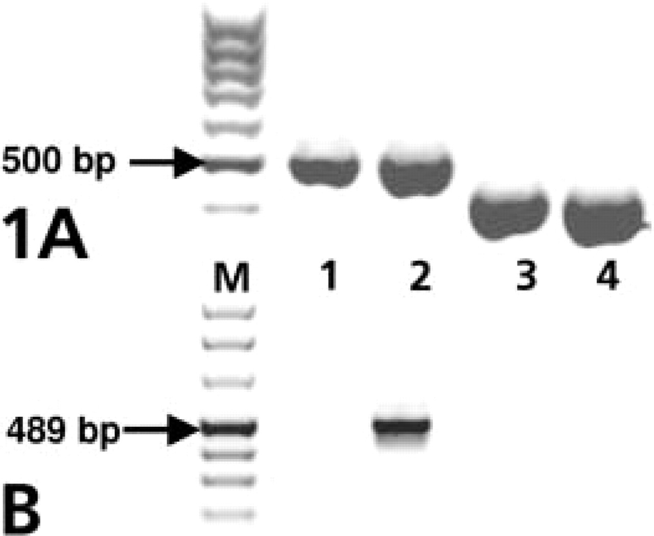
IL-1β, GM-CSF, and INF–γ mRNA expression in KG-1a cells detected by RT-PCR. PCR products for GM-CSF (493 bp, Lanes 1 and 2) and IL-1β (414 bp, Lanes 3 and 4 (

ISH targeting IL-1β, GM-CSF, or INF–γ mRNA by digoxigenin-labeled probes in HOPE-, paraformaldehyde-, or Carnoy-fixed KG-1a cells. Detection was performed with anti-DIG-AP and new fuchsin as a substrate for alkaline phosphatase. Counterstaining was performed with hematoxylin (
Whereas no new fuchsin could be detected in KG-1a cells hybridized with no probe (Figure 2A) or the digoxigenin-labeled pcDNA3.1 fragment (Figure 2B), distinct cytoplasmic staining could be observed within 5 min in cells probed with GM-CSF (Figure 2C) or IL-1β (Figure 2D). Distinct cytoplasmic staining was also detected in SUPLPS-stimulated, HOPE-fixed (Figure 3E) or PFA-fixed KG-1a cells (Figure 3G) probed for INF–γ mRNA, whereas no staining was detected within 30 min in SUPLPS-stimulated KG-1a cells fixed with Carnoy's solution or in unstimulated KG-1a cells fixed with HOPE, PFA, or Carnoy's solution (Figure 3F; and data not shown).
In the next group of experiments we tested whether ICC techniques can be also applied to HOPE-fixed KG-1a cells on cytospin preparations and compared the results, first with flow cytometry data of untreated KG-1a cells and second with data obtained from ICC of PFA- and Carnoy-fixed KG-1a cells.
HOPE-, PFA-, and Carnoy-fixed cells on cytospins were prepared as described above and incubated for 60 min with 20 μg/ml of an FITC-conjugated anti-CD34 antibody (Chemicon International; Hofheim, Germany) or an FITC-conjugated anti-CD86 antibody (BD; Heidelberg, Germany) in PBS. CD86 is not expressed on KG-1a cells and was used as an isotype control antibody (Figure 3A). Slides were washed twice with PBS and stained with 10 μg/ml of an Alexa Fluor 488-conjugated goat anti-mouse antibody (Molecular Probes; Leiden, The Netherlands) for 30 min in the dark. After washing twice with PBS, stained cells were mounted with Mowiol (Calbiochem; Schwalbach, Germany) containing 1,4-diazabicyclo[2.2.2]octane (Sigma; Taufkirchen, Germany). Samples were examined with a Leica TCS SP Spectral Confocal Microscope (Leica; Bensheim, Germany) at an excitation wavelength of 488 nm from an argonion laser. For flow cytometry, cultured KG-1a cells were washed with azide–PBS containing 10% heat-inactivated human serum (HS), followed by staining for 30 min with 20 μg/ml FITC conjugated anti-CD34 or FITC-conjugated anti-CD86 antibody (BD). Finally, cells were washed twice with azide–PBS and analyzed by a FACS-calibur (BD) flow cytometer. KG-1a cells expressed high levels of the stem-cell marker CD34, but no CD86 as determined by FACS analysis (Figure 3A). Consistent with FACS data, strong staining for CD34 was observed by ICC in cytospin preparations of HOPE-fixed KG-1a cells (Figure 3C), whereas incubation of HOPE-fixed KG-1a cells with an anti-CD86 antibody resulted in no staining (Figure 3B). Similar results for CD86 staining were observed in PFA- and Carnoy-fixed KG-1a cells (data not shown). Whereas we observed high staining intensities for CD34 in KG-1a cells fixed with HOPE (Figure 3C) or PFA (Figure 3E), only weak staining was detected in KG-1a cells fixed with Carnoy's solution (Figure 3D).
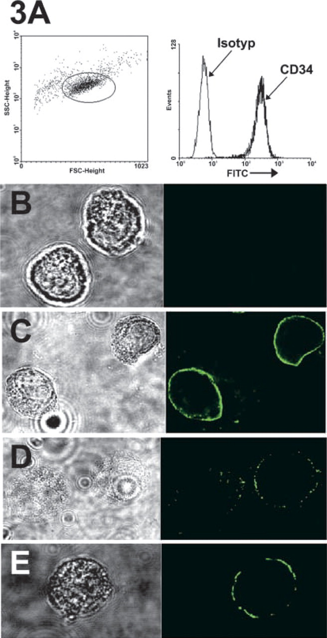
Determination of CD34 expression in KG-1a cells by flow cytometry or ICC using laser scanning confocal microscopy. Cultured KG-1a cells (
HOPE solution has been shown to be an excellent preservative for human soft tissues, providing protection for proteins and nucleic acids in conjunction with well-preserved morphology. We demonstrated that HOPE is suitable not only for tissue sections but also, with slight modifications, for cultured cells on cytospin preparations.
To prevent osmotic shock, we prepared the hybridization mix used for ISH in KG-1a cells with PBS. Our results indicate that PBS did not affect hybridization specificity because no hybridization signal could be detected in KG-1a cells if ISH was performed with digoxigenin-labeled bacterial DNA. Furthermore, no unspecific signals were observed in untreated KG-1a cells probed for INF–γ. Because PFA (Kovacs et al. 2001) and Carnoy's solution (Kanbe et al. 1998) are usually used for ICC methods, we assessed the signals obtained by ISH in HOPE-fixed KG-1a cells in comparison to cytospin preparations fixed by PFA and Carnoy's solution. Whereas in our hands no INF–γ mRNA was detected in SUPLPS-stimulated KG-1a cells fixed with Carnoy's solution, comparable signals for INF–γ mRNA were detected in HOPE-fixed and PFA-fixed cytospin preparations. In contrast to PFA fixation, KG-1a cells fixed with HOPE or Carnoy's solution retained better morphology.
We could also determine the expression of the cell surface marker CD34 by ICC in HOPE-fixed and PFA-fixed KG-1a cells, whereas in our hands Carnoy-fixed cells showed only weak staining for CD34. This is consistent with findings of other groups showing that Carnoy's fixative reduced the number of chymase-positive cells in ICC staining of mast cells, probably by damaging or changing epitopes, whereas PFA did not (Kanbe et al. 1998). Remarkably, ICC in HOPE-fixed KG-1a cells could be conducted with the same anti-CD34 clone (581) used for flow cytometry, although the antibody was not recommended for ICC. This further indicates that antigenic structures and antigen accessibility are retained by HOPE fixation and that the spectrum of antibodies applicable for ICC can be broadened. Furthermore, mRNA expression in HOPE-fixed cells could be quantified in situ by laser scanning confocal microscopy using fluorescent-labeled anti-digoxigenin antibodies or directly labeled probes in the future. Appropriate studies are under way.
In conclusion, we showed that both ISH and ICC can be performed in HOPE-fixed cells on cytospin preparations. This may open the opportunity to combine both procedures, providing a powerful tool to better characterize mRNA-expressing cell populations in situ.
Footnotes
Acknowledgements
Supported by the Deutsche Forschungsgemeinschaft (GRK 288, Project A4).
We thank H. Kühl for excellent technical assistance and T. Scholzen and Maria Manoukian (Department of Immunology and Cellular Biology, Research Center Borstel) for confocal microscopy.