
Abstract
The retinal pigment epithelium (RPE) shows cell heterogeneity in morphology and enzymatic activity. Routine isolation procedures for RPE cells may reduce enzymatic activity and prevent the quantification of regional enzymatic differences in vivo. We developed a new technique for the isolation of RPE cells based on adhesion of the cells to agarose. The morphology of the isolated cells resembled that of RPE cells in vivo. The cells were viable in the dye exclusion test and showed a histochemical staining pattern as RPE cells in vivo. With this technique, quantitative regional differences in the enzymatic activities were detected.
T
RPE cells for quantitative measurements should not contain components originating from Bruch's membrane and from the underlying choroideal connective tissue, such as fibroblasts, endothelial cells, and blood cells. To study quantitative differences between defined RPE cell regions, we developed a technique based on the adhesion of the RPE cells to agarose.
Eyes of adult cows from the local slaughterhouse were used. For isolation of RPE sheets, the anterior eyecup and the vitreous were removed from the posterior eyecup. The neural retina was separated from the RPE by a flush with PBS and cut off at the optic nerve head. Punches of defined size (diameters of 3, 4, 6, 8, 10, and 12 mm) were used to determine the regions of interest. All layers of the posterior eyecup (RPE, choroid, and sclera) were punched out. These punches can be handled more comfortably than punches consisting only of RPE and choroid. In addition, the sclera makes it also easier to discern the side of the RPE in pigmented areas. The RPE was isolated either immediately or the punches were deep-frozen in Dulbecco's minimal medium (Gibco; Grand Island, NY) containing 10% DMSO. The RPE of the punches was isolated either by gentle scraping with the blunt end of a scalpel, by digestion with 0.25% trypsin (Gibco) for 5–45 min, or by adhesion to agarose.
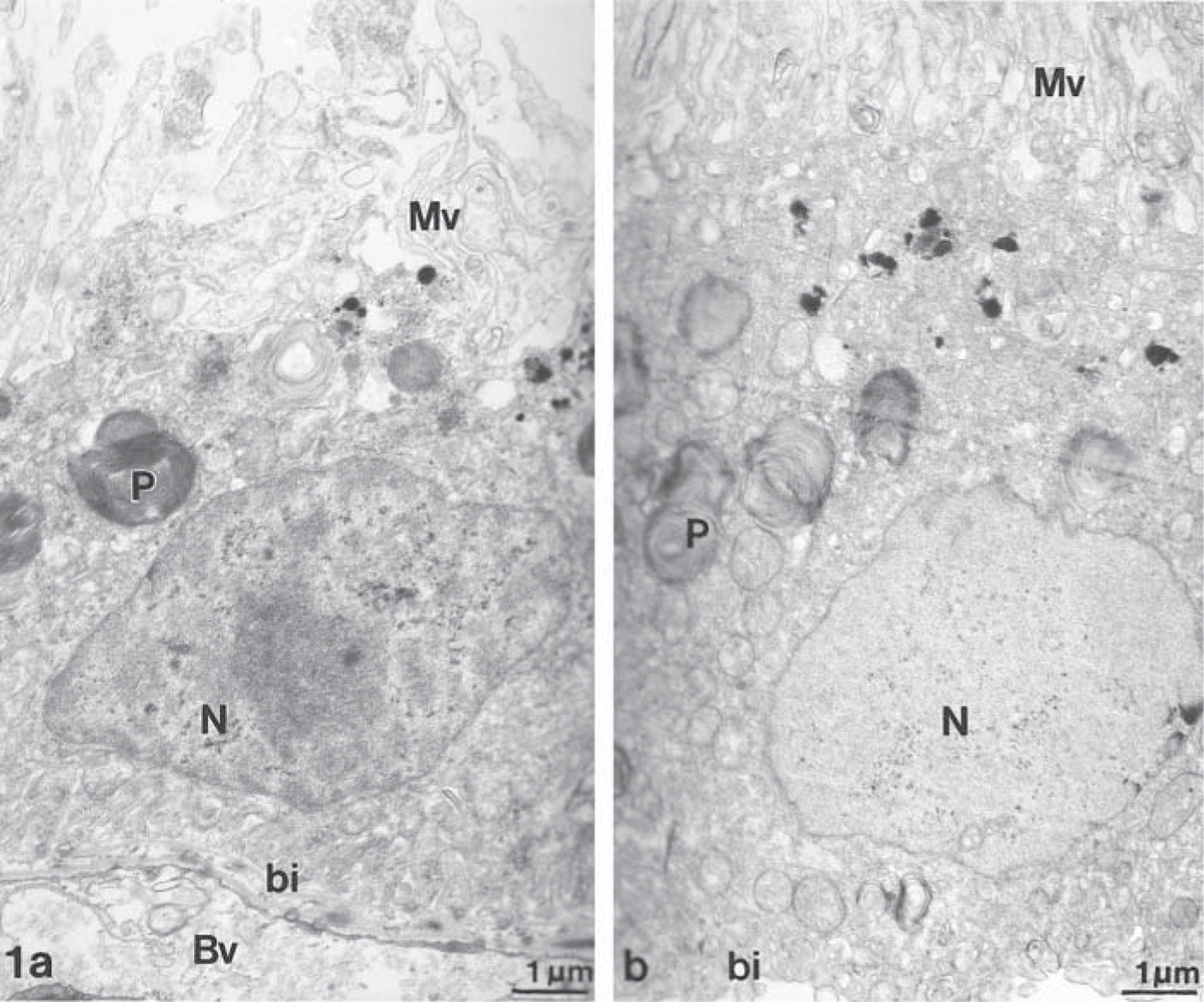
Ultrastructure of RPE cells in vivo (
Enzymatic activity for lactate dehydrogenase (
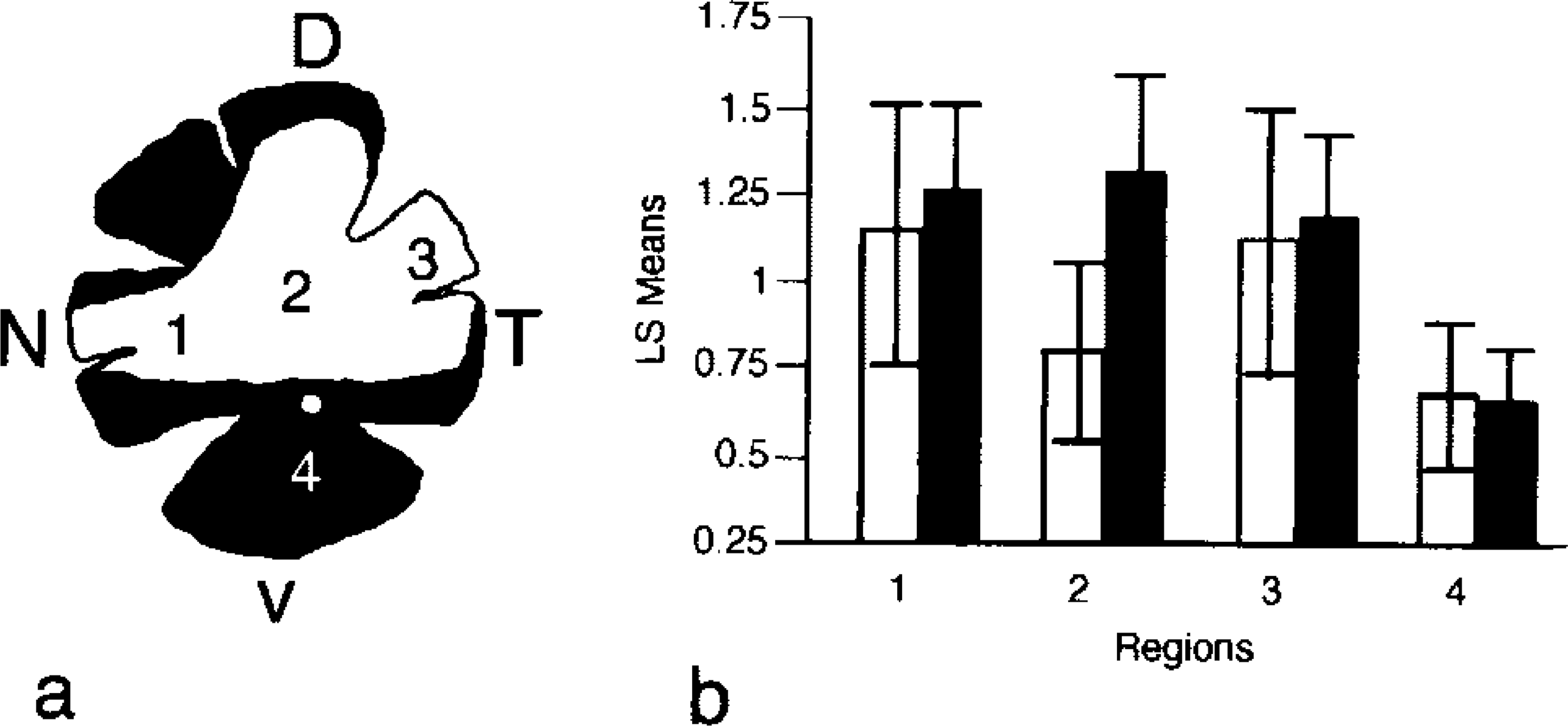
The investigated regions were the following (Figure 3): the unpigmented dorso-peripheral (regions 1 and 3), dorsocentral area (region 2), and the pigmented ventrocentral area (region 4).
Low melting-point agarose (LMP Agarose ultra Pure, electrophoresis grade; BRL Life Technologies, Gaithersburg, MD; 4% in aqua dest) was prepared and poured into a Petri dish. Before complete geling, punches were placed on the surface with the RPE facing the agarose. The Petri dish with the punches was put in the refrigerator for 30 min. Thereafter, choroid and sclera were removed from the agarose and the RPE cells remained attached to the agarose.
After isolation, the isolated RPE cells were stained with hematoxylin and the remaining tissue, consisting of Bruch's membrane, choroid, and sclera, was immersed in a solution of 0.1% DAPI (Sigma; Deisenhofen, Germany) to identify residual RPE cells. The DAPI stain is strongly fluorescent and can be used for whole mounts of RPE-choroid–sclera. Remaining RPE cells can be discerned from fibroblast or endothelial cells by the typical shape of their nuclei.
The following controls were performed for the isolation with agarose. The protein in the isolated RPE cells on agarose was determined (Bradford 1976), because it is another parameter indicating whether similar amounts of RPE cells were isolated with the agarose. The protein was extracted by freezing and thawing in a solution of 0.1% Triton X-100 in aqua dest. To determine the integrity and the ultrastructure of the cells, the isolated RPE cells were studied by routine electron microscopy, by exclusion of the dye Erytrosin (Sigma; 0.1%, 2 min at RT), and by histochemical reaction for lactate dehydrogenase as a marker for cell integrity (Jacobsen 1969). As markers of apically localized enzymes the activities of γ-glutamyltranspeptidase and dipeptidylpeptidase IV were determined. γ-Glutamyltranspeptidase produces the radical scavenger glutathion. Radical scavengers in the eye are especially important because the incidence of light-induced radical oxygen is high (De La Paz and Anderson 1992). These enzymes were localized by histochemistry (Kugler et al. 1985) only in the unpigmented regions (1, 2, and 3) because the pigmentation interferes with detection of the chromogenic substrate. To investigate regional differences in the γ-GT and DPP IV content RPE isolated from regions 1–4 was quantified according to the cleavage of a fluorogenic substrate (Sinha and Gossrau 1984; Sedo et al. 1989). Regional differences were investigated in 14 (γ-GT) and six (DPP IV) eyes. A simple variation analysis (ANOVA) was performed and differences between the regions were evaluated by the Tukey-Kremer Assay.
Schematic drawing of the posterior eyecup of the bovine eye (
By light microscopy, the morphology of all isolated cells resembled that of RPE cells in vivo. However, enzymatic isolation with trypsin abolished the reactivity of the histochemical reaction and markedly reduced reactivity with the fluorogenic substrate. Preparations of RPE cells isolated by scraping with the blunt end of a scalpel contained variable contamination with collagen fibers and cells resembling fibroblasts. After staining with DAPI, various amounts of RPE cells attached to Bruch's membrane could be detected. The samples were also difficult to manipulate. The centrifugations necessary for collection and for washing of the cells severely reduced the reactivity of the cells with the substrates.
Deep-frozen and fresh punches were equally suited for the isolation with agarose. Cells isolated by this method were pure RPE monolayers. RPE sheets of 8 or 10 mm diameter isolated with agarose included 99.0 ± 0.5% of the cells of the selected region. In small punches (3-mm diameter) 78.0 ± 1% of the cells of the selected region were included.
In addition to determination of the remaining cells on the choroid-sclera punch, another (independent) method was used. The protein in the isolated RPE cells on agarose was determined. The standard deviation of the protein content in the RPE patches isolated with agarose was lowest (about 2%) in patches of 10-mm and highest (10%) in the patches of 3-mm diameter.
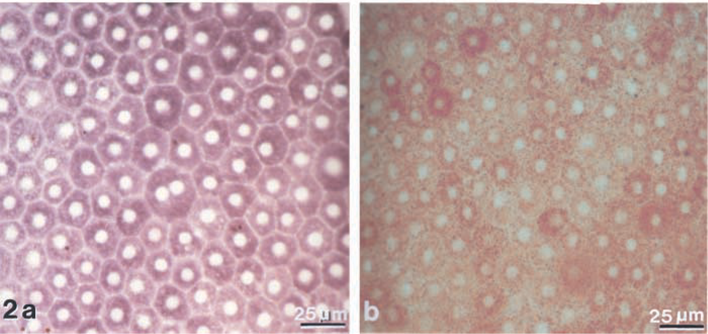
To detect potential ultrastructural damage to the cells and to determine if the isolated material also included Bruch's membrane, the isolated RPE on Agarose was compared to RPE in vivo. Isolated RPE cells show a similar ultrastructure to cells in vivo. They possess an intact apical plasma membrane with many microvilli and a basal plasma membrane that forms basal infoldings. Parts of Bruch's membrane do not adhere to the isolated RPE patches (Figure 1a and 1b). Only a minority of the isolated RPE cells (5 ± 2%) were stained with erythrosin, indicating cell damage. Strong reactivity for lactate dehydrogenase could be detected in almost all cells of the RPE-agarose patches (Figure 2a). The cells also showed reactivity for the apically localized enzymes γ-GT and DPP IV and presented a similar heterogeneous activity pattern as observed in vivo (Figure 2b). Differences in the content of γ-GT and DPP IV between the peripheral and the central regions of the RPE were noted (Figure 3). γ-GT activity was significantly higher in the peripheral regions (1 and 3) than in the central regions (2 and 4). DPP IV activity was significantly higher in the unpigmented dorsoperipheral and dorsocentral regions (1, 2, and 3) than in the pigmented ventrocentral region (4).
The removal of RPE cells with agarose is a mild, highly reproducible, and efficient procedure for isolation of RPE cells of all regions of the posterior eyecup. The consistently low fraction of cells remaining on Bruch's membrane makes protein determination or cell counting in each set of experiment superfluous. For the mechanically isolated RPE samples, cell count or DNA content has to be determined because the fraction of remaining cells is more variable and contaminating Bruch's membrane influences the protein content. The cells isolated with agarose show no ultrastructural damage but high viability, as evidenced by the dye exclusion test and the reactivity with the LDH substrate. The pattern of the histochemical staining for γ-GT and DPP IV reveals intercellular heterogeneity and resembles that of RPE cells in vivo. Using this technique, we detected significant regional differences in the content of γ-GT and DPP IV between peripheral and central areas. Therefore, this isolation method appears to be well suited to obtain pure preparations of viable RPE cells from all regions of the posterior eye-cup for different physiological measurements (investigation of ion channels, quantitative determination of immunoreactive protein, or measurements of enzymatic activities). This method can therefore be used to select RPE cells that are, according to their enzyme content, most suited for transplantation. The procedure can also be used for the contamination-free isolation of other monostratified epithelia, e.g., endothelial or corneal cells.
Footnotes
Acknowledgements
Supported by Fortüne 808–0–0.
We thank Mihnea Nicolescu for the layout and Dr Andreas Mack for suggestions on the manuscript.