
Abstract
To map the mitochondrial capacity to provide adenosine triphosphate (ATP), the activities of cytochrome oxidase (COX) and succinic dehydrogenase (SDH) were respectively evidenced by diaminobenzidine (DAB) and copper ferrocyanide cytochemical techniques in the cerebellar cortex of adult rats. Sampling of the positive mitochondria was carried out by the disector procedure. The ratio (R) overall area of the precipitates due to COX activity within the single mitochondrion/area of the same organelle was automatically calculated to estimate enzyme activity vs mitochondrial size. The number of SDH-positive mitochondria/μm3 of tissue (numeric density, Nv) was morphometrically calculated. Cytochemistry of key enzymes of the respiratory chain enables measurement of the actual capacity of individual mitochondria to provide ATP. This quantitative estimation allows morphofunctional mapping of the mitochondrial metabolic competence in discrete tissue and/or cellular compartments. (
Keywords
T
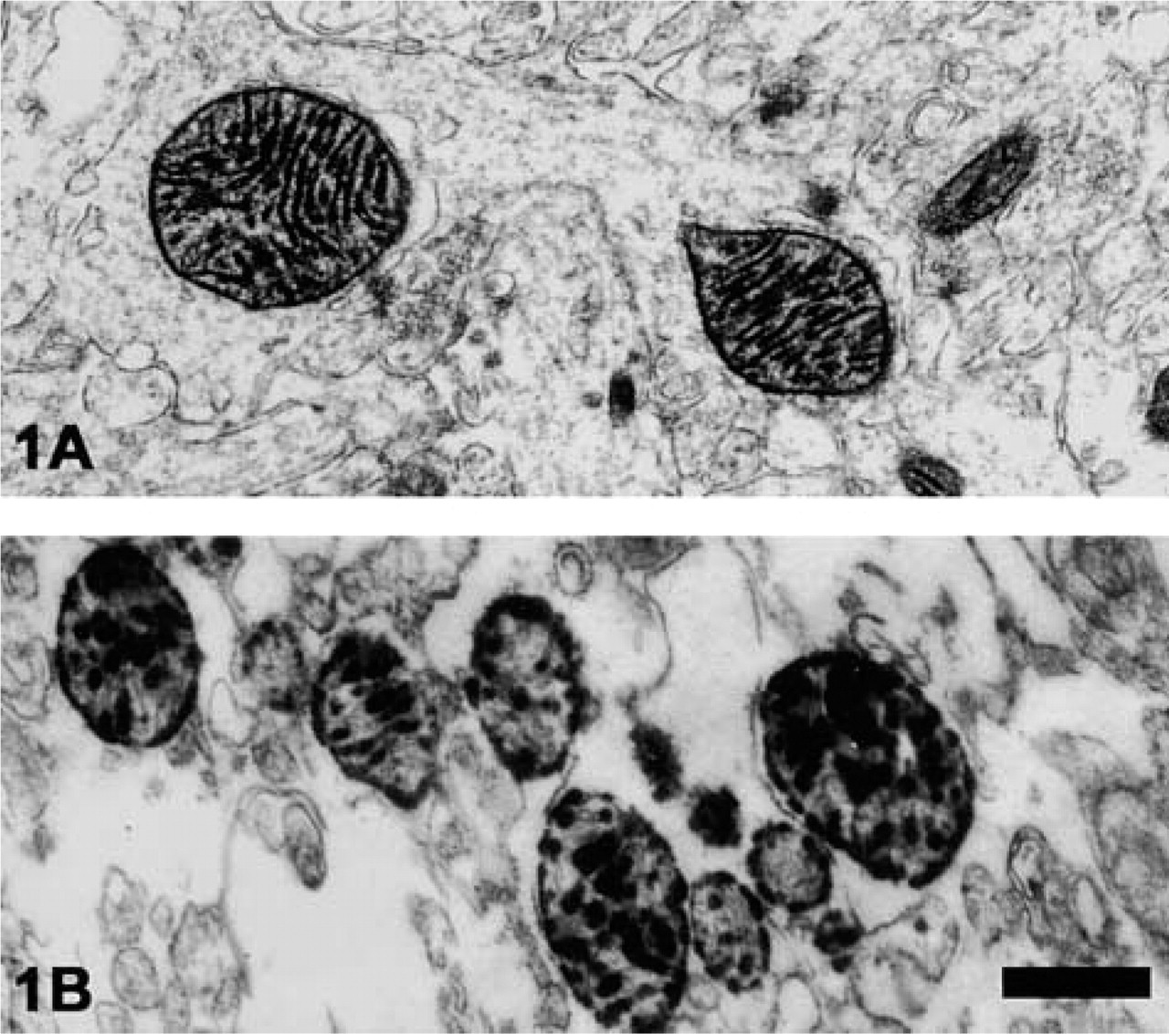
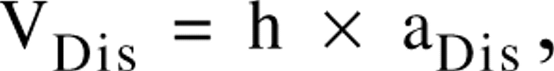
The cerebellar cortex of adult rats (12 months of age) was adopted as a tissue model for our experimental procedures. In the cerebellar glomeruli, COX activity was evidenced by the DAB procedure (Seligman et al. 1968). According to the guidelines of the Italian Ministry of Health regarding the use of laboratory animals, the rats were perfused with 2.5% paraformaldehyde + 1.5% glutaraldehyde in 0.1 M Sorensen buffer, pH 7.4, + 4% sucrose. The tissue samples (40–60-μm thickness) were rinsed overnight in 4% buffered sucrose and then incubated (2–4 hr at 37C in the dark) in the following solution (100 ml): 50 mg DAB, 27 mg cytochrome C, 4 g sucrose. Postfixation in osmium tetroxide was followed by conventional inclusion, sectioning, and contrasting procedures (Figure 1A). The areas of the deposits due to COX activity were easily identified and measured by a computer-assisted image analysis system (Kontron KS300). The ratio (R) total area of the precipitates within a single mitochondrion/area of the same organelle was automatically calculated and reported as enzyme activity/ μm3 of each positive mitochondrion.
In Purkinje cell perikarya, SDH activity was preferentially revealed by the copper ferrocyanide reaction (Hajos and Kerpel–Fronius 1971). Freshly excised cerebellar samples were incubated for 45 min at 37C in a solution (final volume 5 ml) containing 300 mM sodium potassium tartrate in 0.1 M phosphate buffer, pH 7.6, 21 mM copper sulfate, 140 mM sodium succinate, 1.5 mM potassium ferricyanide, and 16 mM phosphate buffer (pH 7.6). Dark spots mark SDH activity at the inner mitochondrial membrane and cristae (Figure 1B). Because the tissue samples were unfixed, only the SDH-positive organelles appeared to be satisfactorily preserved (Figure 1B). The number of mitochondria/μm3 of tissue (numeric density, Nv) was calculated by means of the recently introduced disector method (Sterio 1984). Briefly, pairs of images from serial sections constitute a disector. The mitochondrial profiles found in the first section (reference) are checked in the second one (look up): the organelles found in reference, but not in the look up section, are counted. The volume of the disector (V
(
where h is the disector height (in our case h = 0.2 μm, i.e., each disector consisted of four sections of 0.05-μm thickness), and aDis is the disector area, directly measured by our image analysis system. The number of mitochondria/μm3 (Nv) was calculated by applying the following formula:
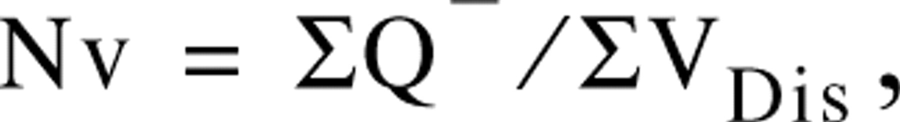
where ΣQ− is the total number of counted mitochondria. The examined area of the reference section (aDis) was 19.35 μm2 and each disector volume (VDis) measured by us was 3.87 μm3 (Bertoni–Freddari et al. 2001).
An example of the data that can be obtained on COX- and SDH-positive mitochondrial populations is shown in Figure 2. The value of R was calculated by sampling 330 COX-positive organelles and can be presumed to account for the percent of mitochondrial surface area involved in actual COX activity. The Nv value of SDH-positive mitochondria was obtained by sampling 12 Purkinje cell bodies from a single animal. Referring this datum to the results of numeric density estimated in tissue samples prepared according to conventional electron microscopic procedures, information on the fraction of SDH-active mitochondria can be obtained.
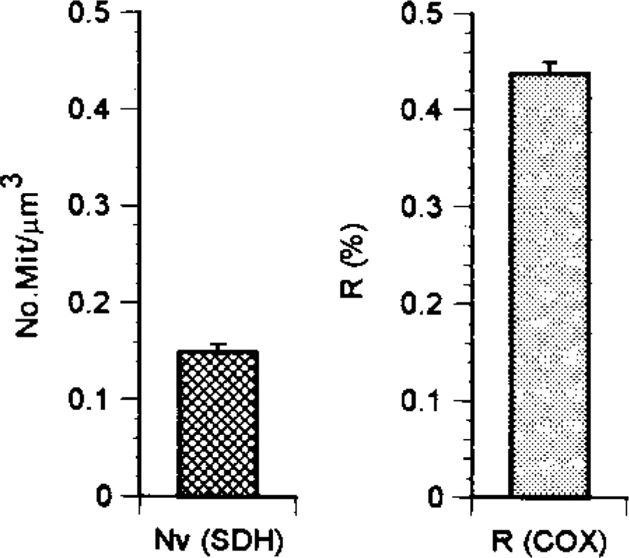
R, ratio area of precipitates due to COX activity/overall area of the single mitochondrion. Mean ± SEM. Nv, numeric density of SDH-positive mitochondria from the cerebellum of an adult rat.
Heteroplasmy, i.e., the mixture of normal and mutated mitochondrial genomes, is a well-documented feature of the mitochondrial population of postmitotic cells, particularly of old individuals. As a consequence, the potential for energy production in these cells is dependent on a mosaic of organelles with different functional capacities (Bertoni–Freddari et al. 2001). Our preliminary data support the notion that an outline of the working mitochondrial fraction can be obtained by quantitative cytochemistry. The precipitates due to COX and SDH molecules constitute reliable correlates of enzyme activities because they demonstrate specific arrays of molecules functionally assembled and arranged in the organelle membrane. Therefore, by preferential cytochemistry, the metabolic competence of the mitochondria considered functional units of cellular bioenergetics can be tested. In this context, morphometric quantitation of COX and SDH deposits may help in identification of early alterations in mitochondrial performance as predisposing conditions leading to impaired organ and system function.