
Abstract
NAD(P)H:quinone oxidoreductase 1 (NQO1; DT-diaphorase; DTD) is a two-electron reductase that efficiently bioactivates compounds of the quinone family, such as mitomycin C. The observation that DTD is overexpressed in many cancerous tissues compared to normal tissues has provided us with a potentially selective target that can be exploited in the design of novel anticancer agents. Because of the relative lack of information on the cell-specific expression of DTD, the purpose of this study was to perform a body mapping of its normal distribution. Tissue samples from various components of the human reproductive system were analyzed by immunohistochemistry. We found strong expression of this enzyme in testicular stromal cells (Leydig cells) and in the epithelium of epididymis, ductuli efferentes, and Fallopian tube. These results suggest that DTD-bioactivated quinones could be responsible for a selective toxicity on these components of the reproductive system and cause clinical problems due to testosterone deficiency and infertility. This observation needs to be investigated in preclinical evaluation of new anticancer quinones and in patients treated with these compounds. (
Q
In our preclinical evaluation of RH1, a new DTD-bioactivated quinone (Winski et al. 1998), we are performing a body mapping of this enzyme. We report our findings concerning DTD expression in different components of human reproductive system. Archival samples of formalin-fixed, paraffin-embedded tissues were analyzed (10 samples of normal testis, five samples of normal epididymis and ductuli efferentes, and 10 samples of normal ovary and Fallopian tube). We performed an immunohistochemical analysis for detection of DTD using anti-DTD monoclonal antibodies (IgG1) derived from a BALB-c mouse immunized with purified recombinant human DTD protein (Siegel et al. 1998). Non-human-reactive monoclonal mouse antibodies (IgG1) produced in tissue culture were used as negative control reagent. Tissue sections (3 μm) were deparaffinized in xylene, rehydrated through graded alcohol, and microwaved. Endogenous peroxidase activity and nonspecific binding were blocked by adding, respectively, peroxidase blocking agent (DAKO EnVision Kit; Carpinteria, CA) and 20% normal rabbit serum. Serial sections were successively incubated with either anti-DTD or control antibodies and then with the secondary antibody (labeled polymer HRP anti-mouse). Immunodetection was performed using a substrate-chromogen solution (hydrogen peroxide and 3,3′-diaminobenzidine chromogen). Slides were counterstained with hematoxylin. The intensity of immunostaining (brown staining) was visually scored as 0 (negative), +1 (very weak), +2 (weak), +3 (strong), and +4 (very intense). There was no immunostaining in control sections when nonspecific antibodies were used (score 0). Normal human testis showed a score 0 staining in seminiferous tubules and in connective tissue stroma, a score +4 staining in interstitial Leydig cells (Figure 1), and a score +3/+4 staining in the endothelial lining of stromal vessels. Epididymis and ductuli efferentes showed a score 0/+1 staining in stroma and a score +4 staining in the columnar ciliated epithelial cells (Figure 2). Ovary showed a score 0 staining in stroma and a score + 1/+2 staining in the mesothelial lining. Fallopian tube showed a score 0 staining in connective tissue stroma and non-ciliated epithelial cells and a score +4 staining in ciliated epithelial cells (Figure 3).
Human male infertility secondary to treatment with cytotoxic chemotherapy has been widely described and is almost entirely a function of damage to the seminiferous tubule germinal epithelium (Meistrich 1993). Alkylating agents other than MMC appear to be the most toxic to the germinal epithelium. Conversely, there is very little evidence of Leydig cell insufficiency after cytotoxic chemotherapy (Howell et al. 1999). The available studies consider the “biological” aspect of Leydig cell insufficiency as defined by an elevation of luteinizing hormone (LH) levels; no histological demonstration has been reported. In our study, DTD was shown to be strongly expressed in various components of the human reproductive system (Leydig cells and epithelium of epididymis and Fallopian tube). A selective toxicity for these structures may be responsible for Leydig cell insufficiency and infertility. If further investigations confirm these observations, patients should benefit from pretreatment counseling about their reproductive function with respect to chance for recovery of spermatogenesis, fertility, semen cryopreservation, and the possibility of need for androgen replacement.
Surprisingly, drugs such as MMC have been used for years, but no studies have focused on this aspect. This raises serious questions as to whether this entity might have previously gone undiagnosed. A prospective evaluation of gonadal function in patients undergoing anti-cancer treatment with quinones is necessary to assess the magnitude of the problem.
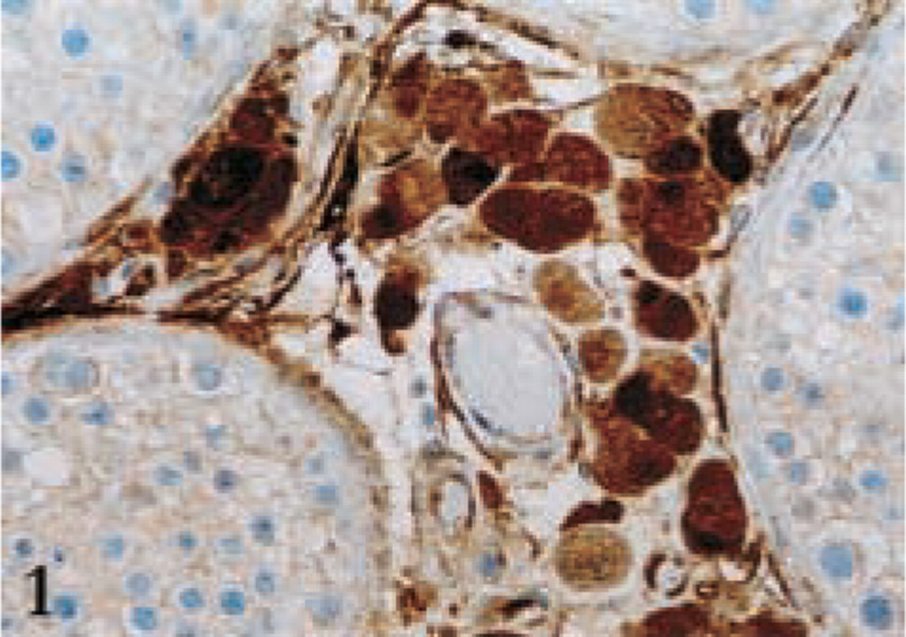
DTD expression in human normal testis.
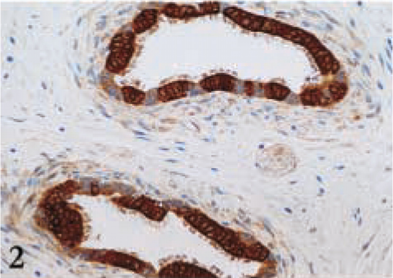
DTD expression in ductuli efferentes.
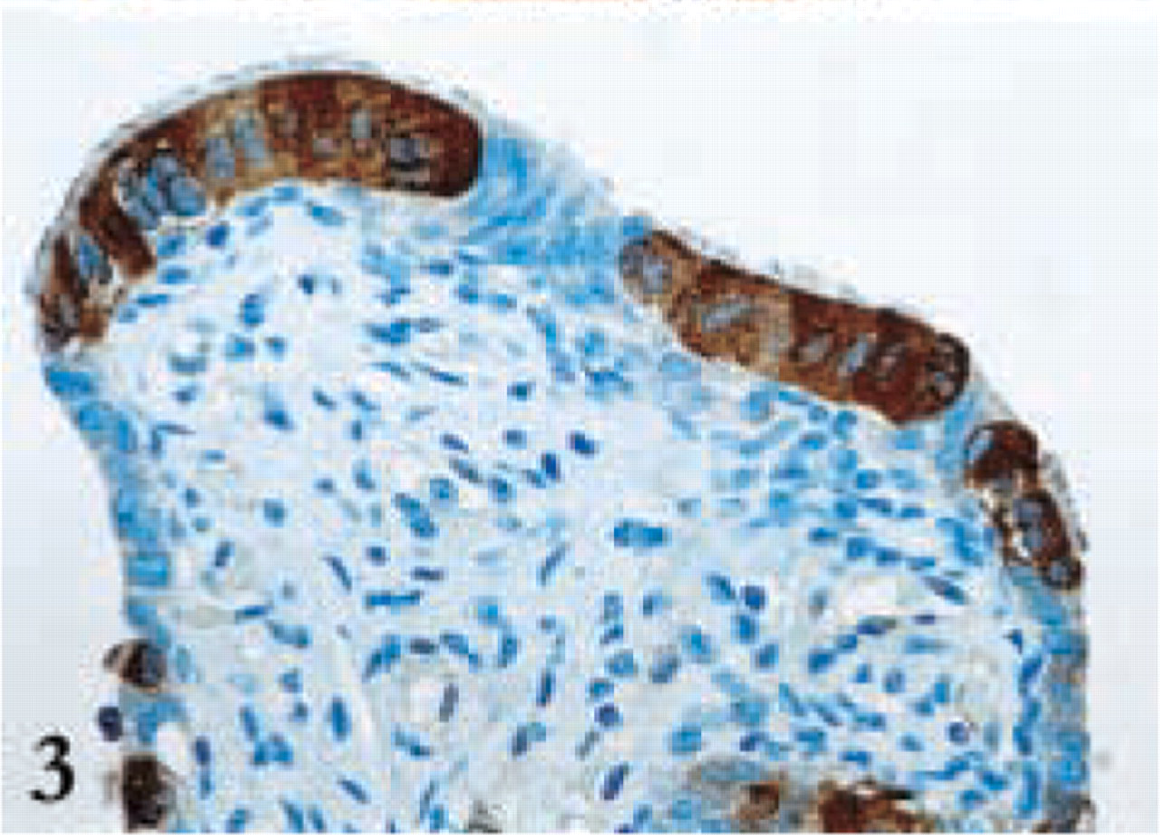
DTD expression in Fallopian tube.
Footnotes
Acknowledgments
FZ received fellowship support from the European Society of Medical Oncology (ESMO).
Anti-DTD antibodies were a kind gift from Professor David Ross (Department of Pharmaceutical Sciences, School of Pharmacy and Cancer Center, University of Colorado Health Sciences Center; Denver, Colorado).