
Abstract
Recently, mast cell tryptase has been identified as another potent proangiogenic factor in tumors, along with fibroblast and vascular endothelial growth factors. Its role has been studied in a number of cancers, including carcinoma of the uterine cervix, with discordant results. Our aim was to study the expression of tryptase and bFGF in mast cells (MCs) during development of neoangiogenesis in premalignant and malignant lesions of the cervix. Biopsy specimens from 21 patients without cancer and from 63 patients with dysplasias and squamous cell carcinomas were used. They were stained with Alcian blue– safranin O (ABSO) and immunostained with specific antibodies against factor VIII, CD105, tryptase, and bFGF. Tryptase-positive mast cells increased with tumor progression and were close to newly formed blood vessels. Vascularization showed a linear increase from dysplasia to invasive cancer. We suggest that MC tryptase may upregulate neoangiogenesis in carcinogenesis of the uterine cervix. (J Histochem Cytochem 49:1061–1062, 2001)
I
Among the many proangiogenic factors identified to date, the fibroblast growth factor (FGF) family (Czubayko et al. 1997) and the vascular endothelial growth factor (VEGF) (Nicosia 1998; Kraft et al. 1999) are the most widely studied in malignant growth. Recently, Kankkunen et al. (1997) found that tryptase, a serine proteinase contained in mast cells, is also a potent proangiogenic factor in malignancies. Although the role of mast cells in the development and progression of cancer has not been elucidated, there is evidence that the number of mast cells in various solid cancers increases with tumor progresssion (Yomita et al. 1999). It has been shown in experimental models that mast cells and their products are capable of inducing tumor angiogenesis (Kessler et al. 1976). The presence of mast cells in cancer of the uterine cervix has been investigated by various authors. Graham and Graham (1966) reported that the number of mast cells in cervical cancer diminishes with cancer progression. Jain et al. (1977) observed an increased number of mast cells only in areas of inflammation around the malignant cells, but a higher number in carcinoma in situ than in invasive cancer. In contrast, Pasternak and Jans (1986) observed an increment of mast cells with cancer progression. The aim of this study was to investigate the expression of FGF and tryptase in mast cells during development of neoangiogenesis in premalignant and malignant lesions of the cervix.
Paraffin-embedded cervical biopsies from 21 patients without cancer, 19 with cervical dysplasia, eight with carcinoma in situ, and 36 with squamous cell carcinoma in different clinical stages were studied. Sections 5 μm thick were mounted on poly-
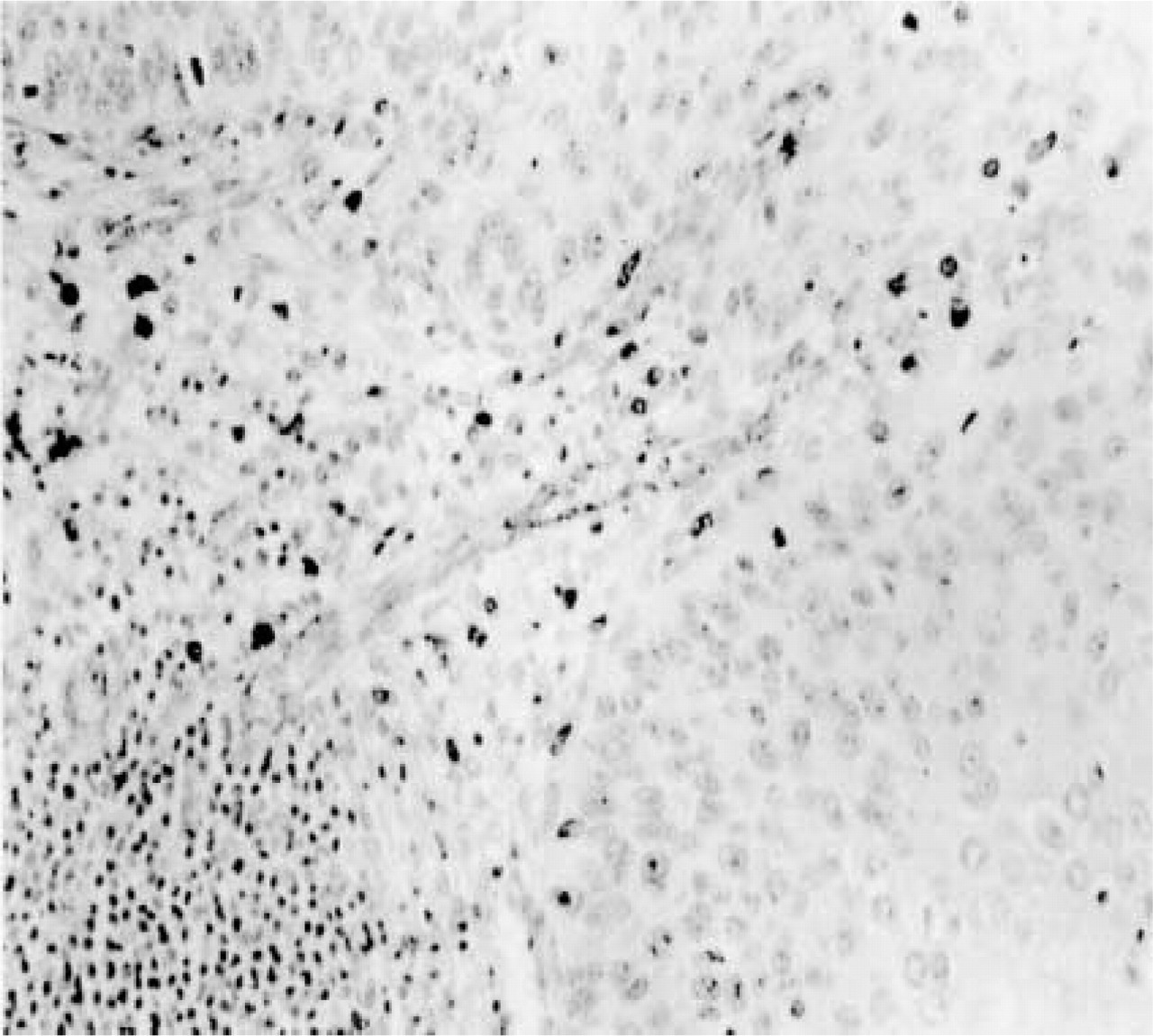
Invasive carcinoma of the cervix with abundant capillaries in the stroma and tryptase-positive mast cells around blood vessels. Double immunocytochemical staining for Factor VIII and tryptase. Original magnification ×400.