
Abstract
Objective:
People with chronic schizophrenia have high rates of physical ill-health such as heart disease. However, there has been less attention to the issue of poor oral health including dental caries (tooth decay) and periodontal (gum) disease, although both have consequences for quality of life and systemic physical health. We therefore measured tooth decay and gum disease in Malaysians with schizophrenia.
Methods:
We recruited long-stay inpatients with schizophrenia from June to October 2014. Four dental specialists assessed oral health using the decayed–missing–filled teeth index, the Community Periodontal Index of Treatment Needs and the Debris Index of the Simplified Oral Hygiene Index. Results were compared with the 2010 Oral Health survey of the general Malaysian population.
Results:
A total of 543 patients participated (66.7% males, 33.3% females; mean age = 54.8 years [standard deviation = 16.0]) with a mean illness duration of 18.4 years (standard deviation = 17.1). The mean decayed–missing–filled teeth was 20.5 (standard deviation = 9.9), almost double that of the general population (11.7). Higher decayed–missing–filled teeth scores were associated with both older age (p < 0.001) and longer illness duration (p = 0.048). Only 1% (n = 6) had healthy gums. Levels of decay and periodontal disease were greatest in those aged between 45 and 64 years, coinciding with the onset of tooth loss.
Conclusion:
Dental disease in people with schizophrenia deserves the same attention as other comorbid physical illness. The disparity in oral health is most marked for dental decay. Possible interventions include oral health assessments using standard checklists designed for non-dental personnel, help with oral hygiene, management of iatrogenic dry mouth and early dental referral.
Keywords
Background
People with schizophrenia are known to have high rates of physical ill-health including heart disease, diabetes and cancer (Kisely et al., 2013, 2015; Montejo, 2010; Monteleone et al., 2009). By contrast, the presence of conditions such as dental caries and periodontal disease receives much less attention (Arnaiz et al., 2011). If left untreated, these conditions can lead to partial or total tooth loss (edentulism), as well as compromising nutrition and general physical health.
This population is at greater risk of dental disease because of poor nutrition and oral hygiene; the heavy consumption of sugary drinks; comorbid substance misuse including tobacco, alcohol or psychostimulants; and financial or other barriers to accessing dental care (Kisely et al., 2014). This is aggravated by dry mouth (xerostomia), which is a common side effect of many psychotropic medications (Bardow et al., 2001).
Dental disease can have important consequences. Aside from social withdrawal and low self-esteem, poor oral health is associated with chronic medical conditions such as myocardial infarction and stroke (Azarpazhooh and Leake, 2006; Chapple, 2009; Cullinan et al., 2009; Desvarieux et al., 2003; Haumschild and Haumschild, 2009; Humphrey et al., 2008; Rai, 2006; Scannapieco, 2005; Shultis et al., 2007; Williams et al., 2008). One explanation is that poor oral hygiene allows oral bacteria to enter the bloodstream. Immune complexes are then formed that, in turn, elicit inflammatory responses in arteries and distal organs (Scannapieco, 2005).
Where studies of oral health in people with schizophrenia have been conducted, they have largely come from Europe, North America, Israel and Australia (Chu et al., 2011; Gurbuz et al., 2010; Kisely et al., 2014; Lalloo et al., 2013; Zusman et al., 2010). Any papers from South East Asia have been restricted to developed countries such as Japan, Hong Kong and Taiwan (Kisely et al., 2011, 2014; Tani et al., 2012; Teng et al., 2011). In addition, the literature has focused primarily on caries rather than periodontal disease, although both contribute to edentulism.
This study therefore examined the oral health status of a large cohort of people with schizophrenia in a developing South Eastern Asian country. Importantly, it included all measures of periodontal disease, dental decay and oral health needs. It was conducted in one of the largest psychiatric hospitals in Asia, the Permai Psychiatric Institution in Malaysia. In contrast to other countries, large psychiatry institutions are the setting where most Malaysians with schizophrenia are treated.
Results were compared with those of the 2010 Malaysian Oral Health survey of the adult population (Ministry of Health, 2010). This is a population-based survey of dental decay and periodontal disease that is carried out every 10 years across Malaysia (Mohd-Dom et al., 2013; Umer and Umer, 2011). In the last survey, 8332 dentate adults were examined (Mohd-Dom et al., 2013). We hypothesized that the rates and extent of dental disease in people with schizophrenia would be significantly greater than those of the general Malaysian population.
Methods
Setting
Malaysia is a developing middle-income country as classified by the World Bank (2015). Mental health care in Malaysia is mainly provided in government-run institutions, although the number of private hospitals is increasing. Despite a growing number of psychiatric units in general hospitals, inpatient care is still largely provided in four large-scale institutions (Parameshvara Deva, 2004). This study was conducted at the Permai Psychiatric Institution, one of the four institutions in Malaysia. Importantly, it took place in an area where fluoride was present in the water supply at a concentration of 0.5–0.7 mg/L. This is only just below Australian guidelines for fluoridation of between 0.6 and 1.1 mg/L to achieve the right balance between the reduction of dental caries and the prevention of fluoride-related harm (National Health and Medical Research Council, 2007).
Subjects
Upon ethical clearance, four dental specialists carried out the dental examinations, over a period of 5 months in late 2014. We included all long-stay patients (18 to 65 years of age) with a diagnosis of schizophrenia (based on the Diagnosis and Statistical Manual of Mental Disorders, 5th edition [DSM-5] and who were not on leave. The diagnosis was by clinical assessment as opposed to a structured interview. For those unable to provide written consent, a surrogate (e.g. the next of kin or legally authorized representative) gave consent on their behalf. Exclusion criteria were patients who were acutely unwell or presenting with a first episode of schizophrenia, as well as those with substance dependence and medical disorders such as epilepsy or cerebrovascular problems. We also excluded patients with comorbid learning disability and those at risk of harming themselves or others. Psychiatric status was determined by clinical assessment of the treating team.
Ethical clearance was obtained from the Ethical Board of the University of Malaya’s Medical Centre (UMMC-MEC, Ref. No.: 926.63) and Ministry of Health (NMRR 13-310-15297). Eight trained research staff with backgrounds in occupational therapy or nursing, and who were fluent in Malay, Chinese or Tamil, approached potential subjects to obtain written consent. Clinical staff assisted in assessing patients’ capacity for informed consent and explaining the dental examination. About 20% of the sample (n = 110) were unable to provide written consent. They came mainly from the long-stay psychogeriatric wards, and in these cases, a surrogate such as a family member or legally authorized representative gave consent on their behalf.
Data collection
Demographic data such as age, gender, ethnicity and duration of illness were obtained from hospital records and medical staff. Psychiatric status was determined by clinical assessment of the treating health team.
Four dental specialists carried out the dental examinations over a period of 5 months in late 2014. Three examiners were calibrated against one specialist who had conducted several national surveys for the country. This was done through repeated examinations of a separate pilot sample using similar indices, followed by meetings to discuss discrepancies and standardize procedures. Kappa scores of 0.9 for inter-rater agreement were achieved. The following three tools were used in this study:
The Decayed–Missing–Filled (DMFT) score. Decay was assessed using this standardized index to evaluate dental caries with scores ranging from 0 to 32. This involved examination for dental caries, dental restorations, fissure sealants, fixed prosthesis, missing teeth and an additional category for traumatized incisors. High scores indicate worse dental health (World Health Organization [WHO], 1987, 2013). Root caries was not assessed in this study.
The Community Periodontal Index of Treatment Needs (CPITN) Index (Ainamo et al., 1982). This measure was used to score periodontal status and assess patients’ treatment needs. The initial sign of periodontal disease is bleeding of the gums. As it progresses, the gum retracts from the base of the tooth to form pockets. The deeper the pocket, the more severe is the periodontal disease. The CPITN is therefore a 5-point scale as follows: no signs of periodontal disease (0), gingival bleeding after gentle probing (1), supragingival or subgingival calculus (2), pathologic pockets 4–5 mm deep (3) and pathologic pockets >5 mm deep (4). Disposable dental mirrors, tweezers, 0.5 mm ball-ended CPITN probes (3.5–5.5 mm) and cotton rolls (to remove plaque) were used.
The Debris Index component of the Simplified Oral Hygiene Index (OHI-S). This tool was used to measure the amount of debris (Greene and Vermillion, 1964). It was scored on six tooth surfaces per participant.
Statistical analyses
We used t-tests to compare findings of this study with the Oral Health survey of adults (Ministry of Health, 2010). A bivariate model was then used to examine the effects of the following variables on the DMFT score: age (divided into 10-year groups), gender, ethnicity and duration of illness. Next, we conducted stepwise multiple linear regression of DMFT and multinomial logistic regression of CPITN with age, gender, race and duration of illness as independent variables to determine factors associated with dental outcome. Pair-wise comparisons were used to compare the effect of subgroups on DMFT scores.
Results
Demographics of the sample
Figure 1 shows the recruitment flow chart. Of the 545 eligible cases who fulfilled the inclusion criteria, 543 (99.6%) participated in the study. Table 1 shows their socio-demographic characteristics. About two-thirds (n = 362) were males and 33.3% (n = 181) females. Of the sample, 42.0% (n = 228) were Malay, 50.5% (n = 274) Chinese, 5.3% (n = 29) Indian, 0.7% (n = 4) indigenous Orang Asli, 0.6% (n = 3) non-Malaysians and 0.9% (n = 5) of undetermined ethnicity. The mean age was 54.8 years (standard deviation [SD] = 16.0), ranging from as young as 17 years of age to as old as 91 years old. The sample was predominately middle aged or older (Table 1). The mean duration of psychiatric illness was 18.4 years (SD = 17.1) with a range of 2–61 years. The average length of stay was 23 years ranging from less than a year to 54 years.
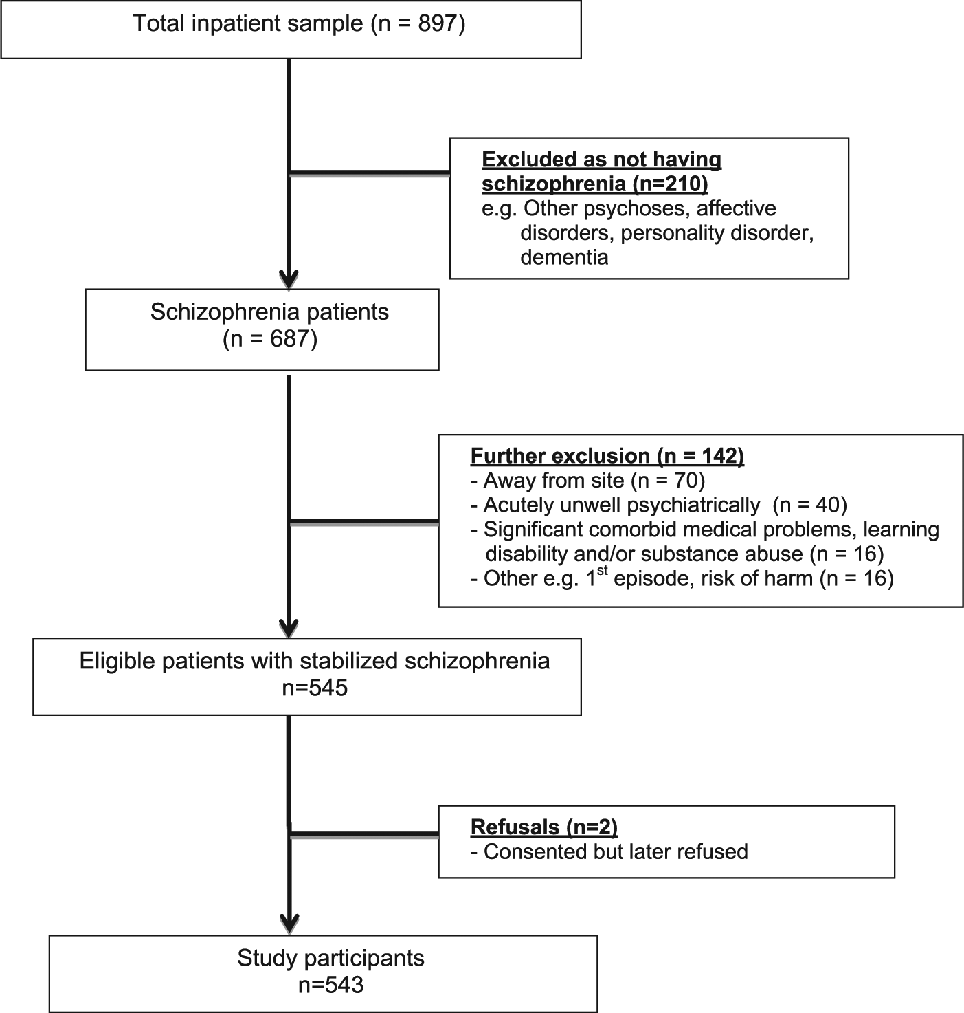
Patient recruitment flow chart.
Demographics of study participants (n = 543).
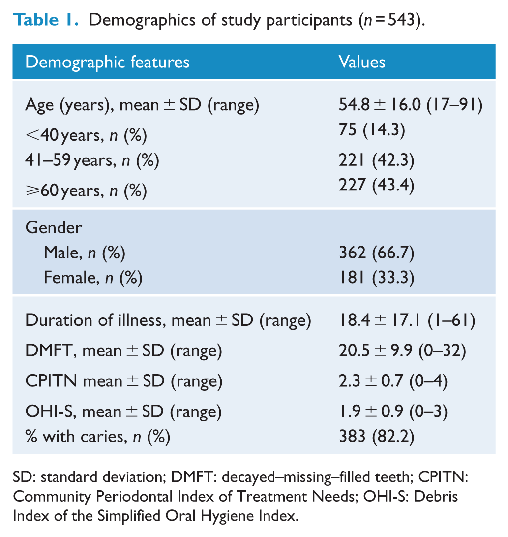
SD: standard deviation; DMFT: decayed–missing–filled teeth; CPITN: Community Periodontal Index of Treatment Needs; OHI-S: Debris Index of the Simplified Oral Hygiene Index.
Dentition status (DMFT scores) and associated demographic characteristics
The mean DMFT score for the 543 participants was 20.5 (SD = 9.9), significantly higher than the scores for the general population of 11.7 (Ministry of Health, 2010). Male patients had a mean DMFT of 19.9 (SD = 10.1), while females had a score of 21.7 (SD = 9.4). Population variances were equal, suggesting homogeneity between gender (p < 0.075). Stepwise multiple linear regression analysis of DMFT using independent variables of age, gender, race and duration of illness showed that gender (p = 0.488) and race (p = 0.380) were not associated with DMFT scores. However, older age (p < 0.001) and longer duration of illness (p = 0.048) were significantly associated with increasing scores. Subgroup analyses showed that DMFT scores were significantly higher than the normal population across most ages (Ministry of Health, 2010) (p = 0.012) (Table 2).
DMFT comparison between study participants and the general population by age.
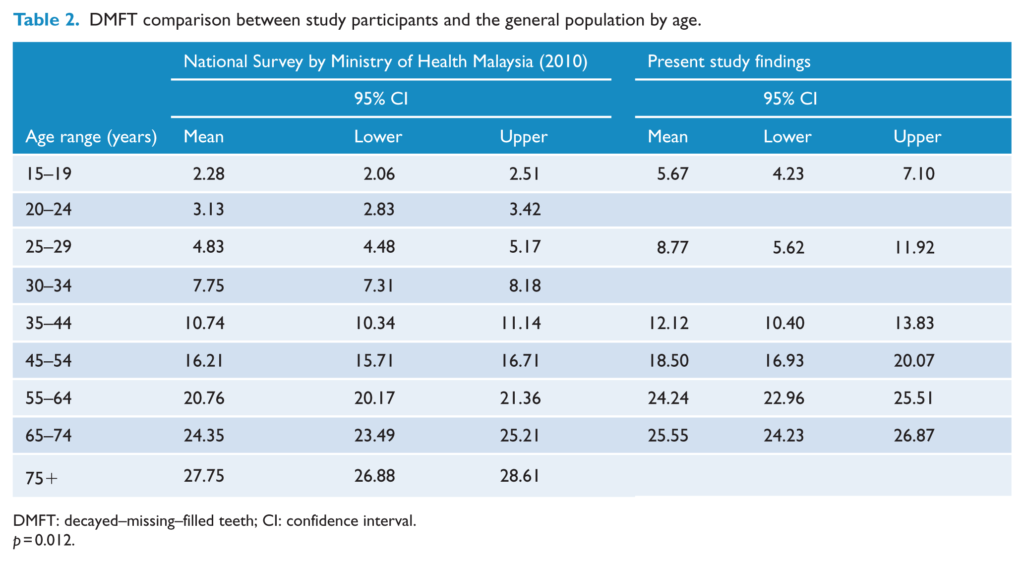
DMFT: decayed–missing–filled teeth; CI: confidence interval.
p = 0.012.
Periodontal disease and debris
Only 1% (n = 6) had healthy gums whereby all six sextants were free of pathology. By comparison, it was 3.2% in the general adult population (Ministry of Health, 2010). Half of the sample (n = 274) had gingival bleeding in at least one sextant. The mean number of sextants with gingival bleeding was 1.5 (SD = 1.9) per person compared with 1.8 in the general population (Ministry of Health, 2000). Of the total sample (n = 543), 47.7% (n = 256) had calculus, 19.2% (n = 104) shallow pocketing of 4–5 mm and 6% (n = 33) deep pockets measuring 6 mm and more. Severity was largely similar to that of the general population, except for the mean number of deep pockets (at 0.10) which were lower in the study population (at 0.37) (Table 3). CPITN scores were also similar to those of the normal population, with p-values ranging from 0.27 to 0.95 for the various age groups (Table 4).
Periodontal severity comparison between study participants and the general dentate population by age.
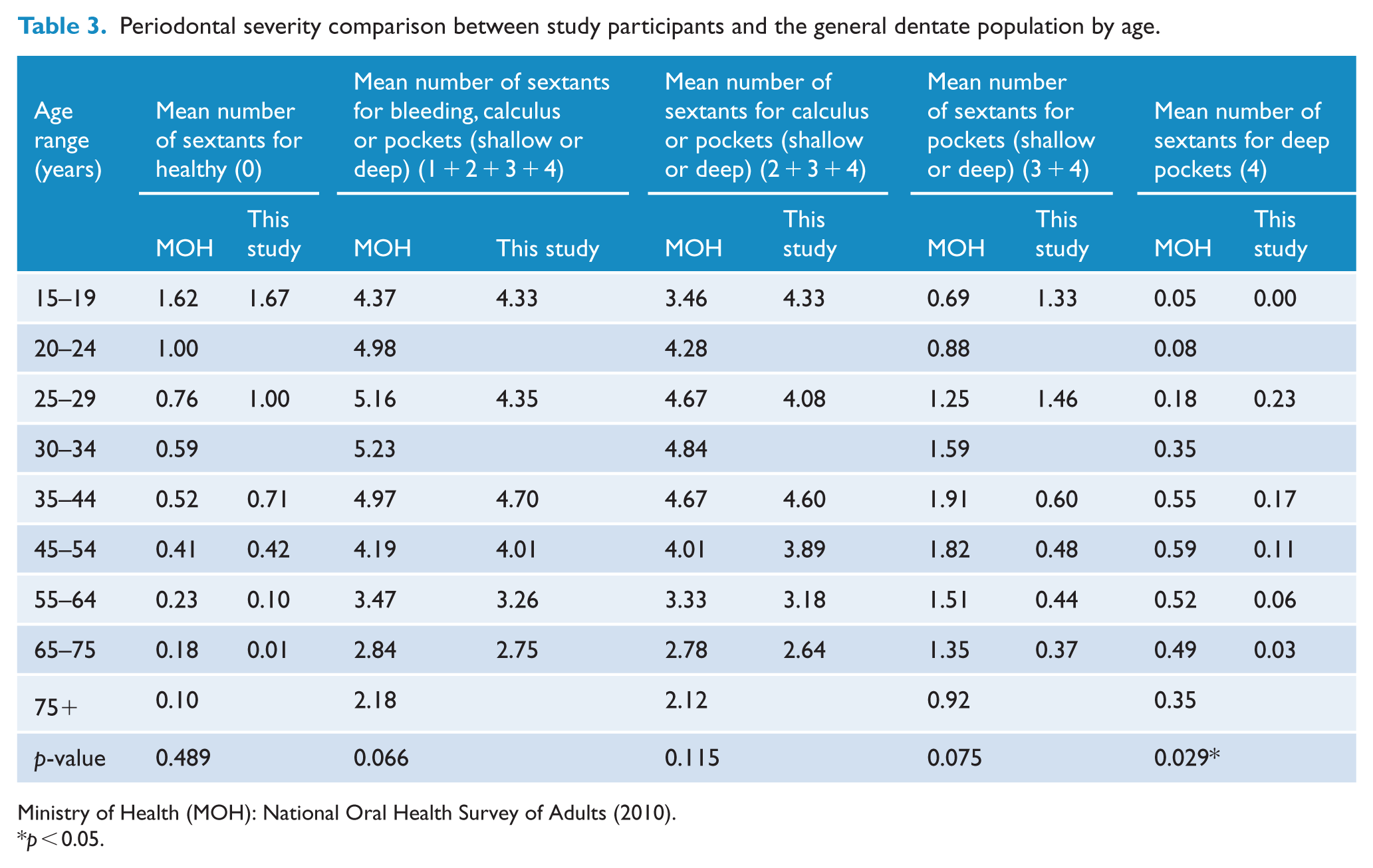
Ministry of Health (MOH): National Oral Health Survey of Adults (2010).
p < 0.05.
CPITN comparison between study participants and the general population by age.
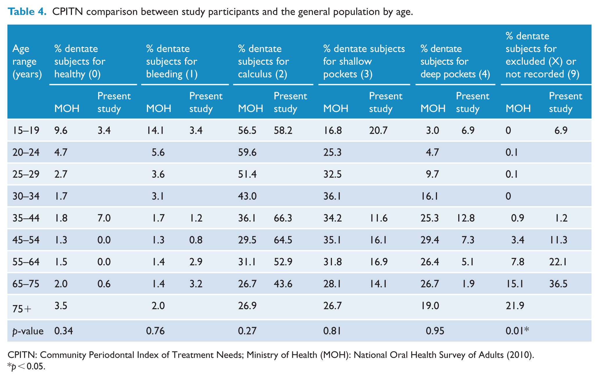
CPITN: Community Periodontal Index of Treatment Needs; Ministry of Health (MOH): National Oral Health Survey of Adults (2010).
p < 0.05.
Multinomial logistic regression analysis showed that patients in the age range of 45–64 years were more likely to have gingival bleeding, calculus or pockets (p < 0.001). In comparison with Malays, Chinese patients were more likely to have bleeding on probing, calculus and pockets (p ⩽ 0.001), while Indians were more likely to have calculus (p ⩽ 0.026) and shallow pockets (p ⩽ 0.003). The mean debris index was 1.9 (n = 441).
Partial and complete edentulism
The mean number of permanent teeth present per person was 15.5 (SD = 11.0), compared to 22.9 of the general population (Ministry of Health, 2010) (p = 0.77). The proportion of edentulism was 14.2% (n = 77) as opposed to 7.3% of the general population (p < 0.0001). The percentage of subjects with at least one natural tooth was 85.8% (n = 466) as compared to 92.7% of the general population (p < 0.0001). In all, 43% of the patients (n = 234) had at least 20 functional teeth compared to 80.6% of the general population, a statistically significant result (p = 0.04) (Ministry of Health, 2010) (Table 5). Of the sample, 70.5% (n = 383) had at least one carious permanent tooth or carious restoration, with the highest prevalence occurring in 45- to 54-year-olds (82.3%) and the lowest in 15- to 24-year-olds (33.3%) (Table 6).
Tooth loss comparison between study participants and the general population by age.
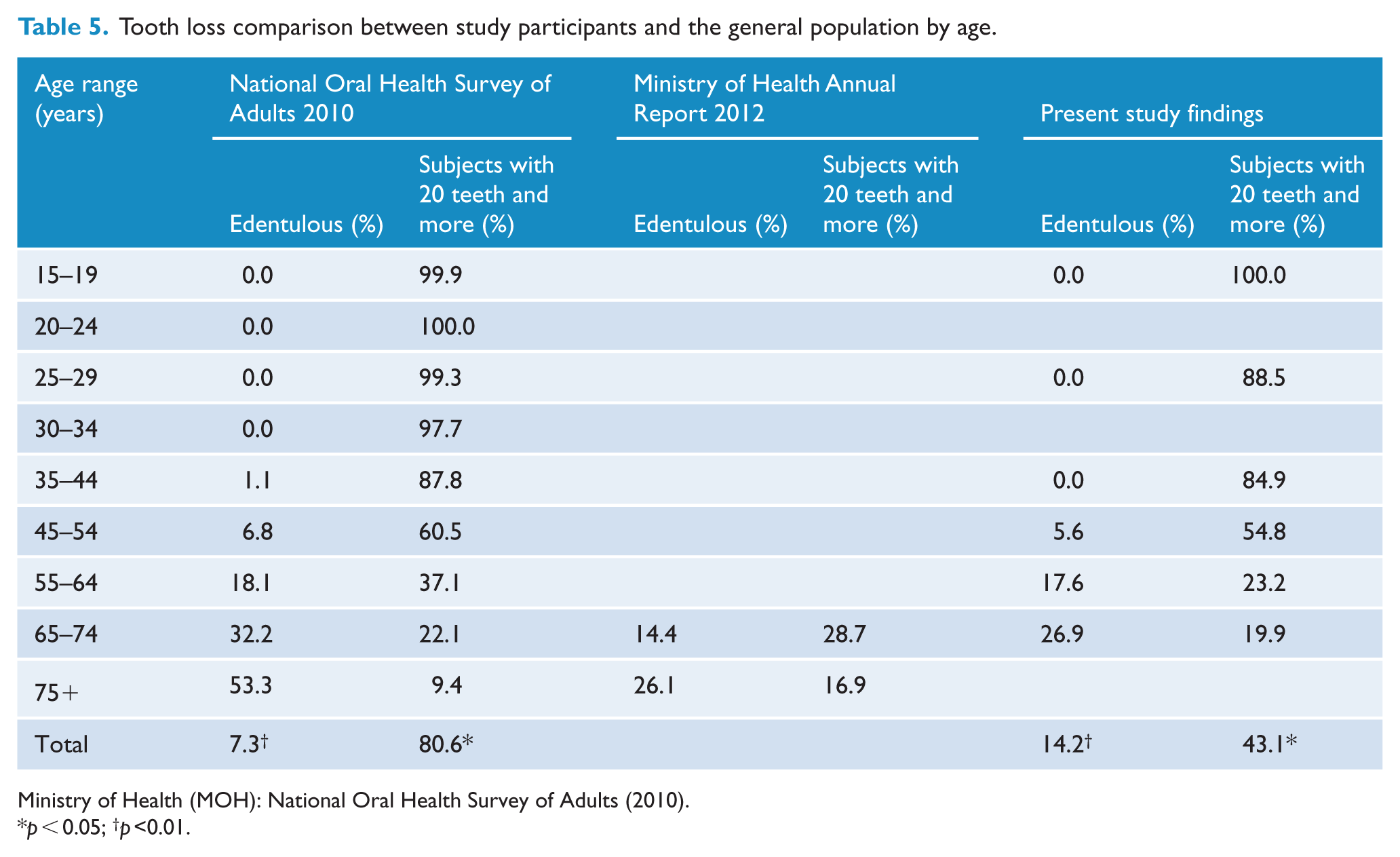
Ministry of Health (MOH): National Oral Health Survey of Adults (2010).
p < 0.05; †p <0.01.
Caries prevalence comparison between study participants and the general population by age.
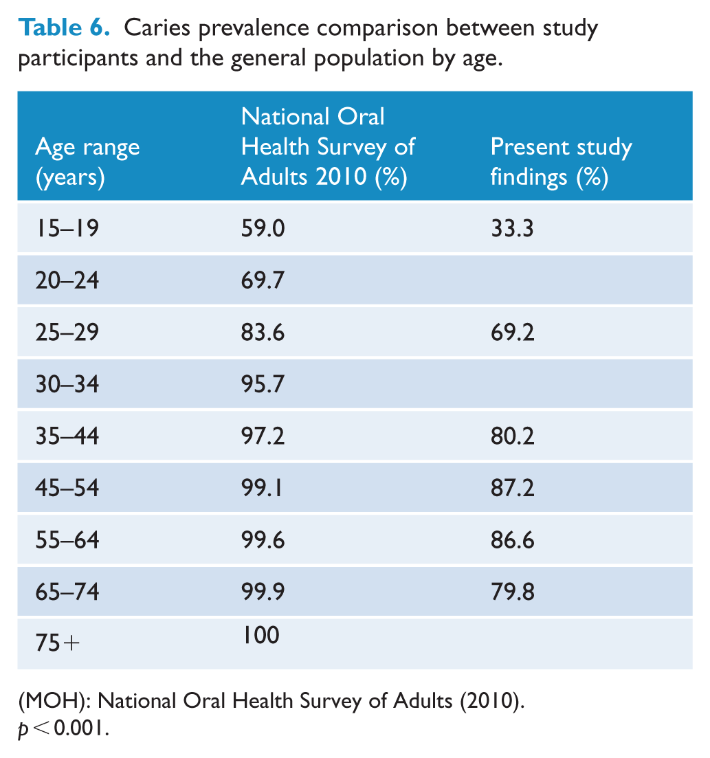
(MOH): National Oral Health Survey of Adults (2010).
p < 0.001.
Discussion
This is one of the few studies to assess the oral health status of people with schizophrenia in a developing country and the first such study in South East Asia. Previous studies from the region have been restricted to Japan, Hong Kong and Taiwan (Kisely et al., 2011, 2014; Tani et al., 2012; Teng et al., 2011).
Malaysia is also a particularly appropriate setting as it conducts 10-yearly oral health surveys of the general population including the measurement of periodontal status (Mohd-Dom et al., 2013; Umer and Umer, 2011). This therefore allows comparisons between people with schizophrenia and the general population for both decay and periodontal disease.
Strengths of the study include the use of standardized instruments by trained dental personnel who were calibrated against an expert. We were also able to stratify results by age in our comparisons between the hospital sample and the population controls. This is important as oral health changes as patients grow older. Although a majority of the hospital sample were male, it is unlikely that differences in the gender distribution between patients and community controls would have affected the results given that the prevalence of dental disease does not vary greatly by gender. Where differences have been reported, females have worse dental disease than males (Australian Institute of Health and Welfare [AIHW] Dental Statistics and Research Unit, 2008a, 2008b; Krustrup and Petersen, 2007; Palmqvist et al., 2000).
There were several limitations. Although psychiatric diagnoses were made using diagnostic criteria, this followed a clinical assessment rather than a structured interview. Use of the Malaysian national oral survey as the control population meant that although it was possible to stratify results by age, we were unable to take into account differences in other socio-demographic characteristics such as education and socio-economic status that could also have an effect on oral health status. In addition, the dental assessors were not blind to patients’ psychiatric status. Data on the prevalence of smoking were not collected. However, signs of tobacco use were commonly observed during the dental examinations, especially in male participants. Finally, the results may have limited generalizability to shorter stay community-based settings.
Dental decay in patients with schizophrenia was much higher than in the general Malaysian community but consistent with findings from other psychiatric populations in developed countries such as the United Kingdom (Lewis et al., 2001), France (Bertaud-Gounot et al., 2013), Italy (Angelillo et al., 1995), Spain (Arnaiz et al., 2011; Velasco-Ortega et al., 2013), Australia (Lalloo et al., 2013) and Israel (Ramon et al., 2003; Zusman et al., 2010) (Table 7). DMFT findings by age groups were also similar to those from Japan (Tani et al., 2012) and Taiwan (Teng et al., 2011). By contrast, these results from Malaysia were much higher than the levels of decay found in psychiatric patients in other developing nations such as Ethiopia (Kebede et al., 2012) and India (Kumar et al., 2006). The prevalence of caries was highest in the age range of 45–54 years, and this corresponded with the onset of edentulism.
International comparisons of DMFT scores.
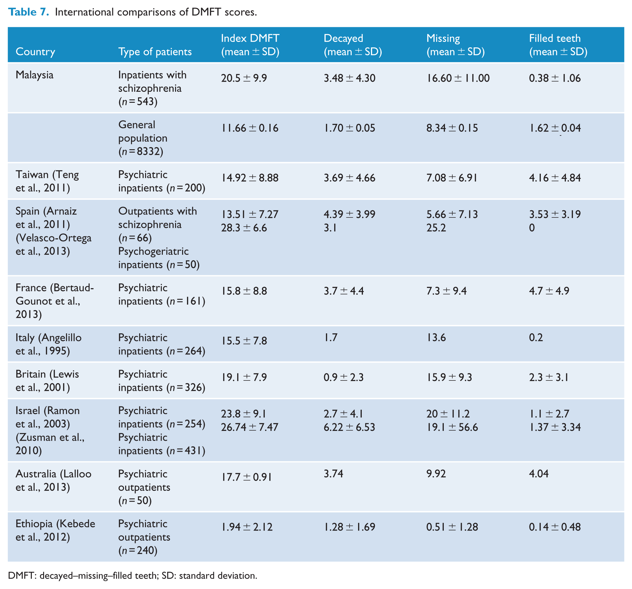
DMFT: decayed–missing–filled teeth; SD: standard deviation.
Explanations for increased levels of decay include poor oral hygiene and the side effects of psychotropic medications like antipsychotics, antidepressants and mood stabilizers. All of these medications induce dry mouth (xerostomia) through reduced salivary flow and can potentially negate the beneficial effects of fluoride (Friedlander and Marder, 2002). As with other aspects of physical ill-health, poor dental health may also be related to poor diet, smoking and poor oral hygiene (McCreadie et al., 2004). It is possible that the increased dental decay in Malaysian patients with respect to other developing countries might be due to differences in diet, access to psychotropic medication, service organization or levels of development. This is discussed further under section ‘Implications’.
These issues are compounded by difficulties with access to dental care, either because of availability or because of fear of pain and dental phobia. Although there is a dental clinic in the hospital, the dentist only attends twice a week to treat those with serious dental caries. With a total patient population of almost 1000, optimal dental care is therefore difficult to achieve and limited to the treatment of established dental disease rather than the promotion of oral health.
By contrast, levels of periodontal disease in this sample were very similar to those in the general Malaysian population and lower than reported among inpatients with schizophrenia from Taiwan (Teng et al., 2011). Where present, it was most likely to occur between the ages of 45 and 64 years. The reasons for this disparity are unclear and might be related to differences in diet or service organization between the two jurisdictions. Differences in smoking levels could also contribute. While smoking is a significant risk factor in the development and progression of periodontal disease overall, it may also diminish gingival bleeding in the short-term through changes in the proportion of small to large blood vessels in the gums (Rivera-Hidalgo, 2003). A further explanation for lower than expected levels of periodontal disease might be that different bacteria are responsible for decay and periodontitis. The former is primarily due to Streptococcus mutans while a wider range of aerobic and anaerobic bacteria are involved in gum disease (Loesche, 1996). It is therefore possible that the preponderance of one group of organisms over the other might affect the degree of decay or periodontal disease.
Implications
This study highlights the poor oral health of long-term patients in one of Asia’s largest psychiatric institutions in terms of dentition (DMFT), periodontal status (CPITN) and debris index. Rates of dental decay in this sample from an upper middle–income developing country were comparable to those of high income jurisdictions such as Australia, Japan, Korea and the European Union and found to be higher than those from low and lower middle countries (Kebede et al. 2011, 2012, 2014; Kumar et al., 2006). Developing countries should not therefore be seen as a homogeneous group when extrapolating results from one country to another.
Our findings also illustrate the need for a greater emphasis on preventative care, rather than solely treating established dental symptoms. Interventions should be initiated well before the onset of tooth loss, which in this population was in 45 years and above. This should be complemented by the promotion of oral hygiene in all ages including the avoidance of sweets and tobacco, as well as learning the correct techniques for brushing teeth. An example from Australia is the ‘Dental as anything’ programme in Victoria (Burchell et al., 2006). This is an assertive outreach collaboration between mental and oral health services where teams take mental and oral health services to ‘hard-to-reach’ settings such as rooming houses and supported housing. Interventions include both education and clinical care. In another example, Queensland’s strategy to improve the physical health of people with severe mental illness (Activate: Mind & Body) includes both the promotion of oral hygiene and regular care from a dentist (General Practice Queensland, 2009).
Oral health is also an important part of comprehensive care for people in long-term residential or institutional facilities. Nursing care plans should include the recording of factors known to cause oral ill-health such as psychotropic medication and tobacco or substance use, as well as the supply of tooth brushes and denture baths. There are also simple assessment tools that can be used by non-dentists. An example is the Oral Health Assessment Tool, which has been evaluated as being reliable and valid in a study of Australian residential care facilities, including those with cognitively impaired residents (Chalmers et al., 2005). This involves assessment of the lips, tongue, gums and teeth, as well as asking about dentures and the presence of dental pain. The contribution of psychotropic medication to xerostomia should also be considered when prescribing.
In conclusion, the oral health of psychiatric patients has been neglected for too long. The management of comorbid dental diseases should attract the same attention and receive the same priority as other comorbid physical illness. This calls for greater collaboration between dental and non-dental staff to improve the quality of life of people with schizophrenia.
Footnotes
Acknowledgements
The authors thank the inpatients who participated in the study. We are indebted to the occupational therapy and nursing staff at the Tampoi Psychiatric Institution and the two dental officers who contributed to the data collection for this study. S.Y.L. and A.K.A.B. conceived the initial study; W.M.C and J.G.D. participated in its design and carried out the oral examinations with the support from S.Y.L. and A.K.A.B; A.K.A.B. and Tampoi’s medical staff assessed the inpatients based on Diagnostic and Statistical Manual of Mental Disorders, 5th Edition (DSM-5) inclusion criteria; W.M.C. performed the statistical analysis; and W.M.C., S.Y.L. and S.K. drafted and refined the manuscript. All authors read and approved the final manuscript.
Declaration of interest
The author(s) declared no potential conflicts of interest with respect to the research, authorship and/or publication of this article.
Funding
The author(s) disclosed receipt of the following financial support for the research, authorship, and/or publication of this article: This study was funded by the University of Malaya (UM.C/625/1/HIR/MOHE/MED/26) with SYL as the PI.