
Abstract
Mesothelioma is an aggressive cancer of pleural and peritoneal cells that is difficult to diagnose and monitor. Numerous studies have attempted to identify a blood- or pleural fluid-based biomarker that could be used in the diagnostic pathway. More recently, there has been interest in the ability of serum/plasma biomarkers to monitor mesothelioma, given the development of newer treatments and limitations of radiological assessment. The majority of research has focused on soluble mesothelin, a soluble glycoprotein expressed by mesothelial cells. Although soluble mesothelin lacks the sensitivity to be used as a standalone diagnostic marker, serial measurements may be informative, with rising concentrations indicating disease progression and poor survival. High concentrations of other soluble glycoproteins, such as osteopontin, fibulin-3 and vascular endothelial growth factor are independently associated with poor prognosis at baseline, although further research is required to ascertain any role outside of clinical trials. More recent literature has focused on the development of novel biomarkers from discovery cohorts. Although many DNA and mRNA biomarkers show promise in the diagnosis or screening of mesothelioma, none have been prospectively evaluated for use in clinical practice. In this review article, we highlight the potential utility of biomarkers and evaluate the existing literature.
Keywords
Background
Malignant mesothelioma is an aggressive and invariably fatal cancer of pleural and peritoneal cells (ratio 4:1), and less commonly of the pericardium and tunica vaginalis. 1 The incidence of mesothelioma is increasing worldwide to the extent that it is now commoner than cancers of the bladder and bone. Mesothelioma is almost exclusively caused by exposure to asbestos, a link that was first published by Wagner et al., 2 a pathologist in South Africa. He noticed that the incidence of pleural mesothelioma, a previously rare cancer, was increasing in areas of the Cape asbestos field which mined Cape Blue (crocidolite asbestos). The direct causal link between asbestos use in industry and mesothelioma allows for the future incidence of the disease to be predicted with some degree of accuracy. Given a mean latency of around 40 years from peak exposure, 3 it is estimated that the incidence of mesothelioma in Europe will rise until between 2015 and 2020. 4 Given ongoing unregulated use of asbestos in countries such as China, India and Russia, mesothelioma will continue to occur, despite unequivocal evidence of its harms. The other rarer causes of mesothelioma are iatrogenic chest wall irradiation (e.g. in treatment of breast cancer or lymphoma) and exposure to erionite (a mineral found in Turkey).5–8
Several different pathogenetic mechanisms have been proposed to explain the link between asbestos exposure and mesothelioma. In the first, long (>5 µm) thin asbestos fibres are inhaled into the lung, penetrating the lung epithelium and entering the pleural space; then, a continuous cycle of pleural irritation, damage and repair eventually results in the mutations that give rise to mesothelioma. In the second (the oxygen free radical hypothesis), phagocytosis of asbestos fibres is associated with release of oxygen free radicals that cause DNA damage and mutations. 9 Third, asbestos fibres are known to penetrate mesothelial cells and interfere with mitosis, as well as inducing phosphorylation and production of various pro-oncogenic protein kinases (mitogen-activated protein and extracellular signal-regulated kinases 1 and 2). Finally, mesothelial cells release inflammatory tumour growth factor-β, platelet-derived growth factor and vascular endothelial growth factor (VEGF), all of which can be utilized by malignant cells for proliferation and angiogenesis. 4 It is likely that host-specific factors also contribute to the pathogenesis of mesothelioma.
There are four main histological subtypes of mesothelioma: epithelioid; sarcomatoid; biphasic or mixed; desmoplastic. These have different microscopic appearances and prognostic implications. Epithelioid is the commonest (accounting for around 70% of cases in most series) and has the most favourable prognosis, with a median survival of 13.1 months.10,11 The sarcomatoid subtype has the poorest prognosis, with a median survival of just four months. The histological subtype determines the treatment offered by oncologists or surgeons; more aggressive subtypes are felt to be not amenable to therapy.
Clinical presentation
The majority of patients with malignant pleural mesothelioma will present with shortness of breath, cough or chest pain. Patients less commonly present with systemic symptoms of weight loss, night sweats and fatigue; if they do, this is a poor prognostic sign, since the disease is likely to be more advanced. 12 An abnormal chest radiograph, performed routinely before an operation, may be the presenting complaint, in patients without respiratory symptoms. A chest radiograph demonstrates a pleural effusion (fluid collection between the lung and chest wall) in 90% of cases; however, this radiological sign is commonly found in many malignant and non-malignant respiratory conditions. 13 However, in patients who present with an abnormal chest radiograph, and known past asbestos exposure, malignant pleural mesothelioma should be suspected. Even if the patient denies previous exposure to asbestos, the diagnosis of mesothelioma should be excluded, given the risk of ‘second-hand’ or non-industrial exposures.
Symptoms from local spread can occur, including obstruction of the superior vena cava, rib destruction and laryngeal nerve palsy. However, clinical manifestations of metastatic spread are uncommon, due to the aggressive nature of the primary disease. 1 At postmortem, most common areas of spread include the thoracic lymph nodes and bone. However, tract metastases, in areas where the chest wall has been operated on for diagnostic or therapeutic purposes, are commoner. These metastases can be disfiguring and/or painful, and research is ongoing into the role of prophylactic radiotherapy following pleural procedures.14,15
Peritoneal mesothelioma presents very differently to pleural disease, commonly with diffuse abdominal pain, abdominal swelling from disease bulk or ascites (fluid accumulation in the abdominal cavity), bowel obstruction, appetite loss or nausea.
Imaging for mesothelioma
At initial presentation, the majority of patients with pleural mesothelioma will have a unilateral pleural effusion on chest radiograph (Figure 1(a)). A minority will have pleural thickening, and fewer still will have evidence of advanced pleural disease with a widespread pleural rind (Figure 1(c)). In most cases, an unexplained unilateral pleural effusion on chest radiograph will lead to a computerized tomography (CT) scan of the chest, abdomen and pelvis with the intention of identifying or excluding malignancy. Diagnosing mesothelioma on CT scan alone is difficult at early stage of disease given the likely presence of pleural fluid, asbestos-related plaques, or folded lung, which can obscure the assessment of the pleura, as well as the difficulty in distinguishing benign pleural thickening from malignancy.
16
Even if the CT scan looks suspicious of malignancy, it can still be challenging to distinguish mesothelioma from pleural metastasis from, e.g. lung, breast, ovary or kidney.
17
CT scanning is currently used to monitor disease following diagnosis, e.g. response to chemotherapy, assessing progression, etc. However, similar challenges apply, and, given that mesothelioma does not grow as a spherical mass, but rather as a pleural or peritoneal rind (Figure 1(b)), it can also be challenging to quantify any change in tumour bulk. Scoring systems that have been modified for mesothelioma’s unique morphology exist but have their limitations.
18
Imaging modalities in mesothelioma. (a) Chest radiograph showing large right-sided pleural effusion. (b) Coronal CT image showing right-sided pleural thickening and nodularity. (c) Chest radiograph showing left-sided pleural thickening and lung volume loss. (d) Horizontal PET image showing right-sided pleural enhancement posteriorly.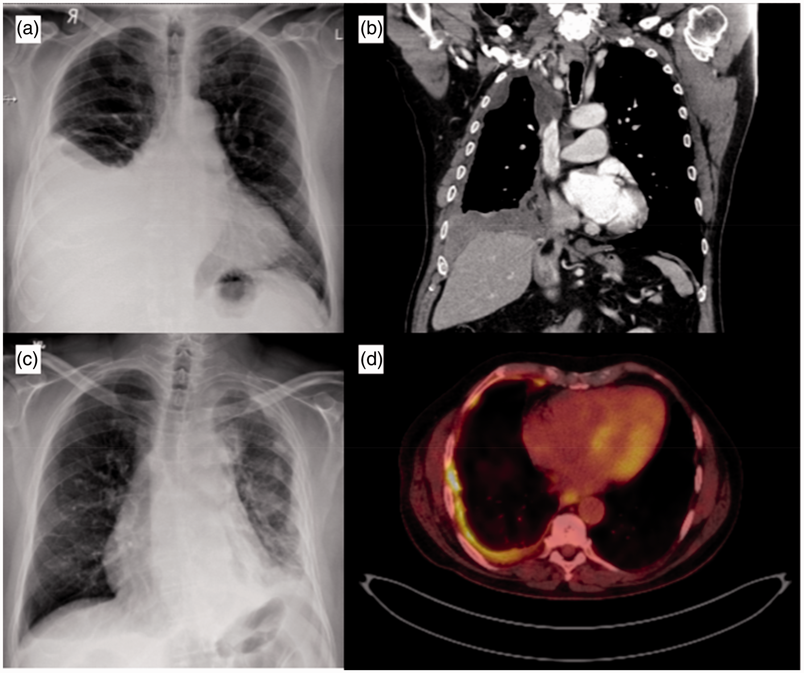
Magnetic resonance imaging (MRI) is developing as a method of diagnosing and staging mesothelioma. Recent studies using diffusion-weighted imaging have shown that MRI is able to differentiate benign from malignant pleural disease with high accuracy. 19 In addition, it may be possible to differentiate between different subtypes of mesothelioma at initial assessment. 20 Similarly, positron emission tomography-computed tomography (PET-CT) imaging is a rapidly developing area in the diagnosis and staging of mesothelioma. It relies on contemporaneously-acquired CT imaging and assessment of uptake of fluorine 18-flurodeoxyglucose (FDG) into tissues. Uptake of FDG is usually higher in more metabolically active malignant tissue and so appears more vivid on imaging (Figure 1(d)). 21 The advantage of PET-CT over standard CT or MRI is the ability to detect local and distant spread of disease, and hence it can be more accurately staged; it is therefore utilized in patients being considered for surgery. 22 However, false positives have been found due to pleural infection, inflammation and prior pleurodesis. In addition, it is not widely available outside specialist centres. 23
Histocytological investigations
For patients referred to a pleural/respiratory clinic with symptoms or signs suspicious of pleural mesothelioma, the diagnostic pathway will be determined initially by the presence or absence of pleural fluid, usually on radiological imaging; the finding of fluid prompts diagnostic or therapeutic aspiration of pleural fluid (thoracocentesis). Typically, aspirated pleural fluid undergoes analysis in biochemistry, microbiology and cytology laboratories, yielding information about protein and glucose content, culture results and predominant cell types. However, the yield of malignant cells seen on pleural fluid cytology is notoriously low for mesothelioma (10–20%). 24 Consequently, most patients will require further invasive diagnostic investigations in order to obtain tissue, either via ultrasound-guided pleural biopsy, direct visualization using thoracoscopy, or open surgical video-assisted thoracoscopic surgery (VATS). This improves the diagnostic yield to over 90%. 25
Immunohistochemistry
Even once a tissue sample is obtained, diagnosis of mesothelioma may be challenging because of the tumour’s wide range of morphological appearances. Additionally, the pleura and peritoneum are common sites for metastases from other malignancies. Differentiating mesothelioma from adenocarcinoma (from metastases of lung or breast cancer) in a tissue biopsy poses particular difficulties. For these reasons, basing the diagnosis purely on microscopic appearance is not recommended, and various immunohistochemical methods are employed. Although methodology varies widely between centres, guidelines generally advocate a combination of at least two positive mesothelial (calretinin, cytokeratin 5/6, Wilms’ tumour 1, D-240) and at least two negative adenocarcinoma (transcription termination factor 1, carcino-embryonic antigen, Ber-EP4) immunohistochemical markers, for a positive diagnosis of mesothelioma. Clinical and radiological context should also be taken into account.
Treatment of mesothelioma
Given the invasive nature of mesothelioma, for most patients, treatment is palliative from diagnosis. Systemic therapies for mesothelioma have changed little in the last decade. A landmark study in 2003 found that a combination of the anti-folate drug pemetrexed and platinum-based drugs (cisplatin, carboplatin) improved survival in a non-placebo-controlled randomized clinical trial (RCT). 26 This led to the standardization of first-line chemotherapy across the UK, although there is considerable variation in the numbers of patients offered chemotherapy nationally. 27 Despite being the only National Institute for Health and Clinical Excellence (NICE)-approved treatment, this combination only adds an average of two months to overall survival, with a response rate of around 30% 28 and a significant side-effect burden. 29 The role of maintenance or second-line chemotherapy is uncertain. Maintenance pemetrexed is safe, but its efficacy is yet to be established. 30 Several second-line agents have been assessed, with positive results using vinorelbine, gemcitabine or re-challenging with pemetrexed, but no national guidance exists on this currently.31–34
In the future, biological therapy will play a greater role following results from the mesothelioma avastin cisplatin pemetrexed study (MAPS), which demonstrated that the addition of bevacizumab to standard chemotherapy had a survival benefit of two months compared with chemotherapy and placebo. 35 This anti-VEGF monoclonal antibody is not yet approved by NICE, but given this finding, and a number of other promising biological studies, the treatment for mesothelioma is likely to become more complex, with increasing cost implications and emphasis on early identification of treatment response.
Radiotherapy for mesothelioma is employed as a palliative measure, to improve symptoms such as chest wall invasion or procedure tract metastases, or as an adjunct to chemotherapy and surgery. Surgery for mesothelioma is highly controversial. Several centres in the UK offer radical surgery to patients with early-stage disease and good performance status, and case series often report excellent survival. However, these series are usually based on highly selected patients, with sparse randomized data. Several surgical techniques exist as do the neo-adjuvant therapies that accompany them. The mesothelioma and radical surgery (MARS) trial (2011) was one of the first randomized trial of surgery for mesothelioma, and compared extra-pleural pneumonectomy (EPP) (where all macroscopically visible tumour is removed in a large open operation), with no surgery. 36 Although there has been considerable disagreement in the interpretation of the data, the trial concluded that EPP should not be offered to patients and might actually be harmful. Another, more recent, RCT of a less invasive surgical technique (video-assisted thoracoscopic partial pleurectomy or VAT-PP) also demonstrated no improvement in survival or quality of life in the surgical arm. 37 There may be a role for pleurectomy decortication in some patients with mesothelioma and this the subject of a current trial in the UK (MARS2), the results of which are awaited with interest.
Potential role of biomarkers
Diagnosis
Using biomarkers to diagnose mesothelioma is an attractive concept for a number of reasons. First, there is a clear ‘at-risk' population – those who have been previously exposed to asbestos. Second, presenting symptoms and radiological findings are difficult to distinguish from other benign and malignant pleural diseases. Third, even with a high index of suspicion, current methods of histocytological diagnosis are invasive and may be inappropriate in a proportion of patients. Fourth, once tissue is obtained, the tumour can still be difficult to distinguish from other malignancies. Consequently, a large body of research has investigated the ability of serum and pleural fluid biomarkers to diagnose mesothelioma both individually and in panels.
Prognosis or treatment monitoring
As discussed above, for most patients, treatment for mesothelioma is palliative. Any intervention therefore must carefully balance improved life expectancy with quality of life. The standard of care with pemetrexed and cisplatin has a response rate of around 30% with a significant side-effect profile and limited impact on symptoms. Oncologists are understandably very interested in selecting out patients who are likely to respond to chemotherapy at baseline or early in treatment. Additionally, in the few centres that offer surgery, there is considerable preoperative assessment to ensure that only patients who are most likely to benefit are put forward for surgery. However, there are very few radiological markers at baseline that can predict prognosis.
In patients who receive chemotherapy, a CT scan is often performed mid-cycle to assess response. However, as discussed above, mesothelioma is difficult to monitor using conventional CT scanning. A biomarker that could measure response to chemotherapy or predict recurrence would offer considerable advantages.
Mesothelin
Soluble mesothelin (SM) is a 40 kDa cell membrane-bound glycoprotein over-expressed by the epithelioid component of malignant mesothelial cells. It is attached to the cell surface by phosphatidylinositol, and although its role is uncertain, it may facilitate cell adhesion and possibly cell–cell recognition and signalling (see Figure 2). It was initially identified in the serum of patients with ovarian cancer, before being found in high concentrations in the serum, plasma, pleural fluid and urine of patients with mesothelioma.
38
The first clinical study of SM, published in 2003, showed that serum concentrations were significantly higher in patients with mesothelioma, compared with healthy controls, asbestos-exposed patients, or those with other inflammatory or malignant lung conditions.
39
It also demonstrated that serum SM concentrations are higher in epithelioid compared with non-epithelioid tumours, and positively correlate with tumour bulk. This paper reported a sensitivity of 84% (95% CI: 73–93) for diagnosing mesothelioma from other pleural diseases with a specificity of 100% (91–100), although numbers were small (44 patients with mesothelioma). This finding led to a number of larger studies of serum SM as a diagnostic and/or screening biomarker. Cui et al.
40
performed a meta-analysis of all studies that had examined the diagnostic ability of serum and pleural fluid SM. There was considerable heterogeneity between studies in terms of patient characteristics (most notably within the control groups), enzyme-linked immunosorbent assay (ELISA) kits and cut-offs used, as well as evidence of publication bias. The majority of studies used the commercial Mesomark™ ELISA, with four using other platforms. The cut-offs used to define an abnormal result ranged from 0.5 nmol/L to 3.3 nmol/L, with most studies using a ‘data-specific' as opposed to a predefined level. From the 28 studies of serum SM included in the analysis, the pooled summary estimates of sensitivity and specificity were 0.61 and 0.87, respectively. For a malignancy which is otherwise difficult to diagnose and has huge implications for the individual, an inability to exclude mesothelioma with a negative result limits its clinical utility. Given that a positive result increases the likelihood of having mesothelioma six-fold there may be a place for serum SM in patients who are unsuitable for or decline more invasive diagnostic procedures if the pretest probability is high. For example, an elderly patient with a significant history of asbestos exposure presenting with a unilateral effusion. The results presented for pleural fluid mesothelin concentrations were superior to serum, with pooled estimates of sensitivity and specificity of 0.79 and 0.85, respectively, but large variation in cut-offs used (3.5 to 24.05 nmol/L).
Maturation of mesothelin protein. The precursor protein is synthesized as a 622-amino acid polypeptide with calculated molecular mass of 77 kDa. The potential signal peptide (SP) and the glycosylphosphatidylinositol anchor signal sequence (GASS) are predicted at the N and C terminus, respectively. The precursor protein has four predicted glycosylation sites (CHO) and a furin cleavage site (RR). Cleavage at the furin site generates membrane-bound mesothelin (green) and the secretory protein megakaryocyte-potentiating factor (red). Reprinted from Hassan et al.
38
with permission from AACR.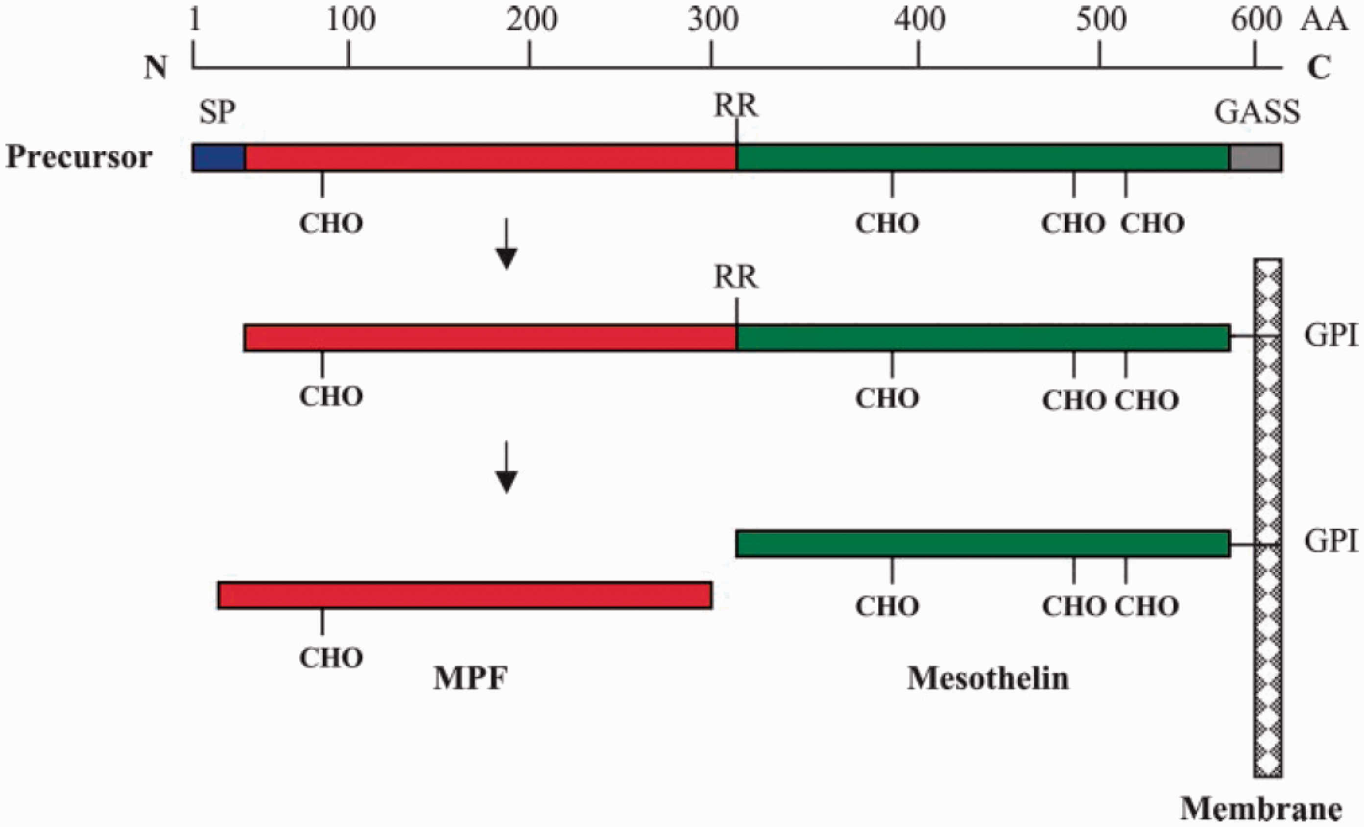
Additional work has been done on a potential role of serum SM in screening at-risk populations (asbestos-exposed workers and their families). In principle, mesothelioma is an attractive screening target with a well-defined at-risk population of asbestos-exposed individuals. A number of studies looked retrospectively 41 at the ability of SM to select out early-stage disease or those at risk of developing mesothelioma, but its accuracy falls below that of an acceptable screening test.
Nine studies assessing the role of serum mesothelin to monitor mesothelioma.
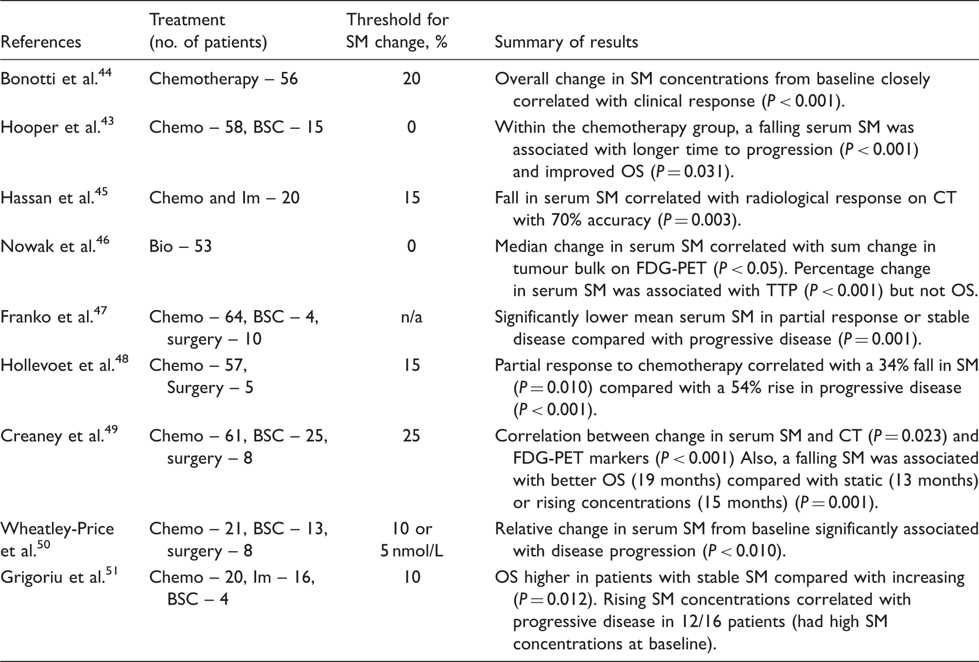
Chemo: chemotherapy; Bio: biological therapy; Im: immunotherapy, BSC: best supportive care; Surg: surgery; Mod RECIST CT: modified response evaluation criteria in solid tumours CT; OS: overall survival; TTP: time to progression; SM: soluble mesothelin; FDG-PET: fluourine 18-flurodeoxyglucose-positron emission tomography.
Megakaryocyte potentiating factor
Megakaryocyte potentiating factor (MPF) is a novel biomarker that is formed from the same precursor protein as SM and has similar expression. There is little additional benefit to SM, with similar limitations due to low concentrations in non-epithelioid disease. Some studies report slightly improved accuracy for disease monitoring, but given only marginal improvement, the focus of future research in this area is likely to be SM.45,48
Osteopontin
Osteopontin (OPN) is a glycoprotein that mediates cell–cell interactions and is over-expressed in many tumours including breast, lung and colon malignancies. 52 Studies have shown that it lacks the sensitivity to be used as a diagnostic test. A meta-analysis of serum and plasma OPN carried out in 2014 found a pooled sensitivity and specificity of 0.57 (95%CI: 0.52–0.61) and 0.81, 95%CI: 0.79–0.84), respectively, with considerable heterogeneity between the nine studies. 53 A thrombin cleavage site impedes reproducibility of measurements in serum; more recent literature has advocated plasma sampling to improve accuracy. 54 In addition, there appears to be little merit in serial monitoring of OPN. A study by Hollevoet et al. 48 showed that, unlike SM and MPF, OPN concentrations did not fall following surgery and did not correlate with treatment response from chemotherapy. Despite this, a potential role for OPN as a baseline predictor of poor prognosis has been demonstrated. Several studies have shown that a high baseline plasma OPN confers a poor prognosis, exclusive of histology, treatment modality or indeed other biomarkers.48,55 Further work is required to ascertain its utility outside of clinical trials, and given the variability in concentrations depending on ELISA used, 56 it is important that any future research adopts a consensus approach.
Fibulin-3
The first major study of fibulin-3 in serum reported a sensitivity of 100% for detecting early-stage mesothelioma in an asbestos-exposed population, with a specificity of 94.1%. 57 These estimates would be high enough for inclusion into the routine diagnostic pathway. However, several follow-up studies using the same commercial ELISA assay were unable to replicate these results, with a variety of estimates for sensitivity. 58 A recent meta-analysis of eight studies gave pooled estimates of sensitivity and specificity of 0.87 (95% CI: 0.58–0.97) and 0.89 (95% CI: 0.77–0.95), respectively. 59 Fibulin-3 is another glycoprotein that was initially discovered in high concentrations in another malignancy (glioma). It is thought to phosphorylate epidermal growth factor and thereby promote tumour growth and invasion. 60 It is particularly over-expressed in pleural fluid from mesothelioma but estimates of sensitivity remain too low to maintain acceptable specificity. Several studies have shown that higher pleural fluid levels at baseline confer a poor prognosis, but it is unclear whether this is primarily due to higher levels being found in more aggressive tumour types or if it offers additional prognostic information.
Hyaluronic acid
Hyaluronic acid (HA) is found in both blood and pleural fluid of patients with mesothelioma, although serum analysis is less useful due to rapid (2.5–5 min) half-life clearance from the systemic circulation by stabilin-2. 61 HA is more stable in pleural fluid in the form of a large fibroblast-formed polysaccharide. As a standalone test, it is not specific for mesothelioma and, given difficulties with its original testing methodology, it has had limited attention since the early studies. More recently, the ability to measure it more reproducibly, and combination with other biomarkers, have re-ignited interest in the pleural fluid analysis of HA. Creaney et al. 62 demonstrated that when combined with pleural fluid SM the area under the curve (AUC) for both biomarkers together improved to 0.92 (C.I: 0.86 to 0.96). Interestingly, the same study followed up the 96 patients with mesothelioma and found that pleural fluid HA was biphasically distributed within the cohort. When dichotomized at 75 mg/L, those with high effusion HA had much better survival (18.0 m) compared with patients with low levels (12.6 m) (P < 0.01). This confirmed the finding of a previous study, 63 and although several explanations have been postulated, the exact pathophysiology is uncertain. Further work is required before the clinical utility of this finding can be fully assessed.
VEGF
Pan-VEGF and its various isoforms have been the focus of many studies of malignant and non-malignant diseases of the lung. VEGF almost certainly has a role in pathophysiology of mesothelioma, although its exact role probably varies depending on the specific isoform. 64 Yasumitsu et al. 65 compared concentrations of pan-VEGF in the serum and pleural fluid of 51 patients with mesothelioma in an asbestos-exposed population. 65 Concentrations were significantly higher in mesothelioma, with highest concentrations in epithelioid disease, but with insufficient accuracy to be included in a diagnostic pathway (sensitivity 70.6% and specificity 88.1%). Notably, VEGF concentration was correlated with the stage of disease and worse survival, which probably relates to its well-documented effects on tumour angiogenesis. It is of particular importance in mesothelioma given the emergence of antiangiogenic VEGF-targeted treatments like bevacizumab that have been shown to improve survival when given in combination with pemetrexed and cisplatin. 35 No studies have demonstrated any ability of serum VEGF to select responders from non-responders for biologic therapy, but this area demands further study given the development of promising but expensive biologicals.46,66
Future biomarkers and biomarker panels
Ongoing research into biomarkers is directed at the validation of existing biomarkers or panels of existing biomarkers and discovery of novel biomarkers. As mentioned above, the combination of mesothelin and HA improved the overall diagnostic accuracy of both. Another study combined two molecular classes of biomarker by analysing plasma mesothelin values with the microRNA miR-103 a-3 p. 67 This improved the sensitivity and specificity of mesothelin alone from 74% and 89% to 95% and 81%, respectively.
Novel approaches involve the proteomic discovery of previously unidentified biomarkers in serum and pleural fluid and their validation in a second cohort. Such a study was performed by Ostroff et al. 68 who demonstrated promising results using a 13-protein panel, reporting AUCs of 0.98 ± 0.04 in blinded verification cohort and 0.95 ± 0.04 in a validation cohort (38 patients with mesothelioma). This proteomic assay is currently being validated in a multicentre prospective trial alongside fibulin-3; it is of note that none of the 13 classifier proteins have previously been associated with mesothelioma. 69 Many studies exist using this technique of protein discovery, but the importance of external validation is paramount. Another study from Morre et al. 70 focused on the presence of two mesothelioma-specific ENOX2 protein transcript variants in the serum of 17 patients pre- and postdiagnosis of mesothelioma. 70 When compared with patients who had been exposed to asbestos but without a diagnosis of mesothelioma, one or both proteins were detectable up to 10 years prior to diagnosis and with a mean of 6.2 years prior to the onset of clinical symptoms. This finding requires further prospective investigation but if useful could be used to detect mesothelioma at an early and more treatable stage.
Conclusion
The role of biomarkers in mesothelioma has been assessed at almost every stage of the disease process including screening, diagnosis, prognosis and monitoring. No single biomarker has sufficient reproducibility in differentiating mesothelioma from other more common benign or malignant conditions. Promising results are being seen using biomarker panels, but none has yet undergone external validation in prospective trials. Results from several studies have supported the use of serum mesothelin as a method of monitoring disease during treatment, although questions remain around its utility in non-epithelioid disease. High plasma OPN at baseline appears to be an independent indicator of poor prognosis, although further studies are required to assess the clinical usefulness of this finding. Given that treatments for mesothelioma are now developing, the pressure to identify patients who will respond to treatment is growing. With the proliferation of biomarker discovery projects and formalized tissue storage for mesothelioma samples (e.g. MesoBank), it is essential that promising biomarkers are investigated beyond the initial detection stage.
Footnotes
Acknowledgements
This article was prepared at the invitation of the Clinical Sciences Reviews Committee of the Association for Clinical Biochemistry and Laboratory Medicine.
Declaration of conflicting interests
The author(s) declared no potential conflicts of interest with respect to the research, authorship, and/or publication of this article.
Funding
DTA is funded by a National Institute for Health Research (NIHR) Academic Clinical Fellowship.
Ethical approval
Not applicable.
Guarantor
Nick Maskell.
Contributorship
DTA and NAM.