
Abstract
Background
Oxidation of lipoproteins is thought to play a crucial role in atherogenesis. Role for triglyceride-rich lipoproteins in atherogenesis is unclear. Thus, we aimed to investigate whether cholesteryl ester hydroperoxides (CEOOH) are present in very low-density lipoproteins (VLDL) and intermediate-density lipoproteins (IDL) by using highly sensitive liquid chromatography/mass spectrometry.
Methods
Total lipids were extracted from the plasma of healthy donors (n = 6) and their fractions of VLDL and IDL. Additional three plasma samples were analysed freshly for CEOOH. Detection and identification of CEOOH was conducted by liquid chromatography/LTQ ion trap mass spectrometry/Orbitrap high mass accuracy mass spectrometry. Authentic standards of CEOOH were used for unequivocal identification on the basis of their mass spectra.
Results
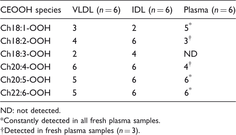
We identified six molecular CEOOH species overall, namely, Ch18:1-OOH, Ch18:2-OOH, Ch18:3-OOH, Ch20:4-OOH, Ch20:5-OOH and Ch22:6-OOH. Of them, Ch18:2-OOH, Ch20:5-OOH, Ch20:4-OOH and Ch22:6-OOH were detected in all IDL samples, while only Ch20:4-OOH was detected in all VLDL samples. All of CEOOH species except for Ch18:3-OOH were detected in plasma, with constant detection of Ch20:5-OOH, and Ch22:6-OOH in all plasma samples.
Conclusion
The presence of CEOOH species in VLDL and IDL was confirmed with the analytical sensitivity of 0.1 pmol, showing the constant appearance of more CEOOH species in IDL than VLDL. This finding might add biochemical evidences of atherogenicity of these lipoproteins. Clinical utility of measuring CEOOH level in these lipoproteins need to be investigated for the risk assessment of the cardiovascular disease.
Introduction
It is widely accepted that oxidation of lipoproteins have immense effect on the development of atherosclerosis. Since lipid hydroperoxides (LOOH) are the major reaction products of lipoprotein oxidation, detection of LOOH in a lipoprotein fraction can indicate its oxidative change and potential role in atherosclerosis. Though oxidized low-density lipoproteins (LDL) are primarily blamed for atherosclerosis, the role of very low-density lipoproteins (VLDL) and intermediate-density lipoprotein (IDL) in their oxidized forms cannot be neglected.1–5
Accumulation of cholesteryl ester hydroperoxides (CEOOH) has been well documented in human atherosclerotic lesions.6,7 Further, CEOOH are the major biologically active components of minimally oxidized LDL, which in turn stimulates wide varieties of cellular and molecular processes involved in atherosclerosis. 8 Moreover, copper-mediated oxidation of LDL as well as high-density lipoproteins (HDL) can induce an increase of CEOOH. 9 Thus, CEOOH can be considered as one of the most relevant lipids to atherosclerosis.
Despite the importance of CEOOH in the atherosclerotic process, the qualitative and quantitative evaluation of CEOOH in lipoproteins has been largely limited, possibly due to structural variability and instability, rapid clearance from circulation, too low concentration for easy detection and the lack of standard CEOOH. Therefore, there is paucity of evidences that show detail chemistry and existence of CEOOH in native plasma lipoproteins, most of all, triglyceride (TG)-rich lipoproteins. VLDL contains cholesteryl ester (CE) as a minor component, and IDL consist only a minor portion of plasma lipoproteins.
Several methods have been reported for the measurement of LOOH in human plasma and specific lipoprotein fractions, namely, colorimetric method by ferrous oxidation using xylenol orange, 10 iodometric measurement, 11 high-performance liquid chromatography (HPLC) with post-column detection based on fluorometry,12,13 chemiluminometry14–16 and electrochemistry.17,18 In recent years, liquid chromatography/mass spectrometry (LC/MS) became a powerful tool for detail study of LOOH. We previously reported highly sensitive LC/MS methods for detection of CEOOH, 9 phosphatidylcholine hydroperoxide (PCOOH) 19 and triacylglycerol hydroperoxide (TGOOH). 20 We have detected and identified several CEOOH species in chemically oxidized lipoprotein fractions using highly sensitive reversed-phase liquid chromatography with a hybrid linear ion trap Orbitrap mass spectrometer (LC/LTQXL Orbitrap, Thermo Fisher Scientific, Waltham, MA, USA) and in-house-built standard compounds. 9 In our previous study, CEOOH were not detected in the isolated LDL and HDL fractions in their native forms while were detected in plasma. 9 This finding led us to a question whether the CEOOH species detected in plasma were carried in VLDL and/or IDL. Thus, we apply our LC/LTQ Orbitrap method for detection and identification of CEOOH in VLDL and IDL.
Materials and methods
Chemicals
We used cholesteryl oleate monohydroperoxide (Ch18:1-OOH), cholesteryl linoleate monohydroperoxide (Ch18:2-OOH) and cholesteryl linolenate monohydroperoxide (Ch18:3-OOH) as standards, which were synthesized chemically in our laboratory reported elsewhere. 21 All other chemicals and solvents used were of analytical grade and obtained from Wako Pure Chemical Industry (Osaka, Japan), unless specified.
Plasma preparation
A fasting EDTA blood (10 mL) was collected from six apparently healthy volunteers (range, 27–36 years). Written informed consent was obtained from all human volunteers. The samples were immediately kept on ice and centrifuged at 2000 g for 10 min at 4℃, within 30 min. A portion of plasma (1.0 mL) was immediately stored at −80℃ until analysed by LC/LTQ Orbitrap, while remaining were used for isolation of VLDL and IDL. Additional plasma sample was collected from three of the six donors and analysed freshly by the LC/LTQ Orbitrap to rule out possible auto-oxidation during storage.
VLDL and IDL isolation
Sequential ultracentrifugation was used to isolate VLDL and IDL from the plasma. 22 Briefly, ultracentrifugation was performed using a near-vertical tube rotor (MLN-80, Beckman Coulter, Fullerton, CA, USA) on a model Optima MAX (Beckman Coulter). Plasma (2.0 mL) mixed with 6.0 mL of d = 1.006 kg/L solution containing NaCl (1.14%, w/v), EDTA-2Na (0.01%, w/v) and 1 M NaOH (0.1%, v/v) was centrifuged at 50,000 rpm for 14 h at 4℃, followed by collection of upper fraction (2.5 mL) as VLDL. The remaining fraction was then adjusted to d = 1.019 kg/L with KBr solution and centrifuged at 40,000 rpm for 20 h at 4℃. Upper fraction (2.5 mL) containing IDL was isolated. The isolated VLDL and IDL were concentrated by ultrafiltration using XM-50 and Millipore Amicon Bioseparations Stirred Cells (Thermo Fisher Scientific, Waltham, MA, USA).
Assessing purity of VLDL and IDL
The purity of isolated VLDL and IDL was checked by determining its chemical composition, apolipoprotein study and characteristic motility in polyacrylamide gel disc electrophoresis (LipoPhor, Jokoh Co, Tokyo, Japan) and agarose gel electrophoresis using universal electrophoresis film (Helena Laboratory, Beaumont, TX, USA).19,23 Total cholesterol (TC), free cholesterol (FC), TG and phospholipid (PL) in the VLDL and IDL were measured by automated enzymatic methods (Kyowa Medex Co., Ltd., Tokyo, Japan). CE concentration was calculated by multiplying the esterified cholesterol concentrations (obtained by subtracting FC from TC) by 1.72. 24 Apolipoprotein study was done in 3–10% sodium dodecyl sulphate–polyacrylamide gel electrophoresis (SDS–PAGE; ATTO Bioscience and biotechnology, Tokyo, Japan) after reduction with 2-mercaptoethanol and stained with SimpleBlue™ SafeStain-Coomassie Brilliant Blue (Invitrogen, USA). Protein content in VLDL and IDL were measured by modified Lowry method. 25
Preparation of sample for LC/LTQ Orbitrap
Lipids from VLDL and IDL were extracted by the previously reported method.9,19 Briefly, 0.1 mL of the lipoprotein, each with total lipid concentration of 0.5 g/L, was mixed with 0.4 mL of freshly prepared 0.005% (w/v) 2,6-di-tert-butyl-p-cresol (as an antioxidant) in acetonitrile and 2.0 mL of chloroform. For extraction of lipid from plasma, 0.2 mL of the plasma was mixed with 0.8 mL of freshly prepared 0.005% 2,6-di-tert-butyl-p-cresol in acetonitrile and 2.0 mL of chloroform. The mixture was then vortex mixed vigorously for 30 s and centrifuged at 3000 g for 10 min at 4℃. The chloroform layer was collected followed by complete evaporation under vacuum, and the residue was dissolved in 300 µL of methanol.
LC/LTQ Orbitrap
Ten microlitres of the lipid extract was injected onto a reversed-phase Syncronis C18 HPLC column (50 mm × 2.1 [i.d.] mm; particle size 1.7 µm; Thermo Fisher Scientific, Waltham, MA, USA) and maintained at 60℃. Gradient elution was performed with a mobile phase composed of 10 mmol/L aqueous ammonium acetate (Solvent A) and 2-propanol (Solvent B). The HPLC gradient elution program was 0.00–1.00 min 50% A and 50% B; 5.01–7.00 min 0% A and 100% B; 7.01–10.0 min 50% A and 50% B at a flow rate of 0.2 mL/min.
High-resolution mass spectrometric analysis was performed using LTQ XL Orbitrap mass spectrometer combined with Surveyor MS pump and an autosampler.9,19 Electrospray ionization tandem mass spectrometry analysis was performed in positive-ion mode, and mass spectra were obtained in Fourier-transform mode, with a target mass resolution of R = 60,000 at m/z 400 under automatic gain control set to 5.0 × 10⁵ as the target value. The ion spray voltage was set at 5.0 kV, with a scan range of m/z 150–1000. The trap fill-time was set at 500 ms. Nitrogen was used as sheath gas (set at 50 arbitrary units). CEOOH were detected as the [M + NH4]+. Extracted ion chromatograms (EIC) were drawn with the mass tolerance set at 5.0 ppm.
Data processing
By use of Qual Browser 2.0 software (Thermo Fisher Scientific), each survey spectrum was converted into a peak list, which reported m/z, relative intensity (normalized to the most abundant peak) and the sum composition for each peak detected with a signal-to-noise ratio above 6. Sum compositions were calculated from the determined precursor masses, assuming the settings: mass tolerance within ±5.0 ppm; even-electron ions. Assumed atomic compositions were restricted to nitrogen, 1–2 atoms per molecule; oxygen, 2–15 atoms and unrestricted carbon and hydrogen.
Results
Based on the findings of chemical composition of VLDL and IDL, apolipoprotein composition determined by SDS–PAGE and characteristic mobility of lipoprotein in LipoPhor, we concluded that our lipoprotein fractions were pure (data not shown).
Detection of Ch18:1-OOH in VLDL and IDL
LC/LTQ Orbitrap in positive-ion mode was used for qualitative analysis of CEOOH in the extracts of VLDL, IDL and plasma. The first step of our approach for hydroperoxide detection was to collect high-resolution spectra. The spectra were acquired for three synthetic CEOOH standards and the extracts of VLDL and IDL.
Figure 1(a) to (c) shows EIC of m/z 700.6238 in positive-ion mode for synthetic Ch18:1-OOH (40 pmol), VLDL and IDL, respectively; their corresponding mass spectra are shown in Figure 1(a') to (c'). For the synthetic Ch18:1-OOH, one peak was observed at a retention time (RT) of 7.07 min (Figure 1(a)); the corresponding mass spectrum obtained is shown in Figure 1(a'), showing [M + NH4]+ at m/z 700.6257 (elemental composition C45H82O4N, theoretical mass 700.6238).
LC/LTQ Orbitrap profiles of cholesteryl oleate monohydroperoxide (Ch18:1-OOH) in positive-ion mode: (a) extracted ion (m/z 700.6238) chromatogram of synthetic Ch18:1-OOH; (a') mass spectrum of peak associated with retention time at 7.07 min in (a); (b) total ion chromatogram (inside square) and extracted ion (m/z 700.6238) chromatogram of VLDL; (b') mass spectrum of peak associated with retention time at 7.00 min in (b) = Ch18:1-OOH; (c) total ion chromatogram (inside square) and extracted ion (m/z 700.6238) chromatogram of IDL; (c') mass spectrum of peak associated with retention time at 7.05 min in (c) = Ch18:1-OOH.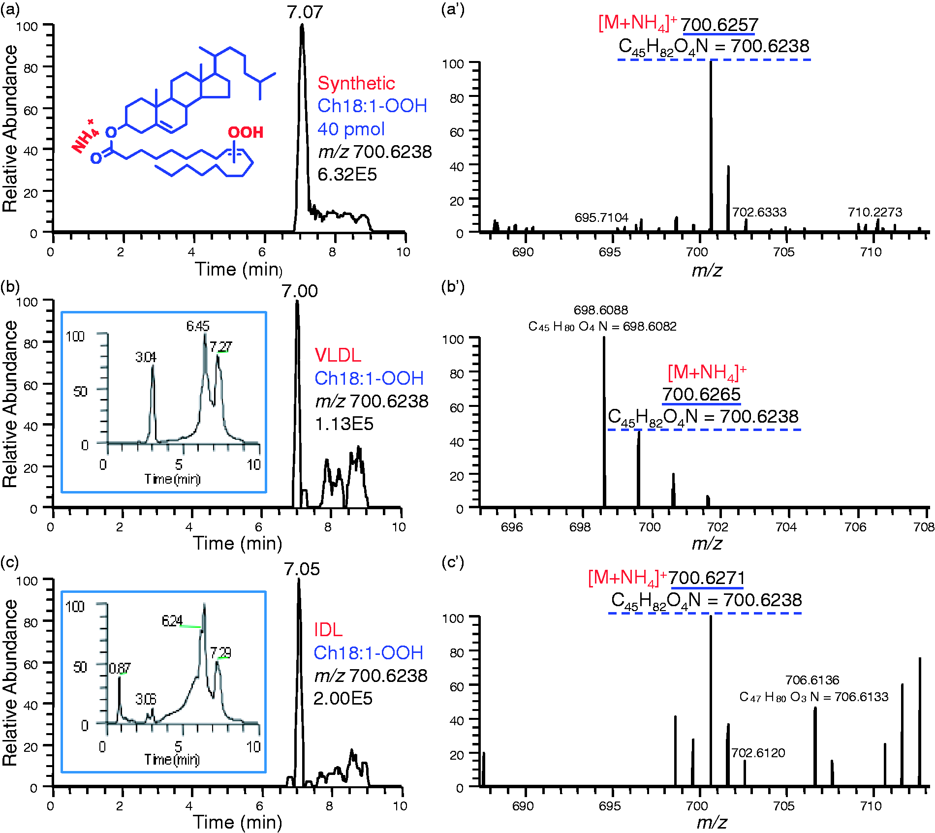
A total ion chromatogram (TIC) of VLDL shows absence of clearly defined HPLC peaks and large over-lapping peaks (Figure 1(b)). However, extraction of a particular signal, m/z 700.6238, from the TIC led to a much better defined chromatogram. One peak was observed at RT 7.00 min, and the corresponding mass spectrum obtained is shown in Figure 1(b'). The peak observed at m/z 700.6265 corresponding to [M + NH4]+, and the ion has the same elemental composition and theoretical mass as the ion from synthetic Ch18:1-OOH, thus the peak at 7.00 min in Figure 1(b) was identified as Ch18:1-OOH in VLDL. In the same way, the peak at 7.05 min in Figure 1(c) obtained from IDL was identified as Ch18:1-OOH.
Detection of Ch18:2-OOH in VLDL and IDL
Chromatogram and mass spectrum of Ch18:2-OOH obtained from standard, VLDL and IDL are shown in Figure 2. Standard Ch18:2-OOH was eluted at the RT of 7.00 min (Figure 2(a)), and corresponding mass spectrum revealed an [M + NH4]+ at m/z 698.6086 (elemental composition C45H80O4N, theoretical mass 698.6082; Figure 2(a')). In Figure 2(b'), the base peak at m/z 698.6088 corresponded to [M + NH4]+ has the same elemental composition and theoretical mass as the ions from standard Ch18:2-OOH (Figure 2(a')), implying the peak at 7.02 min in Figure 2(b) obtained from VLDL is Ch18:2-OOH. Similarly, the peak at 7.01 min in Figure 2(c) obtained from IDL was identified as Ch18:2-OOH.
LC/LTQ Orbitrap profiles of cholesteryl linoleate monohydroperoxide (Ch18:2-OOH) in positive-ion mode: (a) extracted ion (m/z 698.6082) chromatogram of synthetic Ch18:2-OOH; (a') mass spectrum of peak associated with retention time at 7.00 min in (a); (b) extracted ion (m/z 698.6082) chromatogram of VLDL; (b') mass spectrum of peak associated with retention time at 7.02 min in (b) = Ch18:2-OOH; (c) extracted ion (m/z 698.6082) chromatogram of IDL; (c') mass spectrum of peak associated with retention time at 7.01 min in (c) = Ch18:2-OOH.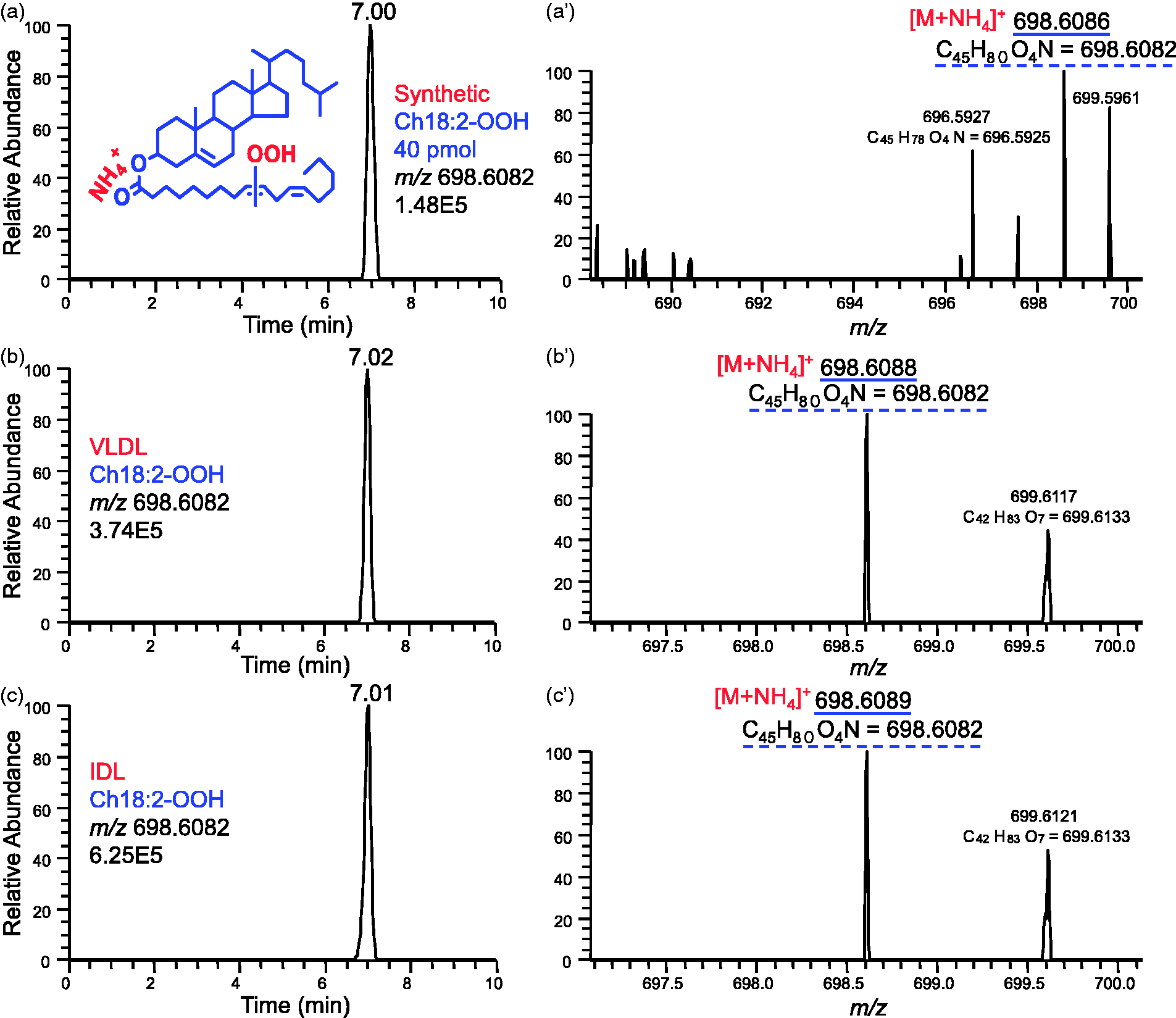
Detection of Ch18:3-OOH in VLDL and IDL
We detected Ch18:3-OOH in VLDL and IDL fractions but not in plasma. Figure 3(a) to (c) shows EIC of m/z 696.5925 for synthetic Ch18:3-OOH (40 pmol), VLDL and IDL, and their corresponding mass spectra are shown in Figure 3(a') to (c'), respectively. Synthetic Ch18:3-OOH was eluted at the RT of 6.96 min (Figure 3(a)); the corresponding mass spectrum is shown in Figure 3(a'), elucidating an [M + NH4]+ at m/z 696.5922 (elemental composition C45H78O4N, theoretical mass 696.5925). In Figure 3(b'), a peak at m/z 696.5936 corresponding to [M + NH4]+ has the same elemental composition and theoretical mass as the ions from synthetic Ch18:3-OOH, indicating the peak at 6.97 min in Figure 3(b) from VLDL is Ch18:3-OOH. Similarly, Ch18:3-OOH was also identified in IDL.
LC/LTQ Orbitrap profiles of cholesteryl linolenate monohydroperoxide (Ch18:3-OOH) in positive-ion mode: (a) extracted ion (m/z 696.5925) mass chromatogram of synthetic Ch18:3-OOH; (a') mass spectrum of peak associated with retention time at 6.96 min in (a); (b) extracted ion (m/z 696.5925) mass chromatogram of VLDL; (b') mass spectrum of peak associated with retention time at 6.97 min in (b) = Ch18:3-OOH; (c) extracted ion (m/z 696.5925) mass chromatogram of IDL; (c') mass spectrum of peak associated with retention time at 6.97 min in (c) = Ch18:3-OOH.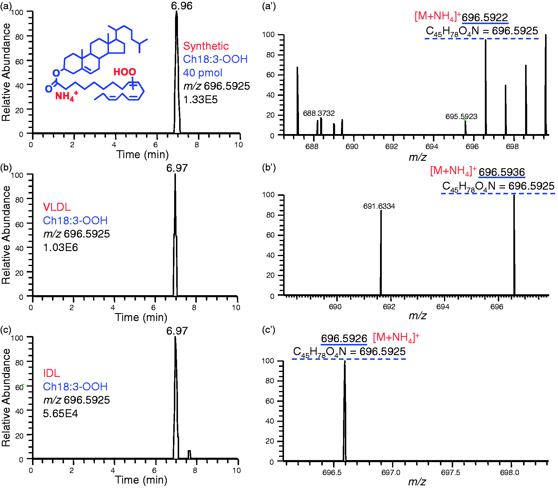
Detection of other molecular species of CEOOH
Diagnostically significant ions of CEOOH in the synthetic standards, VLDL, IDL and plasma obtained from spectra by LC/LTQ Orbitrap in positive-ion mode.
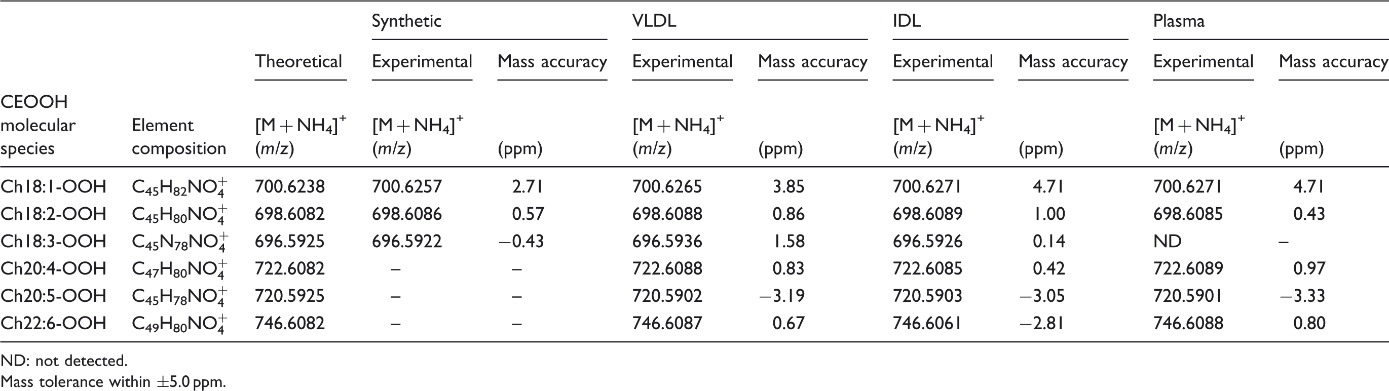
ND: not detected.
Mass tolerance within ±5.0 ppm.
Detection of CEOOH in plasma
We detected five molecular species of CEOOH in plasma. Ch22:6-OOH and Ch20:5-OOH were present in all plasma. Ch18:1-OOH, Ch20:4-OOH and Ch18:2-OOH were found in most of the plasma (Table 2). In contrast, no Ch18:3-OOH was detected in plasma sample. The TIC and spectra of a plasma extract are shown in Figure 4.
LC/LTQ Orbitrap profiles of cholesteryl ester hydroperoxides present in a plasma sample in positive-ion mode: (a) total ion chromatogram of plasma extract; (b) mass spectrum showing m/z 700.6271 = Ch18:1-OOH; (c) mass spectrum showing m/z 698.6085 = Ch18:2-OOH; (d) Ch18:3-OOH (m/z 696.5925) is absent in plasma; (e) mass spectrum showing m/z 722.6089 = Ch20:4-OOH; (f) mass spectrum showing m/z 720.5901 = Ch20:5-OOH; (g) mass spectrum showing m/z 746.6088 = Ch20:6-OOH. Distribution of CEOOH species in VLDL, IDL and plasma. ND: not detected. Constantly detected in all fresh plasma samples. Detected in fresh plasma samples (n = 3).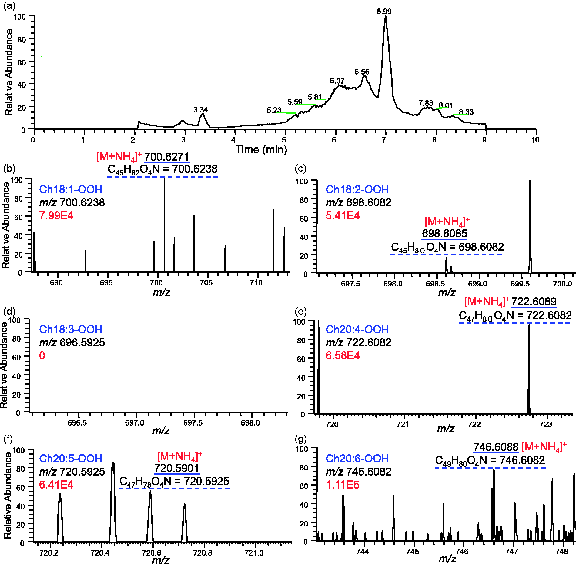
Distribution of CEOOH
We identified six molecular CEOOH species overall, namely, Ch18:1-OOH, Ch18:2-OOH, Ch18:3-OOH, Ch20:4-OOH, Ch20:5-OOH and Ch22:6-OOH (Table 2). Of them, Ch18:2-OOH, Ch20:5-OOH, Ch20:4-OOH and Ch22:6-OOH were detected in all IDL samples, while only Ch20:4-OOH was detected in all VLDL samples. All of CEOOH species except for Ch18:3-OOH were detected in all frozen plasma, with constant detection of Ch20:5-OOH and Ch22:6-OOH. The fresh samples contained Ch18:2-OOH, Ch20:4-OOH, Ch18:1-OOH, Ch20:5-OOH and Ch22:6-OOH overall, of them latter three were constantly detected. Between the fresh plasma samples and the stored plasma samples, no significant difference in distribution of CEOOH was observed.
Discussion
We developed LC/LTQ Orbitrap method that is sensitive enough to detect CEOOH in human plasma and native lipoprotein fractions using in-house-built standards. Use of this method enabled us to identify CEOOH molecular species in VLDL and IDL, namely, Ch18:1-OOH, Ch18:2-OOH, Ch18:3-OOH, Ch20:4-OOH, Ch20:5-OOH and Ch22:6-OOH, on the basis of their mass spectra. The possibility of auto-oxidation during storage was negated since fresh plasma samples showed essentially the same results as the frozen samples.
Our successful demonstration of CEOOH in VLDL and IDL is attributed to several factors. First, we used three authentic standards (Ch18:1-OOH, Ch18:2-OOH and Ch18:3-OOH), which enabled us to develop an unequivocal method for the identification on the basis of their mass spectra and RT on LC. Second, we use LC/LTQ Orbitrap, which can obtain accurate m/z values for adducts of the molecular ions from individual molecules by high mass resolution. It provides high-resolution EIC within ±5.0 ppm relative mass deviation and spectra with selected extraction of ions at ±5.0 ppm accuracy. Most importantly, our method has high analytical sensitivity of 0.1 pmol.
Although previous studies demonstrated the presence of CEOOH in healthy plasma, the molecular species of CEOOH were not specified.12,26,27 More importantly, lipoprotein source of the detected CEOOH was not specified in these studies. In the present report, we focused on VLDL and IDL, because the role for TG-rich lipoproteins in atherosclerosis is poorly understood. Both VLDL and IDL have ability to induce foam cell formation in vitro, and the oxidized forms of these lipoproteins have been identified in atherosclerotic lesions.3,28,29 The detection of CEOOH in VLDL and IDL in the present study might support a possible involvement of these lipoproteins in atherogenic process.
The physiologically relevant mechanisms underlying oxidation of CE in vivo are largely unknown. Although VLDL carry mainly endogenous lipids, possible integration of oxidized lipids from diet into CE during assembly of VLDL in the liver cannot be excluded.30,31 In addition, possibly, the oxidation of CE in VLDL and IDL can occur during systemic circulation. Older plasma lipoproteins are known to be more susceptible to oxidation, suggesting progression of the oxidation during circulation. 32 Relating to this issue, possible contribution of remnant lipoproteins to CEOOH formation in TG-rich lipoproteins is of interest, although the present study is limited to VLDL and IDL. Remnant lipoproteins and bioactive components associated with it are believed to be related to atherogenesis, thus can provide significant predictive value of the cardiovascular risk.33–35 In previous studies, VLDL remnants are related to oxidation and atherogenicity. 36 In our lipoprotein separation method using ultracentrifugation, remnant lipoproteins cannot be isolated. However, previous studies showed that remnant lipoproteins are largely distributed in IDL fraction. 37 In the present study, we found the wider distribution of CEOOH molecular species in IDL in comparison with VLDL, which might suggest a possibility that our IDL fraction contained remnant lipoproteins enriched with CEOOH. It is our next interest to quantify CEOOH in remnant lipoproteins isolated by a reported immunoaffinity technique and compare their levels with those of other TG-rich lipoproteins, LDL and HDL. 38
The present study revealed the molecular species of CEOOH, which in turn become the target of future quantification study. For precise and accurate mass spectrometric quantification, we are synthesizing deuterium-labelled CEOOH with the targeted structures as internal standards.
In summary, we identified six molecular species of CEOOH in human plasma, VLDL and IDL with simple technique of LC/MS. These markers are potentially useful for identification of individuals who are at high risk for future coronary events. Presence of CEOOH in VLDL and IDL might support the atherogenicity of TG-rich lipoproteins. Further work is needed to explore possible underlying pathology of CEOOH in TG-rich lipoproteins and atherosclerosis. We plan on conducting future experiment to develop methodology for CEOOH quantification and its significance in various clinical conditions.
Footnotes
Acknowledgements
We are grateful to the central research laboratory, Faculty of Health Sciences, Hokkaido University for kindly providing working space and equipment.
Declaration of conflicting interests
None declared.
Funding
This work was supported by Regional Innovation Strategy Support Program, Sapporo Health Innovation ‘Smart-H’, of The Ministry of Education, Culture, Sports, Sciences and Technology, Japan and partly by a Grant-in-Aid for scientific research from the Japan Society for Promotion of Sciences, Japan.
Ethical approval
Ethics review board of the Faculty of Health Sciences, Hokkaido University approved this study protocol (approval number 08-57-2).
Guarantor
HC.
Contributorship
RS, SPH and HC researched literature and conceived the study. RS, TS, YT, ST, AY, SJ and HF were involved in sample collection, lipoprotein separation, lipid measurement and data analysis. RS and SPH were involved in mass spectrometric analysis. RS wrote the first draft of the manuscript. All authors reviewed the manuscript and approved the final version.