
Abstract
Background. [18F]FEPPA is a potent TSPO imaging agent that has been found to be a potential tracer for imaging neuroinflammation. In order to fulfill the demand of this tracer for preclinical and clinical studies, we have developed a one-pot automated synthesis with simplified HPLC purification of this tracer, which was then used for PET imaging of neuroinflammation in fine particulate matter- (PM2.5-) exposed rats. Results. Using this automated synthesis method, the RCY of the [18F]FEPPA was
1. Introduction
Neuroinflammation is an inflammatory and adaptive response within the central nervous system (CNS) [1] and is the driving force for disease progression, such as Alzheimer’s disease (AD) [2], major depressive disorder [3], schizophrenia [4], and brain injuries [4]. Activated in response to neuropathologies [5, 6], microglia and astrocytes have been known to be the predominant mediators during the neuroinflammatory process. The transmembrane domain protein, translocator protein-18 kDa (TSPO), is variously expressed throughout the body and has low expression within the brain [7–9]. However, TSPO levels are significantly increased in the brain when microglia and astrocyte are activated [10, 11]. Recently, the results of meta-analyses of TSPO levels in mild cognitive impairment and AD further supported the association of increased neuroinflammation during the progression of mild cognitive impairment and AD, relative to healthy controls [12]. In addition to CNS, an increase in TSPO expression has also been seen in a wide variety of malignant human cells and tissues including brain cancers [13, 14], prostate cancers [15, 16], colon cancers [17–19], breast cancers [20, 21], esophageal cancers [22], endometrial carcinomas [23], ovarian cancers, and hepatic carcinomas [24]. Albeit its lack of specificity to activated microglia, the TSPO levels may at times reflect neuroinflammation in vivo [10].
Exposure to fine particulate matter (PM2.5) has been linked to adverse neurological and behavioral health effects, including increased risk for cognitive decline (AD) [25, 26], Parkinson’s disease [27], ischemic stroke [28], and anxiety or depression [29, 30] in epidemiology studies. Increases in neuroinflammation and oxidative stress have been identified as putative mechanisms by which fine PM2.5 may impair central nervous system function [31, 32], an example of which occurs when exacerbated and unregulated microglial proinflammatory responses play crucial factors involved in brain damage [33]. Chronic activation of the microglia (reactive microgliosis) has been implicated in neuronal injury and neuronal damage after exposure to PM2.5 [34]. One hallmark of microglial activation is the overexpression of TSPO [35, 36]. To the best of our knowledge, the application of a TSPO imaging agent for studying the effects of PM2.5 on human health has not been reported. Thus, we have developed an one-pot automated synthesis of [18F]FEPPA, a potent TSPO imaging agent, and used it to investigate (1) whether PET imaging can noninvasively detect microglia activation after chronic subacute ambient PM2.5 exposure in spontaneous hypertensive rats and (2) whether there is a specific pattern of microglia activation in the brain after chronic subacute ambient PM2.5 exposure.
The first TSPO PET ligand, [11C]PK-11195 was synthesized more than two decades ago [37–39]. However, its utility was limited due to its low brain penetration, high nonspecific binding, high plasma protein binding, and short half-life (for review, see [37, 38]). Therefore, several second-generation 18F-labelled TSPO PET ligands, including [18F]FEPPA [40–43], [18F]FEDAA1106 [44, 45], [18F]DPA-714 [46, 47], and [18F]PBR06 [48, 49] have been developed (for review, see [50, 51]). Recently, a novel third-generation TSPO PET ligand, [18F]GE180 was developed [52–54] and proved to be useful for TSPO imaging [53, 55, 56]. However, its application for neuroimaging is very limited as it suffers from very low brain penetration, similar to that of [11C]PK-11195. Specifically, in humans, the VT of [18F]GE180 is 20-fold lower than that of [11C]PRB28 [57]. Among these tracers, [18F]-N-(2-(2-fluoroethoxy)benzyl)-N-(4-phenoxypyridin-3-yl)acetamide ([18F]FEPPA) shows outstanding properties regarding affinity, stability, lipophilicity, and radiosynthesis [43] and has been used in several preclinical [40, 58, 59] and clinical settings [60–66]. In order to facilitate the usefulness of [18F]FEPPA in both preclinical and clinical studies, it is imperative to have a fully automated, simple, high yield, and reliable manufacturing process method available for the production of this tracer. Thus, we have adopted Wilson’s method [43], with some modifications, to fully automate the synthesis of this potent TSPO imaging agent using a TRACERlab FxFN module (GE Healthcare, Milwaukee, WI) with ethanol and water for purification in high quality and high reproducibility. Vignal et al. reported a similar synthesis using ethanol, water, and phosphoric acid for purification of [18F]FEPPA [42]. We report herein (1) a one-pot automated synthesis of [18F]FEPPA with a simplified HPLC purification, (2) the USP compliant QC and stability tests of [18F]FEPPA, and (3) microPET imaging of [18F]FEPPA in PM2.5-exposed rats.
2. Materials and Methods
The precursor (N-[[2-[2-[[(4-methylphenyl)sulfonyl]oxy]ethoxy]phenyl]methyl]-N-(4-phenoxy-3-pyridinyl) acetamide, TsEPPA, 1) and the nonradioactive authentic sample N-(2-(2-fluoroethoxy)benzyl)-N-(4-phenoxypyridin-3-yl) acetamide (FEPPA, 2) were purchased from ABX Advanced Biochemicals (Radeberg, Germany). All other chemicals and solvents were purchased from either Sigma-Aldrich (Milwaukee, WI, USA) or Acros Organics (Morris Plains, NJ, USA) and used without further purification. [18O]O2H (>98% enriched) was purchased from Rotem Industries (Beer Sheva, Israel). Aqueous [18F]Fluoride was produced in our PET Trace cyclotron (GE Medical Systems, Uppsala, Sweden) via 18O(p, n)18F nuclear reaction. All Sep-Pak® cartridges were purchased from Waters Associates (Milford, MA, USA).
2.1. Automated Radiosynthesis
The [18F]FEPPA (2) was produced by automated synthesis via fluorination of the TsEPPA (1) with K[18F]/K2.2.2 followed by purification with HPLC, to give [18F]FEPPA (2) as previously reported (Scheme 1) [43].

Radiosynthesis of the [18F]FEPPA (2).
The [18F]FEPPA (2) was produced by automated synthesis using a modified TRACERlab FxFN module (GE Healthcare, Milwaukee, WI; Figure 1). Briefly, fluorination of TsEPPA (1) with K[18F]/K2.2.2 in anhydrous MeCN at 70°C for 20 min gave the crude product (1) (Scheme 1). After dilution with H2O, the crude product (1) was purified with a semipreparative HPLC (Waters Xterra RP-18, 10 μm,

Modified TRACERLab FxFN module for the [18F]FEPPA (2) radiosynthesis.
2.2. Quality Control (QC) and Stability Tests of the [18F]FEPPA (2)
The radiochemical purity, chemical purity, and molar activity of 2 were analyzed with an HPLC system (Agilent 1100 series) equipped with a Bioscan FC3300 flow count radioactivity detector (
Other items of QC test and corresponding criteria of 2 were set according to the U.S. Pharmacopoeia (USP) for radiopharmaceuticals [67], which included visual inspection, pH, half-life of radionuclide, radionuclidic purity, radiochemical purity, chemical purity, residual K2.2.2, residual solvents, bacterial endotoxins, filter integrity, and sterility test.
The stability of 2 at
2.3. MicroPET Imaging of the [18F]FEPPA (2) Injection in Rats Exposed to Ambient Fine Particulate Matter (PM2.5)
2.3.1. Animals
Male spontaneously hypertensive rats (7-week-old, average weight of 350 g,
At the end of exposure, [18F]FEPPA microPET/CT brain scan was performed for each rat to assess the degree of neuroinflammation. During scanning, rats were anesthetized by passive inhalation of a mixture of isoflurane (5% for induction and 2% for maintenance) in oxygen, followed by a bolus tail vein injection of
2.4. Immunohistochemical Analysis
After PET image acquisition, all rats were sacrificed. The brain tissues were processed using an automated tissue processor (Shandon Excelsior, Thermo Scientific, UK), embedded in paraffin, and cut at a thickness of 3-5 μm for immunohistochemical (IHC) staining. Ionized calcium binding adaptor molecule (Iba1) is a macrophage-specific calcium-binding protein, participating in membrane ruffling and phagocytosis in the activated microglia. For Iba1 staining, brain tissues were stained against the activated microglial marker Iba1 (GeneTex, San Antonio, TX, USA) [72]. The percentage of areas occupied by the stained nuclei of activated microglia at 40x high power field (0.26 mm/pixel) in the hippocampal region of the rats was calculated using a digital slide scanner (MoticEasyScan, Motic®, Canada). The brain tissues were examined by a histopathologist blinded to the exposure data.
2.5. Statistical Analysis
Data was expressed as
3. Results
3.1. A One-Pot Automated Synthesis of the [18F]FEPPA (2) Using a Modified FxFN Module
In this paper, we presented the detailed automated production of [18F]FEPPA along with a full set of quality control specifications and results in USP compliance. Using this modified purification method, we were able to routinely produce 2 with a FxFN module in
The QC test results of 2 are tabulated in Table 1. Moreover, the stability test showed that 2 synthesized by this method was stable at room temperature (
The QC tests of the [18F]FEPPA (2).
Stability of three consecutive productions of the [18F]FEPPA (2) (
3.2. MicroPET Imaging of the [18F]FEPPA (2) Injection in Rats Exposed to Ambient Fine Particulate Matter (PM2.5)
The mean mass concentration of PM2.5 during the exposure period was
Typical whole-brain biodistribution of [18F]FEPPA in the PM2.5 and FL rats was depicted in Figure 2. Increased tracer activity of [18F]FEPPA in selected brain regions was shown in Table 3 and Figure 3 and expressed as SUV mean.

Representative imaging of coronal, sagittal, and axial sections of rat brain exposed to ambient PM2.5 ((a) PM2.5, left panel) versus filtered air ((b) FL, right panel).

MicroPET Imaging of the [18F]FEPPA (2) in ambient PM2.5-exposed (
Abbreviations: FA: filtered air; NS: not significant; PM: particulate matter.

Tracer distribution of the [18F]FEPPA (2) in the hippocampus (a) and retrosplenial region (b) of the temporal lobes.
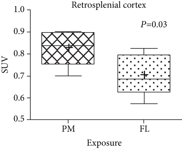
Specifically, significant increased [18F]FEPPA tracer activity was observed in the temporal lobe of the PM2.5 compared to that of the FL rats (
3.3. Immunohistochemical (IHC) Staining of the Rat Brain
The IHC staining of the hippocampus showed that the % of area with Iba1 staining at 40x high power field were

The immunostaining (brown-colored nuclei, red arrows) in the hippocampus of the PM rats (a), FL rats (b), and the differences between these two groups (c).

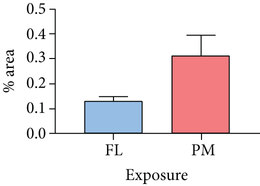
There is a trend for higher Iba1 staining (brown-colored nuclei, red arrows) in the hippocampus of PM rats (a) compared to the FL group (b), but the difference is not statistically significant (staining area of
4. Discussion
[18F]FEPPA is a potent TSPO imaging agent that has been found to be a potential tracer for imaging neuroinflammation. In order to fulfill the demand of this tracer for preclinical and clinical studies, we have developed a one-pot automated synthesis with simplified HPLC purification of this tracer, which was then, for the first time used for PET imaging of the effects of PM2.5 in rats.
[18 F]FEPPA (2) has been synthesized by several methods in various radiochemical yields and radiochemical purities [42, 43, 58, 73]. Initially, 2 was synthesized manually by nucleophilic fluorination of the corresponding tosylate-precursor (TsEPPA, 1) with [18F]Fluoride, followed by purification with HPLC, to give 2 in 71~85% yield (EOB) (Scheme 1) [43]. Later, several automated syntheses of 2 were reported [42, 58, 73] (Table 4).
Synthesis of the [18F]FEPPA (2) via various methods.
However, most of these automated syntheses used toxic MeCN as the solvent for semipreparative HPLC purification, and thus, reformulation was necessary. As a result, we and others [42] have chosen to use aqueous ethanol as the mobile phase for purification of 2 with HPLC. The HPLC purification conditions of 2 have been optimized by using a Waters Xterra RP-18 column (10 μm,

Representative semipreparative HPLC purification chromatogram of the [18F]FEPPA (2).
However, a small amount of [18F]Fluoride was detected in the eluate, which may have had an impact on PET image quality [74]. Thus, the eluate was further passed through an Alumina-N cartridge and a PTFE sterile filter in series to remove the residual [18F]Fluoride and resulted in the pure and sterile 2 in
PET imaging in rats showed that the whole-brain [18F]FEPPA tracer activity of the PM2.5 rats was significantly higher than that of their age-matched FL rats. In addition, the brain tracer activity of [18F]FEPPA was found to be regional-specific, and the difference was more pronounced in the temporal and insular lobes. In the temporal lobe, only the hippocampus and retrosplenial cortex showed statistical higher [18F]FEPPA activity in the PM2.5 rats when compared to that of the FL rats (Table 3, Figure 3). A trend of increased tracer activity was found at both insular and frontal regions in the PM2.5 rats but did not reach a statistically significant difference.
The IHC staining results showed that although there was not a statistically significant difference in the hippocampus, a trend of increased Iba1 staining in the microglia cells was observed in the PM2.5 group compared to that of the filtered air group, which suggested that microglial activation and inflammation, especially in the temporal lobe, may play important roles in the response of the brain to traffic-related PM.
Taken collectively, we have developed an improved method to automatically produce [18F]FEPPA in high quantity, high quality, and high reproducibility, and for the first time using it to noninvasively elucidate the microglial changes in different brain regions of rats after subchronic real-world exposure to ambient PM2.5. Our study confirmed that chronic subacute ambient PM2.5 exposure can lead to diffuse microglial activation. The brain area that was especially vulnerable to PM2.5 effects was the temporal lobe, particularly the hippocampus and retrosplenial cortex. These regions played crucial roles in memory, learning, and navigation, especially in the hippocampus. However, there are several limitations of the present rodent study. First, the limited sample size may influence the overall power of the test. On the other hands, this also suggests that with larger sample sizes, perhaps more brain regions will be affected, as we did observe a trend of increased [18F]FEPPA activity (not reaching statistical significance) in the insular and frontal regions of PM2.5 rats compared to their controls. Second, hypertensive rats were used in the current experiment, a subgroup that may be more vulnerable to fine PM exposure than normotensive rats. Spontaneous hypertensive rats were chosen since cardiovascular diseases are recognized as risk factors for developing neurodegenerative disease in humans and therefore may be more susceptible to traffic-related PM2.5 exposure. Third, blocking experiment using a nonradioactive FEPPA or other nonradioactive TSPO ligand was not performed in the current study. Nonetheless, [18F]FEPPA is a relatively popular ligand that had been used in many preclinical and clinical studies [75, 76], including and not limited to psychiatric disorders [62, 77]. Various studies have also demonstrated that [18F]FEPPA reliably binds to TSPO in the brain [78–80]. Lastly, not all regions of the brain were analyzed for Iba1 staining. Nevertheless, the areas of increased Iba1 staining were consistent with the [18F]FEPPA PET findings.
5. Conclusions
With this one-pot automated synthesis and improved purification of the [18F]FEPPA (2) using a FxFN Module, [18F]FEPPA (2) can be produced with high quality, quantity, and reproducibility. PET imaging in rats exposed to low-level PM2.5 demonstrated that pulmonary exposure to fine PM may exert health effects on specific areas of the brain, including the hippocampus. The microglial activation and inflammation can be noninvasively evaluated and followed by [18F]FEPPA PET imaging. The role of microglial response after low-level fine PM2.5 exposure warrants further investigation in humans. The applications of [18F]FEPPA (2) for studying inflammation in the peripheral organs of animals and humans are in progress.
Footnotes
Data Availability
The datasets used and/or analyzed during the current study are available from the corresponding authors on reasonable request.
Ethical Approval
The animal experiments adhered to the Guide for the Care and Use of Laboratory Animals and were approved by the Laboratory Animal Center at National Taiwan University (Taipei, Taiwan).
Conflicts of Interest
The authors declare that they have no competing interests.
Authors’ Contributions
MFC, TJC, YLG, HMW, WSH, and CYS were responsible for the study conception and design. TJC, HMW, and WSH were responsible for the grant support. YYH and CHC performed the radiopharmaceutical synthesis and quality control. MFC did the animal experiments. MFC and RFY interpreted the microPET images and performed the data analysis. MFC and YYH were major contributors in writing the manuscript. CYS critically reviewed and edited the manuscript for intellectual content. All authors read and approved the final manuscript. Ya-Yao Huang, Wen-Sheng Huang, and Chyng-Yann Shiue contributed equally to this work.
Acknowledgments
We are grateful to Ms. Kwanyu Lin, Ms. Yu-Ning Cheng, Mr. Pei-Yau Lin, and Mr. Chi-Han Wu, for their technical support. This work was supported by the Ministry of Science and Technology of Taiwan (MOST 103-2314-B-002-040-MY3, MOST 105-2314-B-075-060-MY3, and MOST109-2314-B-002-101) and National Taiwan University Hospital (NTUH 109-S4475).