Migraine is a common, disabling, neurological problem whose acute management would benefit from the development of purely neurally acting therapies. The trigeminocervical complex is pivotal in nociceptive signaling in migraine, and is an accepted target for putative antimigraine agents. Whole-cell patch-clamp or extracellular recordings were made of trigeminal neurons identified in rat brainstem slices. Bath application of the large conductance calcium-activated potassium (BKCa) channel opener NS1619 caused a dramatic decrease of cell firing that could be reversed by the co-application of iberiotoxin. NS1619 hyperpolarized the resting membrane potential and reduced the frequency of spontaneous action potentials in these neurons. These data suggest the presence of BKCa channels in the trigeminocervical complex. In vivo in cat L-glutamate-evoked firing was facilitated in nociceptive neurons, also responding to stimulation of the superior sagittal sinus, in the trigeminal nucleus caudalis by the BKCa peptide antagonists, iberiotoxin and slotoxin. Of units tested, 70% responded to microiontophoretic application of the blockers, identifying a subpopulation of trigeminal neurons expressing toxin-sensitive BKCa channels. NS1619 inhibited 74% of cells tested, and this was reversed by slotoxin, suggesting that the action of NS1619 in these cells was mediated through BKCa channels. These data are consistent with the presence of BKCa channels in the trigeminal nucleus caudalis that are potential targets for the development of antimigraine treatments, and may also offer insights into receptor mechanisms involved in sensitization and thus allodynia, in migraine.
Migraine is a common (1) and disabling (2) neurological disorder involving activation, or the perception of activation, of trigeminal neurons (3). It has been estimated to be the most costly neurological disorder in the European Community at more than €27 billion per year (4) and to cost the US economy $19.6 billion per year (5). Large conductance calcium-activated potassium channels, the BKCa or previously MaxiK channels, are ubiquitously expressed intrinsic membrane proteins that regulate many physiological functions (6). It has been postulated that BKCa channels constantly monitor the electrical and metabolic state of the cell based on their unique gating mechanism, being activated by a combination of membrane potential and intracellular calcium. BKCa channels are important in the nervous system (7, 8), where they act as regulators of neuronal excitability and neurotransmitter release (9, 10). Given the general concept of migraine pathophysiology including dyshabituation of normal sensory traffic (11), could BKCa channels be a target for antimigraine therapeutic development that may allow a suitable reduction in dural trigeminovascular traffic and thus be useful in migraine? Moreover, given the still substantial unmet need for new therapies in migraine of a non-vasconstrictor type, we have studied BKCa channels since they have both neural actions and no vasoconstrictor liability.
BKCa channels are very sensitive to the selective inhibition of the ‘pore-blocking’ peptides iberiotoxin (12) and slotoxin (13). Conversely, NS1619, a benzimidazolone analog, is an organic small molecule modulator of neuronal BKCa channels, and acts as a channel opener or activator (14); other novel substituted benzanilides have also been discovered (15). Compounds of this class have a potentially powerful influence in the modulation and control of numerous consequences of muscular and neuronal hyperexcitability. NS1619 activates BKCa channels in rat cortex (16, 17), and the effects of iberiotoxin and NS1619 on action potential firing of small and medium-sized sensory neurons from L6 and S1 dorsal root ganglia of adult rats have been examined in vitro(18). Iberiotoxin increases the firing frequency of the neurons in whole-cell patch-clamp studies, whereas NS1619 reversibly suppresses the action potential firing. These studies suggest that BKCa modulation can alter the firing properties of neurons in central and peripheral nociceptive pathways. Given the receptor-based targets of current therapies, such as serotonin 5-HT1B/1D receptor agonists, triptans (19), or calcitonin gene-related peptide receptor antagonists (20), a channel target would be a radically different approach to migraine therapy.
We made whole-cell patch-clamp and extracellular recordings from trigeminal nucleus caudalis (TNC) neurons identified in rat brainstem slices, with bath perfusion of NS1619 and iberiotoxin to examine the influence of BKCa channels on membrane potential and spontaneous action potential (sAP) firing in TNC neurons. The model of superior sagittal sinus (SSS) stimulation was also employed as it has proved useful in predicting agents that are effective in the acute treatment of migraine, such as the triptans (19), and has predicted lack of efficacy in other putative antimigraine agents, such as substance P/neurokinin 1 receptor antagonists (21). We explored whether modulation of BKCa activity in the cat trigeminocervical complex would effect nociceptive signaling. The effects of the selective BKCa peptide inhibitors iberiotoxin and slotoxin, as well as the activator NS1619, were tested using a combination of microiontophoresis and in vivo electrophysiology in the whole system cat SSS stimulation model.
Methods
Cat studies were conducted in accordance with a project licence issued by the Home Office of the UK under the Animals (Scientific Procedures) Act 1986 and all protocols were approved by the Institutional Animal Care and Use Committee of Amgen Inc. in accordance with the National Institutes of Health's Guide for the Care and Use of Laboratory Animals.
In vitro electrophysiology
Brain slice preparation
Transverse TNC slices were prepared from 10 Sprague-Dawley rats (2–4 weeks old; Hollister, CA, USA). The rats (35–110 g) were deeply anesthetized with an intraperitoneal injection of a pentobarbital sodium (60 mg/kg) and euthanized by decapitation, the brain and brainstem were rapidly removed and transferred to an ice-cold dissection solution containing 140 mm NaCl, 5 mm KCl, 0.4 mm KH2PO4, 0.34 mm Na2HPO4, 1.26 mm CaCl2·H2O, 1 mm MgSO4·7H2O, 5 mmN-2-hydroxyethylpiperazine-N′-2-ethanesulfonic acid (HEPES), 10 mm d-glucose, pH 7.4 with N-methyl-d-glucamine (NMG). Coronal sections (250 µm) were cut from the brainstem using a Vibroslice apparatus (World Precision Instruments, Sarasota, FL, USA). Consecutive sections containing the TNC (Sp5C, running caudally from approximately 14 mm posterior to bregma) were transferred into oxygenated artificial cerebrospinal fluid (ACSF) and incubated in a modified chamber (Warner Instruments, Hamden, CT, USA) at room temperature for at least 1 h before recording. The composition of this ACSF included 125 mm NaCl, 2.5 mm KCl, 1.25 mm KH2PO4, 2.5 mm CaCl2·H2O, 2.0 mm MgSO4·7H2O, 10 mm d-glucose and 25 mm NaHCO3. During experiments, slices were fully submerged and continuously perfused with ACSF saturated with 95% O2 and 5% CO2, at 31°C.
In vitro recording of trigeminal nucleus caudalis neurons in brain slices
TNC neurons were visualized by infrared differential interference contrast video microscopy (Axioskop 2; Zeiss, Oberkochen, Germany) using a C2741-60 Enhanced Charge-Coupled Device Camera C. Whole-cell patch-clamp and extracellular electrophysiological recordings were made using a Multiclamp 700B amplifier (Axon Instruments, Union City, CA, USA). Data were acquired using pClamp/Digidata 13200 (Axon Instruments). Patch pipettes had resistances of 3–5 MΩ for whole-cell recordings and 1–3 MΩ for extracellular recordings.
Whole-cell recordings of spontaneous excitatory post-synaptic currents (sEPSCs) were recorded from post-synaptic neurons in voltage-clamp mode. The external solution was ACSF. The holding potential was −57 mV with an internal solution containing 140 mm Cs-gluconate, 9 mm CsCl, 10 mm HEPES, 1 mm hydroxyethylenediaminetriacetic acid (HEDTA), 1 mm Mg-ATP, 0.5 mm Na2-GTP, pH 7.2 with NMG+. The addition of cesium and HEDTA to the internal solution allows the recording of only post-synaptic electrical activity. To ensure high-frequency sEPSCs, the composition of external solution ACSF was adjusted with KCl concentration from 2.5 to 10–15 mm and NaCl concentration was reduced correspondingly. Under these experimental conditions the reversal potential for Cl- was about −70 mV. Membrane potential values were corrected for the liquid junction potential of about 13.0 mV. Whole-cell and extracellular recordings for sAPs were recorded from the trigeminal neurons in current-clamp mode. The pipette solution for whole-cell patch recording of sAP contained 140 mm KCl, 10 mm HEPES, 10 mm HEDTA, 1 mm Mg-ATP, 0.5 mm Na2-GTP, pH 7.2 with KOH. The pipette solution for extracellular recordings was 3 m NaCl.
Application of drugs
All drugs were applied at known concentrations via a motorized perfusion system (403U/VM3; Watson, Wilmington, MA, USA) at a speed 2 ml/min. Stock solutions of the BKCa channel blocker iberiotoxin and slotoxin (Alomone Labs, Jerusalem, Israel), the AMPA/kainate antagonist 6-cyano-7-nitroquinoxaline-2,3-dione disodium (CNQX; Tocris Bioscience, Ellisville, MO, USA) and N-methyl-d-aspartate receptor antagonist CGS19755 (cis-4-[phosephomethyl]-piperidine-2-carboxylic acid; Tocris Bioscience) were obtained by dissolving the drugs in water. The agonists NS1619 (1-(2'-hydroxy-5'-trifluoromethylphenyl)-5-trifluoromethyl-2(3H)benzimidazolone; Sigma, St Louis, MO, USA) was dissolved in dimethylsulphoxide (DMSO) before using. The perfusion solution of iberiotoxin and slotxin contained 0.1 mg/ml albumin (Sigma) to prevent non-specific peptide binding to tubing. The final concentration of DMSO in the working solution was 0.3% (v/v). At this concentration, DMSO alone did not significantly modify the resting membrane potential or the frequency of spontaneous action potentials or sEPSCs in trigeminal neurons.
Data analysis
Data were acquired with pClamp-9.2 and analyzed with Clampfit 9.2 (Axon Instruments) for spontaneous action potentials and Minis Analysis program (Synaptosoft, Decatur, GA, USA) for sEPSCs. The sEPSC events were counted if their amplitude was above the noise (5 pA) or significantly visible. These data are presented as mean ±s.e.m. and were compared for their statistical significance using Student's t test or one-way repeated measures analysis of variance (anova) from control with Dunnett post hoc comparisons of means (Originpro v. 7; OriginLab, Northampton, MA, USA).
In vivo electrophysiology
Surgical preparation
Cats of either sex (n = 15; six female, nine male) weighing 3.2 ± 0.4 kg (mean ±s.d.), were anesthetized with α-chloralose (60 mg/kg i.p., Sigma) that had been recrystallized (22) to increase its purity, which was confirmed by liquid chromatography-electrospray ionization tandem mass spectrometry (23). Isoflurane (Merial Animal Health, Harlow, UK; 0.5−3.0% in a 40% oxygen/air carrier gas mixture) was administered from an anesthetic machine (Ohmeda-BOC Healthcare, Steeton, UK) during surgical procedures and then discontinued during experimental protocols. In each subject a catheter (4 FG × 74 cm; Portex, Hythe, UK) was placed in the left femoral artery and inserted about 15 cm from the groin up into the aorta for arterial blood sampling and continuous measurement of arterial blood pressure (DTXplus transducer, Ohmeda, Madison, WI, USA; PM-1000 amplifier, CWE, Ardmore, PA, USA). A similar catheter was placed in the left femoral vein and connected to a 150-ml buret and intravenous giving set (Soluset; Abbott, Donegal, Ireland), allowing fluid (saline for intravenous infusion BP 0.9% w/v; Baxter Healthcare, Thetford, UK) and drug administration. Amoxicillin trihydrate (150 mg; Fort Dodge Animal Health, Southampton, UK) was given by subcutaneous injection to control postoperative infection prophylactically during the experiments. Cats were intubated after local anesthesia with 2% (w/v) lidocaine hydrochloride (2–5 mg; Intubeaze, Arnolds, Shrewsbury, UK or Xylocaine Spray, AstraZeneca, Macclesfield, UK). The animals were fixed in a heavy-duty stereotaxic frame (Kopf Instruments, Tujunga, CA, USA) after 5% lidocaine and prilocaine cream (EMLA Cream; AstraZeneca) had been applied to their ear canals. Jackson urethral catheters [3 FG (toms)/4 FG (queens); Rocket Medical, Washington, UK; SIMS Portex) were inserted to drain their bladders, providing more even temperature regulation, more stable control of blood pressure through control of bladder distension, and evaluation of urine output. Corneal desiccation was prevented by coating their corneas with ocular lubricant ointment (Lacri-lube; Allergan Pharmaceuticals, Westport, Ireland).
Arterial blood gases and other biochemical parameters, including pH, calcium and lactate concentrations, were monitored using an automated analyzer (Instrumentation Laboratory, Lexington, MA, USA). Core temperature was monitored and maintained between 37 and 39°C using a rectal thermistor probe and a low-electromagnetic-noise-emitting homeothermic heater blanket system (Harvard Apparatus, Holliston, MA, USA). Cats were ventilated with a 40% oxygen in air mixture (Model 6025 ventilator; UgoBasile, Comerio, Italy), and end-tidal CO2 was continuously monitored and maintained between 2.5 and 4.0% (Capstar-100 carbon dioxide analyzer; CWE) by adjustment of respiratory rate and depth to maintain arterial blood pH within physiological limits (pH 7.35–7.45). Heart rate was monitored by electrocardiography (CT-1000; CWE), and also derived from blood pressure changes. Depth of anesthesia was monitored periodically throughout the experiment by testing for sympathetic (pupillary and cardiovascular) responses to noxious stimulation and withdrawal reflexes in the absence of neuromuscular blockade. Supplementary doses of α-chloralose in 2-hydroxypropyl-β-cyclodextrin (Sigma) were given intravenously as required, at a rate of 5–10 mg kg−1 h−1(24).
CNS surgery
A midline craniotomy (about 20 mm diameter) and C1–C2 laminectomy were performed in each cat, allowing access to the SSS and the area for recording neuronal activity in the TNC. To isolate the SSS, the adjacent dura mater and falx cerebri were dissected over a distance of about 15 mm. A small polyethylene sheet was inserted under the isolated sinus, laid over the outlying dura mater and tucked under the edges of the craniotomy. To prevent dehydration and to provide additional electrical insulation to the cortex, a circular polypropylene dam was sealed to the bone around the craniotomy with polymethylmethacrylate-based dental cement (Vertex, Zeist, the Netherlands) and filled with liquid paraffin (BDH Laboratory Supplies, Poole, UK). The likelihood of possible artefacts from arterial pulsation and respiratory movement (such as movement of the electrode from recording sites) was reduced by: bilateral pneumothoraces, kept patent with polypropylene tubes; immobilization of the spine by clamping a thoracic spinous process (1780 spinal unit; Kopf); clamping the C1 transverse processes, and clamping the remaining caudal portion of the dorsal C2 spinous process.
In vivo stimulation and recording
The isolated SSS was gently lifted onto a bipolar platinum hook electrode pair connected to a stimulus isolation unit (SIU5A; Grass Instruments, West Warwick, RI, USA). To activate primary trigeminal afferents and provide the initial search stimulus for responsive cells in the TNC, the SSS was supramaximally stimulated with stimulus-isolated (Grass SIU) square wave pulses from a Grass S88 stimulator (80–150 V, ∼50 µA; 250 µs, 0.3–0.5 Hz) after neuromuscular blockade with gallamine triethiodide (Concord Pharmaceuticals, Dunmow, UK; initially 5–10 mg/kg intravenously and maintained with 5–10 mg kg−1 h−1). The dura mater above the recording regions on the surface of the spinal cord was reflected after a midline incision and held to the bony edges of the laminectomy with N-butyl-cyanoacrylate, further stabilizing movement of the cord in this sling-like arrangement. Extracellular recordings and microiontophoretic delivery of test compounds were made using custom-built carbon-fiber containing multibarrelled-microiontophoresis combination electrodes (Carbostar 7S; Kation Scientific, Minneapolis, MN, USA). Recording electrode impedances were typically 1–3 MΩ when measured at 1 kHz in 0.9% saline (Impedance check module; FHC, Bowdoinham, ME, USA). After careful local removal of the pia mater, which was dissected away from a small area of the underlying spinal cord at the sulcus above the dorsal root entry zone, the electrodes were lowered into the cord substance around the C2 roots in the area of the dorsal root entry zone. The point of contact of the electrode tip with the pial surface was taken as the zero reference point. The electrodes were advanced or retracted in the cord substance in discrete 5-µm steps using an ultra-low-drift (<1 µm/h) LSS-100 microelectrode positioner system consisting of a piezoelectric motor (IW-711; Lateral Stability Option ± 0.2 µm lateral motion; Burleigh Instruments, Victor, NY, USA) and ultra-low-noise controller (6000ULN) attached to a heavy-duty micromanipulator (Kopf 1760–61). Tissue-culture grade agar (Sigma) 3% w/v in pyrogen-free saline (Baxter Healthcare) was set over the exposed cord after electrode insertion to further reduce cardiovascular-related movement of the spinal cord. Signal from the recording electrode attached to a high-impedance headstage preamplifier (NL100AK; Neurolog, Digitimer, Welwyn Garden City, UK) was fed via an AC preamplifier (Neurolog NL104, gain ×1000) through filters (Neurolog NL125; bandwidth about 300 Hz to 20 kHz) and a 50-Hz noise eliminator (Humbug; Quest Scientific, North Vancouver, BC, Canada) to a second-stage amplifier (Neurolog NL106) providing variable gain (to ×100). This signal (total gain about ×20 000 to ×95 000) was fed to a gated amplitude discriminator (Neurolog NL201) and analog-to-digital converter (Cambridge Electronic Design, Cambridge, UK), and to a microprocessor-based personal computer (DELL Latitude C800-PP01X; Bracknell, UK) where the signal was processed and stored. Filtered and amplified electrical signals from action potentials were fed to a loudspeaker via a power amplifier (Neurolog NL120) for audio monitoring, and were displayed on analog- and digital-storage oscilloscopes (Goldstar, LG Precision, Seoul, Korea and Metrix Electronics, Chauvin Arnoux, Paris, France, respectively) to assist the isolation of single unit activity from adjacent cell activity and noise.
To record the response of single units to SSS stimulation, post-stimulus time histograms were constructed on-line with Spike2 software (Cambridge Electronic Design) using 0.5-ms bins and saved to disk. During the experiments electrophysiological data, blood pressure, heart rate, core temperature and end-tidal CO2 were processed and recorded on VHS magnetic tape (Pulse Code Modulator; Vetter, Rebersburgh, PA, USA) and magnetic hard disk for documentation and later review.
Animals from which data are reported had cardiorespiratory parameters that were normal for α-chloralose anesthetized cats. Arterial blood gas and biochemical parameters were measured at intervals throughout experiments and were within normal limits (mean ±s.d.): arterial blood pH 7.39 ± 0.03; Pco2 2.89 ± 0.49 kPa; Po2 34.01 ± 3.16 kPa; Ca+2 1.23 ± 0.05 mmol/l; lactate 0.71 ± 0.39 mmol/l. Urine production was 4.5 ± 2.4 ml/h.
Identification of receptive fields
Cells responding to SSS stimulation were characterized as receiving low threshold mechanoreceptor input if they responded to innocuous input, such as brushing or stroking of cutaneous receptive fields on the face or forepaws with a cotton pledget on a wooden shaft. They were characterized as nociceptive specific if they responded to noxious mechanical stimuli, such as pinching with toothed forceps or pricking with a needle, or wide dynamic range (WDR) if they responded to both [see Fig. 4B, (25)]. These cells usually had an increased firing rate in response to noxious stimuli.
Localization and classification of neurons recorded in vivo. (A) A transverse section through the spinal cord at the level of C2 is represented. Although the positions of the neurons are mapped to only one side of the cord in the figure, they represent results obtained from both the left-hand and right-hand side of the spinal cord. The scale bars represent a distance of 1 mm in both directions. Solid circles indicate sites where recording sites were marked with Pontamine Sky Blue (PSB; C.I. 24410) by microiontophoresis (−1.00 µA for 20–25 min) and identified histologically. Open circles indicate the positions of unmarked recording sites, or sites where marks could not be recovered, which were identified by reference to the position of dye marks at other recording sites or at the end of electrode tracks and electrode tip coordinates. (B) Representative rate histogram showing the responses evoked by mechanical stimulation of cutaneous VI receptive field on the head of the cat by innocuous stimuli (brush with a cotton pledget) and ‘noxious’ stimulus (pinch with toothed forceps). This representative cell was characterized as receiving wide dynamic range (WDR) input. (C) Representative bipolar compound action potential evoked by l-glutamate microiontophoresis from a single trigeminocervical neuron linked to electrical superior sagittal sinus stimulation.
Microiontophoresis
Micropipettes used for microiontophoresis had orifices in the range 1–3 µm and were filled with 200 mm monosodium l-glutamate (Sigma), pH 8.0; 100 µm slotoxin or iberiotoxin (Alomone Labs) in helium-sparged 50 mm sodium acetate buffer, pH 4.5 to reduce oxidation of disulphide bonds in their native structure and ionize them as cations; 15 mm NS1619 (Sigma) in 100 mm NaOH; saline; 50 mm sodium acetate buffer, pH 4.5 or 100 mm NaOH as vehicle controls; and 2.5% Pontamine Sky Blue [‘Gurr’ 6BX dye (C.I. 24410), BDH Laboratory Supplies, Poole, UK] in 100 mm sodium acetate, pH 8.0, for marking recording positions. A microiontophoresis current generator (Model 6400A; Dagan Corp., Minneapolis, MN, USA) provided the current for ejecting test substances from the barrels. Retaining and balancing currents were used routinely (26). If cells were responsive to l-glutamate ejection, the current was adjusted to between −8 and −80 nA so that firing activity of neurons was around 15 Hz during pulses of application. The application pulses were typically 10 s duration alternated with 5 s retention, so that inhibition of cell activity, or facilitation of firing by the test substances, could be determined and distinguished from random firing and noise. Responses were quantified on rate histograms using 1-s bins. The l-glutamate ejection current required to produce a stable baseline response across at least five epochs of l-glutamate application demanded for cell testing were established empirically for each neuron. l-glutamate, NS1619, and Pontamine Sky Blue were ionized as anions and retained with small positive currents (∼3–5 nA) to restrain passive diffusion from barrels between ejection periods. Recombinant slotoxin and iberiotoxin were ionized as cations and were retained in the pipette barrels with small negative retaining currents (∼−3 to −5 nA). Ejection currents (15–400 nA) in directions opposite to the retaining currents were used. Experimental controls were based on passing currents of similar magnitude through barrels containing vehicle alone. Current balancing was provided through a barrel containing 200 mm NaCl.
After filling, electrode barrels passing useful (> 10 nA) iontophoretic currents at ±135 V compliance had resistances of 24–200 MΩin situ and impedances of 5.6–26.2 MΩ tested at 10 nA peak-to-peak at 1 kHz in saline. In particular, respective resistances and impedances for barrels containing 200 mm l-glutamate Na+, pH 8.0, were 48 ± 24 and 14 ± 2 MΩ, n = 15; 100 µm iberiotoxin in 50 mm sodium acetate, pH 4.5, 94 ± 46 and 15 ± 2 MΩ, n = 10; 100 µm slotoxin in 50 mm sodium acetate, pH 4.5, 95 ± 42 and 16 ± 4 MΩ, n = 15; 15 mm NS1619 in 100 mm NaOH, 118 ± 76 and 15 ± 7 MΩ, n = 5; saline; 50 mm sodium acetate, pH 4.5, 86 ± 32 and 17 ± 4 MΩ, n = 10; 100 mm NaOH, 120 ± 79 and 15 ± 7 MΩ, n = 5; 2.5% Pontamine Sky Blue in 100 mm sodium acetate, pH 8.0, 26 ± 18 and 12 ± 3 MΩ, n = 15; 200 mm NaCl, impedance 13 ± 2 MΩ, n = 15.
Identification of in vivo recording sites
The position of the recording electrodes was controlled by use of a heavy-duty stereotaxic micropositioner (Kopf 1760–61) with reference to the mid-point of the C2 dorsal roots. Together with the depth of the recording electrode tip with respect to the surface of the spinal cord at the dorsal root entry zone, as determined by the distance traveled display on the ULN6000 piezoelectric motor controller (Burleigh Instruments), this provided the coordinates of the recording sites. The location of selected recording sites and the end of electrode tracks were marked with microiontophoretically delivered Pontamine Sky Blue dye using a −1.00-µA current for 20–25 min. After euthanasia of the animals at the end of experiments with sodium pentobarbital (400 mg), followed by KCl (10% w/v; 5 ml), the section of spinal cord containing the recording sites was resected, fixed with neutral buffered 10% formalin, stored in phosphate buffered 30% sucrose, pH 7.4, until sunk and sectioned (40 µm; HM500 OM cryostat microtome; Microm Laborgeräte, Walldorf, Germany). Pontamine Sky Blue marks were counterstained with neutral red Nissl stain or nuclear fast red, procedures that allowed identification of the laminae of the gray matter. The positions of the recording sites within the cord were determined from histologically identified dye marks, and unmarked recording sites were located by reference to other dye marks, for example marking the end of recording tracks, and the stereotaxic coordinates of the electrode tip at the recording site.
Statistical analyses
Neuronal firing was distinguished from noise using an amplitude discriminator. Statistical evaluations were made for each unit using the mean rate of firing (Hz) evoked during each epoch of microiontophoretic l-glutamate application. The background neuronal discharge was calculated by averaging the period of ongoing basal activity immediately preceding each period of l-glutamate-evoked excitation and subtracting this value from the evoked responses. Stable baseline response to l-glutamate application was determined by examining the response to each of at least five epochs of l-glutamate application at the same current to check that there was no significant difference across the responses (27) before applying test compounds. For each unit a minimum of five paired baseline-response data were collected. To assess whether there was an effect on firing by a test compound in any individual cell we applied the critical ratio test (28–30), which uses a function of the baseline or control firing, the firing under test conditions and the standard normal deviation of this firing (31). In practice, a critical ratio of 1.96 closely approximates a 30% change from baseline or control firing evoked by l-glutamate or SSS stimulation and is significant at P < 0.05. Neuronal responses were, therefore, considered inhibited if there was a reduction in response ≥ 30%, facilitated if there was an increase in response ≥ 30%, or not affected if there was < 30% change.
Microiontophoretic currents with a magnitude of at least 80 nA, and up to 400 nA for toxins, where ‘blocking’ did not occur at lower currents, were applied for at least 10 epochs of l-glutamate application (∼2.5 min; or 5 min in the case of stimulated SSS evoked firing) before a cell was considered unresponsive. A significant change in mean firing rate was confirmed using a paired samples two-tailed t test vs. baseline, or ANOVA from control with Dunnett post hoc comparisons of means as appropriate, where significance (P) was assessed at the 0.05 level (spss v. 11.5; SPSS Inc., Chicago, IL, USA).
Results
BKCa channels modulate the firing properties of trigeminal nucleus caudalis neurons post-synaptically
Whole-cell recordings in current-clamp mode were performed on TNC neurons in freshly sectioned slices of native rat brainstem. Individual neurons were visible under infrared differential interference contrast video microscopy. Spontaneous action potentials were verified by their firing properties, i.e. shape and size (Fig. 1A), and the effect of the sodium channel blocker tetrodotoxin (1 µm).
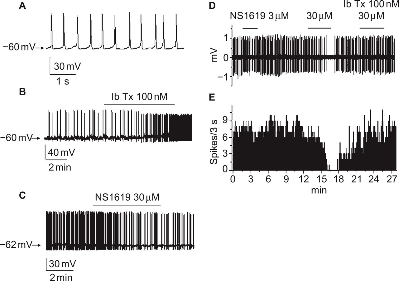
Effects of iberiotoxin and NS1619 on the excitability of individual neurons from the trigeminal nucleus caudalis (TNC) in a slice preparation. (A) Expanded time-base view of spontaneous action potentials recorded from a single TNC neuron using the whole cell patch-clamp technique in current-clamp mode. (B,C) Representative traces illustrating the effect of 100 nm iberiotoxin (IbTx) and 30 µm NS1619 on the frequency of spontaneous action potentials and the change in membrane potential in single TNC neurons (n = 13, n = 10 respectively). (D,E) Representative trace and the corresponding frequency histogram of an extracellular recording illustrating that 100 nm iberiotoxin can prevent the reduction in spontaneous firing by NS1619 (n = 15). The bin size in (E) is 3 s.
The impact of BKCa channels on the membrane potential and spontaneous action potentials was first examined by bath perfusion of the selective antagonist iberiotoxin. Bath application of iberiotoxin caused a significant depolarization (> 2 mV) in 10 of 18 neurons (56%). In seven out of 13 neurons (54%), iberiotoxin significantly increased the firing rate of spontaneous action potentials (Fig. 1B). Typically it took 1–3 min for a robust increase in firing frequency to occur, but in a couple of neurons the effect was very fast and occurred in as little as 15 s after drug perfusion was initiated. We believe the differences in time to reach peak effect were due to how deep the neurons were in the slice preparation. Interestingly, two of the seven neurons that showed positive modulation in firing in the presence of iberiotoxin exhibited decreased firing with long-term exposure (Supplemental Fig. S1). This effect could be analogous to the tachyphylaxis effect observed in the in vivo recordings (Supplemental Fig. S2). As seen in Fig. S1A, these neurons exhibited strong depolarization in the resting membrane potential in response to iberiotoxin. Presumably, the reduction in firing was due to sodium channel inactivation.
We then tested whether activating BKCa channels would have the opposite effect to iberiotoxin on the membrane potential and spontaneous action potentials. Indeed, 30 µm NS1619 hyperpolarized the resting membrane potential and reduced the frequency of spontaneous action potentials in five out of 10 neurons measured by whole-cell patch-clamp recordings (Fig. 1C). To confirm this result and allow us to run longer experiments, we tested the effect of NS1619 using an extracellular recording technique. Consistent with the whole-cell patch results, eight out of 15 neurons showed a reduction in the resting membrane potential and the spontaneous action potential firing frequency (Fig. 1D,E). These data indicate that 3 µm NS1619 caused a negligible effect on firing, but 30 µm caused a dramatic decrease that could be reversed by the co-application of iberiotoxin.
Combined perfusion of CNQX and CGS19755 did not alter the firing frequency or amplitude of the spontaneous action potentials (Supplemental Fig. S3A,B).
BKCa channels also contribute to presynaptic glutamatergic neurotransmitter release
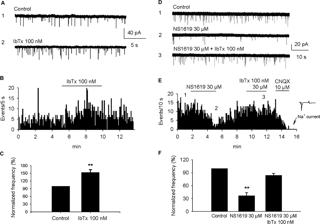
To evaluate the possibility that BKCa channels localized on presynaptic terminals may modulate transmitter release, sEPSCs were recorded before and after perfusion of iberiotoxin (Fig. 2). To prevent the influence of post-synaptic BKCa channels, the potassium channel blocker cesium (149 mm) was included in the internal patch-pipette solution. Figure 2A,B illustrates representative traces and the corresponding frequency histogram. These data show that the frequency, but not the amplitude of sEPSC events was increased in the presence of 100 nm iberiotoxin. As shown in the histogram, the frequency of sEPSCs started to rise approximately 1 min after iberiotoxin perfusion and reached a peak after 3 min perfusion. After iberiotoxin was removed the effects were reduced, but remained above baseline for the rest of these experiments. Data were summarized from eight neurons and showed that the frequency of sEPSC events was significantly increased to 153.65 ± 10.56% in the presence of iberiotoxin (Fig. 2C).
Effects of iberiotoxin and NS1619 on the frequency of spontaneous excitatory postsynaptic currents (sEPSCs) in individual neurons from the trigeminal nucleus caudalis in a slice preparation. (A) Traces illustrating sEPSCs and the effect of 100 nm iberiotoxin (IbTx) on a single neuron under whole cell patch-clamp recording in voltage-clamp mode. For this particular neuron the holding potential was −57 mV with 149 mm Cs+ and 9 mm Cl- in the internal (pipette) solution and 130 mm Cl- in external solution. (B) Frequency histogram graph of sEPSCs from the full trace data from the neuron in (A). Bin size is 5 s. (C) Histogram showing the mean effect of 100 nm iberiotoxin on the frequency of sEPSCs (n = 8 neurons). Error bar indicates s.e.m. (∗∗P < 0.01, t test). (D) Spontaneous EPSCs in control conditions (D1) and the effect of 30 µm NS1619 in the absence (D2) and presence (D3) of iberiotoxin (IbTx). Data were collected using whole cell patch-clamp in the voltage-clamp mode. Again the holding potential was −57 mV with 149 mm Cs+ and 9 mm Cl- in the internal (pipette) solution and 130 mm Cl- in the external solutions. (E) The frequency histogram of the sEPSCs from the full trace shown in (D). Bin size is 10 s. The effect of glutamatergic AMPA/kainite receptor antagonist 6-cyano-7-nitroquinoxaline-2,3-dione disodium (CNQX) was tested toward the end of experiment. At the conclusion of these experiments a sodium current was measured to verify that the neuron was still viable. (F) Histogram illustrating the mean reduction in the spontaneous EPSCs by 30 µm NS1619 and the reversal of this effect by iberiotoxin (n = 12). Error bars indicate s.e.m.[∗∗P < 0.01 compared with the control group, one-way repeated measures analysis of variance (anova) followed by a Dunnett post hoc multiple comparisons test].
Effects of NS1619 were also tested on the frequency of sEPSCs in TNC neurons (Fig. 2D–F). The raw traces and corresponding frequency histogram illustrate that 30 µm NS1619 reduced the frequency of the sEPSCs. Like the post-synaptic finding, 100 nm iberiotoxin reversed the effect of NS1619. No significant effects on the amplitude of the sEPSCs were detected after applying the BKCa modulators. Summarized data from 12 neurons show that the average frequency of sEPSCs was significantly reduced by 63 ± 7% in the presence of 30 µm NS1619 and that this effect was reversed to near baseline conditions in the presence of iberiotoxin. To verify that these sEPSC events were synaptic currents generated from the release of the excitatory neurotransmitter glutamate from the presynaptic terminal, the glutamatergic AMPA/kainate receptor antagonist CNQX was applied at the end of every experiment (Fig. 2E). Application of 10 µm CNQX completely blocked spontaneously occurring synaptic events, indicating that they were EPSCs and were mediated by glutamatergic AMPA/kainate receptors.
BKCa channel blockers facilitated TNC firing in vivo
Extracellular recordings were made from 72 neurons in the trigeminocervical complex of cats (32). Neurons responding to the SSS search stimulus that were also activated by l-glutamate were located +1 mm rostral to −4 mm caudal to the midpoint of the C2 rootlets, ±150 µm to the dorsal roots entry zone at a depth of 820–3145 µm below the dorsal surface of the spinal cord. The median depth was 1903 µm and 90th the percentile range was 1074–2984 µm (Fig. 4A). Cells responded to electrical SSS stimulation with latencies of 8–10 ms, consistent with the velocity expected from Aδ fiber input (Fig. 5). Cells received wide dynamic range or nociceptive-specific mechanoreceptor input from cutaneous VI receptive fields on the head, or cutaneous receptive fields on forepaws, or both (Fig. 3B). The cells responding to forepaw inputs were 1587 ± 298 µm deep (n = 10), median depth 1665 µm, range 1185–2115 µm as broadly reported (33).
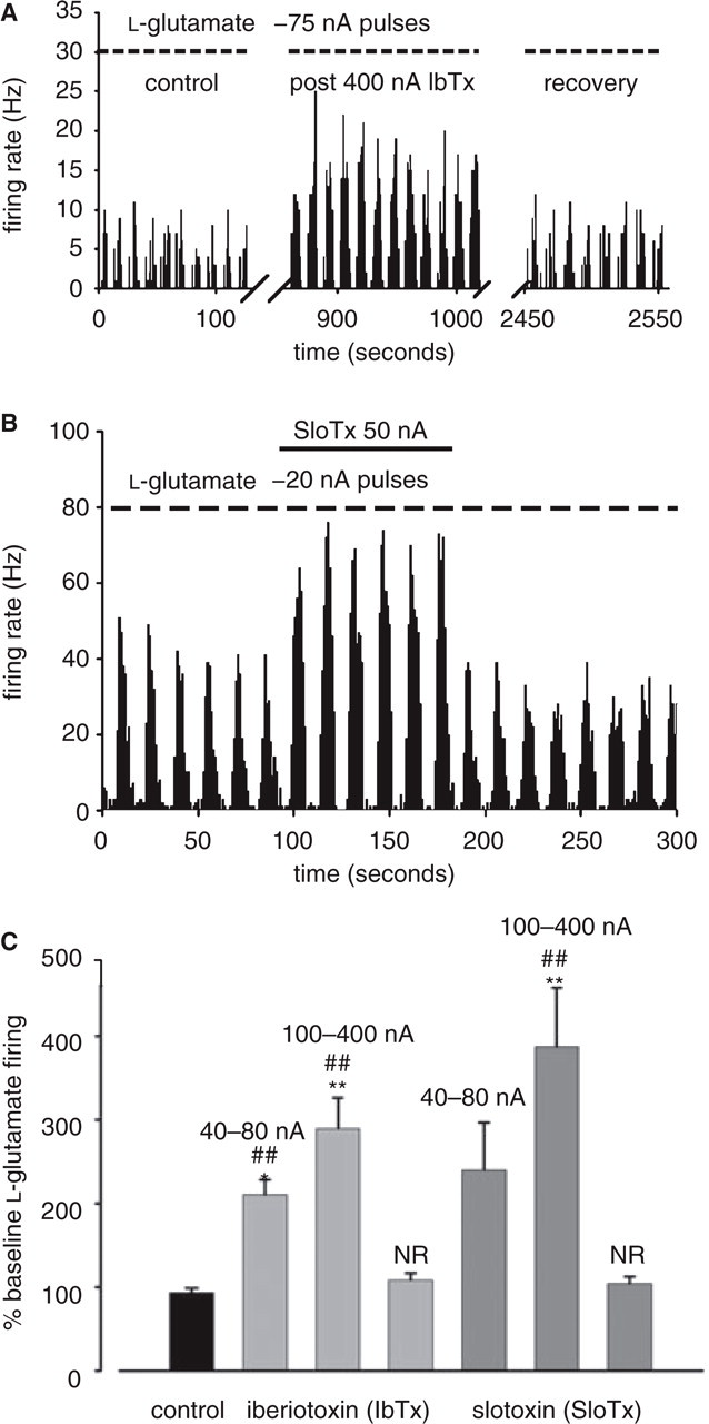
Effects of iberiotoxin and slotoxin on l-glutamate-evoked superior sagittal sinus-linked trigeminal nucleus caudalis neuron firing in vivo. (A,B) Representative rate histograms illustrating the facilitation of l-glutamate-evoked action potential firing by iberiotoxin and slotoxin. l-glutamate was pulsed for 9 s every 15 s, as indicated by the bars. Signal obtained from amplitude-discriminated spikes passing a threshold above noise level was pooled in 1-s bins displayed across the horizontal axis to give a firing rate in Hz indicated on the vertical axis. (A) Iberiotoxin (IbTx) was applied at 400 nA continuously for about 11 min between 100 and 900 s after steady-state l-glutamate responses were reached. Following facilitation of l-glutamate responses, they recovered to control levels after about 20 min. (B) Slotoxin (SloTx) was applied continuously for approximately 90 s after steady-state l-glutamate responses were reached. (C) Bar graph illustrating the dose–effect relationship of iberiotoxin and slotoxin. NR represents the average firing of neurons that were non-responsive to toxin application (80–400 nA, P > 0.2). Data are presented as mean ±s.e.m. (n = 12, 7, 17, 5, 6, 24 and 10 from left to right). (∗P < 0.05; ∗∗P < 0.001, two-tailed one-sample t test vs. baseline; ##P < 0.005, Dunnett's t test vs. vehicle control).
Effects of iberiotoxin and slotoxin on electrically evoked superior sagittal sinus (SSS)-linked trigeminal nucleus caudalis (TNC) neuron firing in vivo. (A,B) Representative post-stimulus histograms illustrating that direct electrical stimulation of the SSS recruits units (baseline controls) facilitated by iberiotoxin (IbTx) (A; 400 nA for 5 min) and slotoxin (SloTx) (B; 400 nA for 5 min). The histograms represent the cumulative frequency of action potential firing of cells in the TNC in response to 50 successive electrical stimuli of the SSS at latencies up to 50 ms divided into 0.05-ms bins. The stimulus artefact can be seen on the most left-hand side of each histogram. Supramaximal electrical stimuli were applied to the SSS via bipolar platinum hook electrodes carefully placed under the sinus. The dashed vertical lines represent cursor positions used for calculation of percentage change between various conditions and the bold figures indicate mean firing per bin between the cursors. (C) Mean effects of iberiotoxin and slotoxin on electrically stimulated SSS-evoked firing of cells showing significant facilitation by both BKCa channel blockers [P < 0.05, two-tailed one-sample t test vs. baseline (100%, dashed line), n = 5 and 5]. There was no significant difference in facilitation between the two toxins (t4 = −0.88, P > 0.43, paired-sample two-tailed t test).
Sixty-five cells were reversibly excited by microiontophoretic application of l-glutamate (–8 to −80 nA), although many more unresponsive units that were also linked to SSS stimulation were actually observed. It is likely that these units corresponded to axons or dendrites of neurons. The excitatory effect of l-glutamate identified recordings as being made from cell bodies, as characterized by their biphasic action potential morphology [Fig. 4C, (34)]. l-glutamate was ejected in pulses until the firing had reached steady state over five epochs, after which toxin was continuously ejected (100 or 50 nA) over five or six l-glutamate epochs. The firing rate of TNC neurons was 15.8 ± 1.2, 14.9 ± 1.1, 15.2 ± 1.2, 14.4 ± 1.2 and 14.8 ± 1.2 (Hz, mean ±s.e.m.) for each of five consecutive applications of l-glutamate (15.0 ± 9.8 Hz at −23 nA median; interquartile range −15 to −35 nA; n = 52). There was no difference between the responses (F3.41,268 = 2.03, P = 0.564) and their reliability across time was excellent, with a Cronbach's α of 0.98.
For neurons that had identified cutaneous receptive fields, which were always ipsilateral and mostly on the head above the ear or on the forepaw, iberiotoxin and slotoxin facilitated l-glutamate-evoked firing in seven of 10 (70%) neurons (data not shown).
In cats, microiontophoresis of iberiotoxin and slotoxin significantly facilitated l-glutamate-evoked firing in 19 of 27 (70%) and 29 of 41 (71%) TNC neurons tested, respectively (Fig. 3).
Interestingly, tachyphylaxis of the facilitation from iberiotoxin and slotoxin occurred in about one-third of the responding neurons [slotoxin six of 19 (32%); iberiotoxin 11 of 29 (38%)]. Typically the tachyphylaxis occurred at current ejections > 100 nA, at which the initial increase in firing rate was often large. Cells were considered to respond ‘tachyphylactically’ if the Pearson correlation across the first five epochs of l-glutamate application was < −0.5. The statistical significance of toxin facilitation from baseline firing was generally lost after the first or second epoch (P < 0.05, two-tailed t test). The tachyphylaxis was particularly pronounced when l-glutamate firing was evoked by very low currents, indicating that the microiontophoretic pipette tip and the neuron were in close proximity (Supplemental Fig. S2).
Facilitation of the l-glutamate-evoked firing rate by both toxins occurred in a dose-dependent manner based on ejection current magnitude (Fig. 3C). The vehicle control effect (50 mm sodium acetate, pH 4.5) was not significantly different from baseline firing when applied at similar ejection currents (40–400 nA). The l-glutamate-evoked firing rate of cells resistant to effects of the toxins (non-responders) showed insignificant change during toxin application and was unaffected by increased current ejections of the toxins.
BKCa channel blockers facilitated electrically evoked SSS TNC firing in vivo
We then sought to determine if the toxins could facilitate neuronal firing in the TNC that was evoked by an electrical stimulus. In those neurons whose l-glutamate firing was facilitated by the toxins, firing was evoked directly by electrical stimulation of the SSS. Supramaximal stimuli were applied to the SSS via bipolar platinum hook electrodes carefully placed under the sinus. Application of iberiotoxin and slotoxin (at 400 nA) for 5 min nearly doubled the neuronal firing in response to the electrical stimuli (Fig. 5). The histograms represent the cumulative frequency of action potential firing of cells in the TNC in response to 50 successive electrical stimuli of the SSS at latencies up to 50 ms divided into 0.5-ms bins (the stimulus artefact just after 0 ms latency can be seen on the most left-hand side of the histograms). Cell firing frequency returned to baseline control levels about 30 min after toxin application was stopped.
NS1619 decreased l -glutamate-evoked firing in SSS-linked TNC cells in vivo
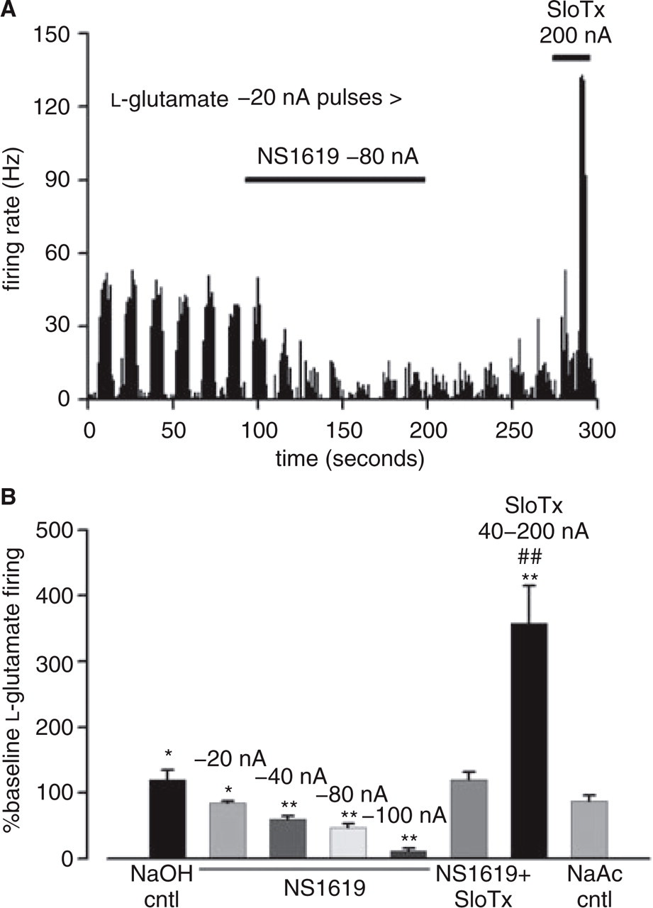
The effects of the BKCa selective toxins suggested that BKCa is expressed in the nociceptive pathway of the trigeminocervical complex, and that BKCa inhibition increases the firing frequency of most neurons that receive nociceptive input. Therefore, we hypothesized that BKCa agonism would reduce firing in a toxin-sensitive manner. Indeed, NS1619 significantly inhibited l-glutamate-evoked firing in 14 of 19 (74%) cells tested. The inhibition was persistent across at least five epochs of l-glutamate application in each cell, and the inhibition slowly reversed after NS1619 application was stopped. In 10 of 10 (100%) cells tested, this inhibition was reversed to baseline firing or above by the application of slotoxin (Fig. 6). A clear dose–effect relationship of NS1619 was observed on neurons where increasing ejection currents were used (from −20 to −100 nA). Activity of current-matched control ejections from barrels containing vehicle controls had a much less and usually insignificant (P > 0.05) effect on l-glutamate-evoked firing compared with the test compounds. These results suggest that the action of NS1619 in the neurons receiving nociceptive input in the trigeminocervical complex is mediated, at least in part, through BKCa channels.
NS1619 inhibits l-glutamate-evoked firing of superior sagittal sinus-linked trigeminal nucleus caudalis neurons in vivo. (A) l-glutamate was ejected in pulses (–20 nA for 9 s of every 15 s) as indicated by the bars. When l-glutamate-evoked firing had reached a steady state over five epochs, the potassium channel opener NS1619 was microiontophoretically co-ejected (–80 nA) over a period of seven l-glutamate pulses as indicated by the solid horizontal bar. This effect reversed slowly and was sensitive to slotoxin (SloTx) when microiontophoretically ejected (200 nA). (B) Mean effects of microiontophoretic application of NS1619, NS1619 co-ejected with slotoxin, and slotoxin alone. The vertical axis represents the mean response to the test substances over five l-glutamate pulses, where the firing response to l-glutamate had reached a steady state, or for the first l-glutamate pulse where tachyphylaxis was sometimes observed at high (SloTx) currents, expressed as a percentage of baseline l-glutamate-evoked firing. NS1619 inhibition of l-glutamate-evoked action firing was dose dependent from −20 to −100 nA (∗P < 0.01, ∗∗P < 0.001, two-tailed one-sample t test vs. baseline). Slotoxin delivered (+40 to +80 nA) in the presence of NS1619 (–40 to −80 nA) reversed the suppression of l-glutamate-evoked action potential firing to around control levels (t9 = 1.68, P = 0.127, two-tailed one-sample t test). Slotoxin alone significantly facilitated l-glutamate-evoked firing [##P < 0.01, anova from sodium acetate, pH 4.5, vehicle control (NaAc ctrl) with Dunnett post hoc comparison]. Ejection of the 100-mm sodium hydroxide vehicle control (NaOH cntl) did not produce significantly different change from baseline l-glutamate-evoked firing (t4 = 1.4, P > 0.23, two-tailed, one-sample t test; n = 5, 5, 9, 7, 7, 10, 30 and 6 from left to right).
Discussion
These data provide direct evidence that large conductance calcium-activated potassium (BKCa) channels modulate the firing properties of neurons with dural trigeminovascular nociceptive input. Three independent lines of evidence support this interpretation. First, the BKCa selective peptide inhibitors, slotoxin and iberiotoxin, dramatically facilitated the firing evoked by microiontophoretic application of l-glutamate in vivo and iberiotoxin facilitated the spontaneous activity of TNC neurons in slice preparations. Second, the archetypical BKCa activator NS1619 reversed TNC neuron firing in a concentration-dependent and toxin-sensitive manner both in vivo and in slice recordings. Third, slotoxin and iberiotoxin facilitated the firing evoked by direct electrical stimulation of the SSS, a pain-producing intracranial structure used to model nociceptive processing in migraine. The data suggest that the BKCa channel may offer a non-vasoconstrictor novel therapeutic target for the management of migraine.
BKCa modulation of nociceptors
The data demonstrate that BKCa channel modulators affect neuronal activity in response to excitatory chemical and electrical stimuli within the trigeminovascular nociceptive pathway. However, we are unable to determine in the in vivo preparation whether the modulation occurs presynaptically, on the invading axon from the primary sensory afferent emanating from the trigeminal ganglion, or post-synaptically, on the second-order neuron within the TNC itself, or at both sites. Since combined perfusion of CNQX and CGS19755 did not alter the firing frequency or amplitude of the spontaneous action potentials in the brain slice preparation, this suggests that the spontaneous action potentials from TNC neurons in that preparation are generated from intrinsic activity of the post-synaptic neurons. In patch clamping experiments, cesium was used to block post-synaptic BKCa channels to evaluate the possibility that BKCa channels localized on presynaptic TNC terminals may modulate neurotransmitter release. NS1619 reduced the frequency of sEPSCs and iberiotoxin reversed the effect of NS1619 under these conditions, indicating that BKCa channels also contribute to presynaptic neurotransmitter release. The complete block of sEPSCs by CNQX at the conclusion of these experiments indicates that they were mediated by glutamatergic AMPA/kainate receptors after presynaptic glutamate release under the influence of BKCa channels.
Strong evidence supports the expression and physiological role of toxin-sensitive BKCa channels on nociceptors. Small and medium-sized neurons (putative nociceptors) from primary cultures and thin slice preparations of rat dorsal root ganglion express toxin-sensitive BKCa channels (8, 18). In these in vitro settings, voltage-clamp experiments of single BKCa channels, as well as current-clamp recordings of whole neurons, indicate that the agonists NS1619, ethanol, and trichloroethanol, as well as the pore-blocking peptide iberiotoxin, have profound effects on the single channel kinetics, the action potential shape, as well as intrinsic firing properties. Similarly, NS1619 reduced single unit firing of Aδ and C-fibers recorded from guinea pig trachea and blunted the citric acid aerosol cough response in an iberiotoxin-sensitive manner. Several of these studies reported that a majority of the neurons tested (∼55–70%) express iberiotoxin-sensitive BKCa channels. This is in direct agreement with our results, where ∼70% of the TNC neurons electrically linked to the SSS (Aδ fiber input) were toxin sensitive and more than half of the TNC neurons tested in slice preparations significantly depolarized or increased their rate of firing in response to bath application of iberiotoxin. Little has been reported about BKCa channel expression or modulation on second-order neurons within the dorsal horn of the spinal cord or in the TNC. However, given the ubiquitous nature of BKCa channel expression within the central nervous system, it would be surprising if BKCa channels were not also expressed in these neurons.
BKCa channels have different functional roles depending on their subcellular localization. On the cell body, for example, BKCa channels reduce repetitive firing by shortening the action potential duration, speeding repolarization, and contributing to the fast after-hyperpolarization. Alternatively, at the presynaptic terminal, BKCa channels contribute to neurotransmission by counteracting calcium influx, thereby limiting vesicle release. Therefore, it is entirely possible that different mechanisms underlie the BKCa modulatory effects found in the l-glutamate paradigm, which uses local excitatory chemical stimuli in the TNC, and the electrical stimulation paradigm, which activates the peripheral afferent nerve ending. The electrophysiological data using extracellular and whole-cell recordings from rat TNC neurons in slice preparations suggest that BKCa modulation affects both the synaptic activity of the invading sensory neuron and the firing properties of the post-synaptic second-order neuron.
Potential impact of regulatory β-subunits
BKCa channels can be formed by homotetramers of α-subunits or by complexes with at least four families of β-subunits (β1–4). The coexpression of β-subunits, which have two transmembrane domains and a large extracellular loop, modifies the BKCaα-subunit kinetic properties, intracellular calcium sensitivity, intrinsic modulation, and pharmacology such as toxin sensitivity. Slotoxin and iberiotoxin specifically and reversibly inhibit the BKCa channel pore-forming α-subunit as ‘pore-blockers’(35). However, slotoxin is capable of differentiating three types of BKCa channel complexes, α, α + β1, and α + β4 (13). Slotoxin inhibits the homomeric α-subunit with very low potency (IC50∼1–10 nm) and the block is reversible upon wash-out. When α + β1 is the predominate BKCa channel complex, which is common in smooth muscle, but rare in neurons, where β1 expression is very low, slotoxin inhibits with very low potency (IC50∼1–10 nm), but the inhibition is irreversible. However, when α + β4 is the predominating complex, which is possible in neurons because β4 expression is high, slotoxin is impotent (activity shifted ∼1000-fold lower), the on-rate is extremely slow (of the order of minutes), and the inhibition is irreversible. Given that in our experiments the facilitation of firing by slotoxin was rapid (within tens of seconds) and reversible, the results indicate that either the homomeric α-subunit is the predominant channel complex or that the α + β4 complex is toxin-sensitive in these trigeminal neurons. In addition, it is possible that the two other β-subunits that form a complex with the α-subunit (α + β2 and α + β3), of which less in known about their physiological role, could contribute to the pharmacological effects or potentially even underlie the mechanism of the tachyphylaxis observed in some neurons.
Clinical relevance
To explore a possible therapeutic potential for BKCa modulators we employed the model of SSS stimulation. Stimulation of dural structures is painful in humans (36), and in animal models, stimulation of dural sites, including the SSS and middle meningeal artery, results in neuronal activation in the trigeminovascular system. This activation is significantly inhibited by acute antimigraine drugs (37–45). Moreover, compounds found to be ineffective in acute migraine, such as substance P/neurokinin 1 receptor antagonists (21) and plasma protein extravasation inhibitors (46), are also ineffective in blocking trigeminocervical transmission in this model [e.g. (47, 48)]. This model compares favorably with investigation of the aura homologue phenomenon cortical spreading depression [CSD, (49)], which has not been predictive at all of the likely utility of acute antimigraine treatments (50, 51). The CSD model may be more useful in predicting effectiveness of preventive medicines (52, 53). Inhibition of trigeminal nerve activation is therefore helpful in evaluating new targets for the treatment of acute migraine. Another interesting aspect of the BKCa channel in migraine may be sensitization, the clinical manifestation of which is likely to be allodynia (54, 55). Our data indicate that should the BKCa channel be dysfunctional, as modeled by channel blocker treatments, trigeminal neurons become sensitized. Since not all migraineurs have allodynia, might it be that allodynia represents an additional functional burden in that group mediated by cosegregation of migraine biology and BKCa, or a similar channel, dysfunction? Finally, the widespread distribution of these channels on smooth muscle in vessels and in the lung may provide both challenges and opportunities. As dilators they might be predicted to reduce blood pressure or even have therapeutic actions in airways disease, although one might observe that simple is best in therapeutics and widespread actions may produce unwanted side-effect issues at some point (56).
Concluding remarks
BKCa channels provide a negative feedback mechanism for cells to pass strong hyperpolarizing potassium currents during times of elevated firing frequencies that cause prolonged depolarizations and elevated intracellular calcium levels. Substances such as slotoxin and iberiotoxin, which block these channels, remove the BKCa-dependent negative feedback loop to membrane polarization and cause neuronal hyperexcitability. Conversely, BKCa activators, such as NS1619, cause membrane hyperpolarization and reduce neuronal excitability and therefore have a potentially powerful influence in blunting the repetitive firing seen in many disease states. One such example of an important disease state where such mechanisms could be conceived would be migraine. Our results are consistent with the presence of large conductance, calcium-activated potassium channels on nociceptive trigeminovascular neurons. These channels may provide a completely novel target for drugs aimed at the treatment of primary neurovascular headaches, such as migraine and cluster headache.
Competing interests
D.C.I. and R.Y. are employees of Amgen.
Acknowledgements
The authors thank Paul Hammond and Michele Lasalandra for their excellent technical assistance. Dr Ken Hunter, Elizabeth Sharp, and Dr Michael Taylor of the Scottish Agricultural Science Agency for analysis of chloralose samples.
References
1.
LiptonRBStewartWFDiamondSDiamondMLReedM. Prevalence and burden of migraine in the United States: data from the American Migraine Study II. Headache2001; 41:646–57.
2.
MenkenMMunsatTLTooleJF. The global burden of disease study: implications for neurology. Arch Neurol2000; 57:418–20.
3.
GoadsbyPJLiptonRBFerrariMD. Migraine—current understanding and treatment. N Engl J Med2002; 346:257–70.
4.
Andlin-SobockiPJönssonBWittchenHUOlesenJ. Cost of disorders of the brain in Europe. Eur J Neurol2005; 12 (Suppl.1):1–27.
5.
StewartWFRicciJACheeEMorgansteinDLiptonR. Lost productive time and cost due to common pain conditions in the US workforce. JAMA2003; 290:2443–54.
6.
GribkoffVKStarrettJEJrDworetzkySI. The pharmacology and molecular biology of large-conductance calcium-activated (BK) potassium channels. Adv Pharmacol1997; 37:319–48.
7.
RobitailleRGarciaMLKaczorowskiGJCharltonMP. Functional colocalization of calcium and calcium-gated potassium channels in control of transmitter release. Neuron1993; 11:645–55.
8.
ScholzAGrußMVogelW. Properties and functions of calcium-activated K+ channels in small neurones of rat dorsal root ganglion studied in a thin slice preparation. J Physiol1998; 513:55–69.
9.
VogalisF. Potassium channels in gastrointestinal smooth muscle. J Auton Pharmacol2000; 20:207–19.
10.
VolkKAMatsudaJJShibataEF. A voltage-dependent potassium current in rabbit coronary artery smooth muscle cells. J Physiol1991; 439:751–68.
GribkoffVKStarrettJEJrDworetzkySI. Maxi-K potassium channels: form, function, and modulation of a class of endogenous regulators of intracellular calcium. Neuroscientist2001; 7:166–77.
13.
Garcia-ValdesJZamudioFZToroLPossaniLD. Slotoxin, alphaKTx1.11, a new scorpion peptide blocker of MaxiK channels that differentiates between a and a+β (β1 or β4) complexes. FEBS Lett2001; 505:369–73.
14.
OlesenS-PMunchEMoldtPDrejerJ. Selective activation of Ca2+-dependent K+ channels by novel benzimidazolone. Eur J Pharmacol1994; 251:53–9.
15.
BiagiGGiorgiILiviONardiACalderoneVMartelliASynthesis and biological activity of novel substituted benzanilides as potassium channel activators. V. Eur J Med Chem2004; 39:491–8.
16.
LeeKRoweICMAshfordMLJ. NS 1619 activates BKCa channel activity in rat cortical neurones. Eur J Pharmacol1995; 280:215–9.
17.
SellersAJAshfordMLJ. Activation of BKCa channels in acutely dissociated neurones from the rat ventromedial hypothalamus by NS 1619. Br J Pharmacol1994; 113:659–61.
18.
ZhangX-FGopalakrishnanMShiehC-C. Modulation of action potential firing by iberiotoxin and NS1619 in rat dorsal root ganglion neurons. Neuroscience2003; 122:1003–11.
19.
GoadsbyPJ. The pharmacology of headache. Prog Neurobiol2000; 62:509–25.
20.
GoadsbyPJ. Calcitonin gene-related peptide (CGRP) antagonists and migraine—is this a new era?Neurology2008; 70:1300–1.
21.
MayAGoadsbyPJ. Substance P receptor antagonists in the therapy of migraine. Expert Opin Investig Drugs2001; 10:673–8.
22.
WhiteAHixonRM. Structure of the chloraloses, alpha and beta-glucochloraloses. J Am Chem Soc1933; 55:2438–44.
23.
HunterKTaylorMJSharpLMMeltonLMBouhellecS. Determination of chloralose residues in animal tissues by liquid chromatography-electrospray ionization tandem mass spectrometry. J Chromatogr B2004; 805:303–9.
24.
StorerRJButlerPHoskinKLGoadsbyPJ. A simple method, using 2-hydroxypropyl-β-cyclodextrin, of administering α-chloralose at room temperature. J Neurosci Methods1997; 77:49–53.
25.
HuJWDostrovskyJOSessleBJ. Functional properties of neurons in cat trigeminal subnucleus caudalis (medullary dorsal horn). I. Responses to oral-facial noxious and nonnoxious stimuli and projections to thalamus and subnucleus oralis. J Neurophysiol1981; 45:173–92.
26.
BloomFE. To spritz or not to spritz: the doubtful value of aimless iontophoresis. Life Sci1974; 14:1819–34.
27.
StorerRJAkermanSGoadsbyPJ. Characterization of opioid receptors that modulate nociceptive neurotransmission in the trigeminocervical complex. Br J Pharmacol2003; 138:317–24.
28.
MandelbrodIFeldmanSWermanR. Inhibition of firing is the primary effect of microelectrophoresis of cortisol to units in the rat tuberal hypothalamus. Brain Res1974; 80:303–15.
29.
MandelbrodIFeldmanSWermanR. Mediobasal hypothalamic neurons are excited by the iontophoretic application of sodium. Brain Res1983; 273:35–44.
30.
NaglerJConfortiNFeldmanS. Alterations produced by cortisol in the spontaneous activity and responsiveness to sensory stimuli of single cells in the tuberal hypothalamus of the rat. Neuroendocrin1973; 12:52–66.
31.
ArmitagePBerryG. Statistical methods in medical research, 3rdedn.Oxford: Blackwell Science1994.
32.
StorerRJGoadsbyPJ. Microiontophoretic application of serotonin (5HT)1B/1D agonists inhibits trigeminal cell firing in the cat. Brain1997; 120:2171–7.
33.
CarstensETrevinoDL. Anatomical and physiological properties of ipsilaterally projecting spinothalamic neurons in the second cervical segment of the cat's spinal cord. J Comp Neurol1978; 182:167–84.
34.
FusseyIFKiddCWhitwamJG. The differentiation of axonal and soma-dendritic spike activity. Pflugers Arch1970; 321:283–92.
35.
MacKinnonRMillerC. Mechanism of charybdotoxin block of the high-conductance, Ca2+-activated K+ channel. J Gen Physiol1988; 91:335–49.
36.
RayBSWolffHG. Experimental studies on headache. Pain sensitive structures of the head and their significance in headache. Arch Surg1940; 41:813–56.
37.
HoskinKLGoadsbyPJ. Comparison of more and less lipophilic serotonin (5HT1B/1D) agonists in a model of trigeminovascular nociception in cat. Exp Neurol1998; 150:45–51.
38.
HoskinKLKaubeHGoadsbyPJ. Central activation of the trigeminovascular pathway in the cat is inhibited by dihydroergotamine. A c-Fos and electrophysiological study. Brain1996; 119:249–56.
39.
HoskinKLKaubeHGoadsbyPJ. Sumatriptan can inhibit trigeminal afferents by an exclusively neural mechanism. Brain1996; 119:1419–28.
40.
CumberbatchMJHillRGHargreavesRJ. The effects of 5-HT1A, 5-HT1B and 5-HT1D receptor agonists on trigeminal nociceptive neurotransmission in anaesthetized rats. Eur J Pharmacol1998; 362:43–6.
41.
CumberbatchMJHillRGHargreavesRJ. Rizatriptan has central antinociceptive effects against durally evoked responses. Eur J Pharmacol1997; 328:37–40.
42.
CumberbatchMJHillRGHargreavesRJ. Differential effects of the 5HT1B/1D receptor agonist naratriptan on trigeminal versus spinal nociceptive responses. Cephalalgia1998; 18:659–63.
43.
KaubeHHoskinKLGoadsbyPJ. Inhibition by sumatriptan of central trigeminal neurones only after blood–brain barrier disruption. Br J Pharmacol1993; 109:788–92.
44.
KaubeHHoskinKLGoadsbyPJ. Intravenous acetylsalicylic acid inhibits central trigeminal neurons in the dorsal horn of the upper cervical spinal cord in the cat. Headache1993; 33:541–4.
45.
LambertGALowyAJBoersPMAngus-LeppanHZagamiAS. The spinal cord processing of input from the superior sagittal sinus: pathway and modulation by ergot alkaloids. Brain Res1992; 597:321–30.
46.
PeroutkaSJ. Neurogenic inflammation and migraine: implications for therapeutics. Mol Interv2005; 5:304–11.
47.
GoadsbyPJHoskinKLKnightYE. Substance P blockade with the potent and centrally acting antagonist GR205171 does not effect central trigeminal activity with superior sagittal sinus stimulation. Neuroscience1998; 86:337–43.
48.
GoadsbyPJHoskinKL. Differential effects of low dose CP122,288 and eletriptan on Fos expression due to stimulation of the superior sagittal sinus in cat. Pain1999; 82:15–22.
49.
LauritzenM. Pathophysiology of the migraine aura. The spreading depression theory. Brain1994; 117:199–210.
50.
HansenAJLauritzenMTfelft-HansenP. Spreading cortical depression and antimigraine drugs. In: AmeryWKNuetenJMVWauquierA, eds.Pharmacological basis of migraine therapy. London: Pitman1982; 139–54.
51.
KaubeHGoadsbyPJ. Anti-migraine compounds fail to modulate the propagation of cortical spreading depression in the cat. Eur Neurol1994; 34:30–5.
52.
AkermanSGoadsbyPJ. Topiramate inhibits cortical spreading depression in rat and cat: impact in migraine aura. Neuroreport2005; 16:1383–7.
53.
AyataCJinHKudoCDalkaraTMoskowitzMA. Suppression of cortical spreading depression in migraine prophylaxis. Ann Neurol2006; 59:652–61.
54.
SelbyGLanceJW. Observations on 500 cases of migraine and allied vascular headache. J Neurol Neurosurg Psychiatry1960; 23:23–32.
55.
BursteinRCutrerMFYarnitskyD. The development of cutaneous allodynia during a migraine attack. Brain2000; 123:1703–9.
56.
EichhornBDobrevD. Vascular large conductance calcium-activated potassium channels: functional role and therapeutic potential. Naunyn Schmiedebergs Arch Pharmacol2007; 376:145–55.