
Abstract
Ophthalmoplegic migraine (OM) is a rare condition with an incidence of approximately 0.7 per million (1). It is characterized by recurrent attacks of unilateral migrainous headache followed by ophthalmoplegia. In most cases the oculomotor nerve (CNIII) is affected (1), but the abducent (CNVI) or the trochlear nerve may also be involved (2–5). Although the diagnostic criteria for OM include ‘migraine-like headache’, this disease has recently been reclassified by the Headache Classification Subcommittee of the International Headache Society (IHS) as a form of neuralgia instead of migraine (6, 7).
The pathogenesis of OM is still unclear (8, 9), especially in those cases in which the CNVI is involved. Reversible contrast enhancement of the cisternal segment of CNIII has been observed in almost all cases of OM with involvement of the CNIII (10–13), suggesting an inflammatory aetiology for this form of OM. Possible pathoaetiologies being considered are recurrent inflammation similar to the Tolosa–Hunt syndrome (10, 13), recurrent demyelinating neuropathy (9), microvascular origin (14) and schwannoma (15). Magnetic resonance imaging (MRI) of OM with CNVI palsy is typically reported to be completely normal (2, 5, 11). Thus, the pathogenesis of OM with abducens nerve palsy is still completely uncertain and enigmatic.
We report on a woman who developed adult-onset OM with recurrent CNVI palsy and showed compression and distortion of the CNVI at its point of exit from the brainstem by the basilar artery and the anterior inferior cerebellar artery (AICA). We propose that neurovascular compression is the underlying cause of OM.
Case report
A 51-year-old woman presented with a history of ophthalmoplegic migraine of 11 years' duration. In 1996 she experienced the first episode of a severe right-sided, initially hemicranial, later on retrobulbar headache lasting for about 4 days. This was followed by horizontal double vision during right gaze as a consequence of a nearly complete abducens nerve paresis that fully recovered after some days. During the following years she had up to four such attacks per year. The duration of the sixth nerve palsy increased up to 2 months. The headache phase lasted 2–4 days, and the headache was migraine-like with some degree of phono- and photophobia as well as nausea. Sumatriptan as well as ibuprofen and acetylsalicylic acid were of no benefit.
Furthermore, the patient had had migraine with visual aura since her second decade with one attack every 2 months. These attacks normally responded to ibuprofen and she reported on a familial history of migraine (sister).
Two days after onset of an attack, the woman presented at the out-patient clinic of our Neurology Department. She suffered from right-sided, retrobulbar headache. An orthoptic examination revealed normal vision in both eyes, but indicated a slight deficiency of abduction with reduced motility of the right eye, and a slight gaze-evoked nystagmus during maximal gaze to the right. She reported double vision during lateral gaze of more than 30° to the right. There was no further neurological deficit.
Blood count, sedimentation rate, blood glucose, as well as other laboratory parameters were normal. Earlier cerebrospinal fluid (CSF) analysis, including angiotensin converting enzyme, intrathecal immunoglobulin production, syphilis and Lyme serology as well as herpes virus, Epstein–Barr virus (EBV) and cytomegalovirus serology were normal. Serum immunological tests showed antibodies against herpes simplex and EBV. There were no signs of intrathecal immune responses to herpes virus or EBV.
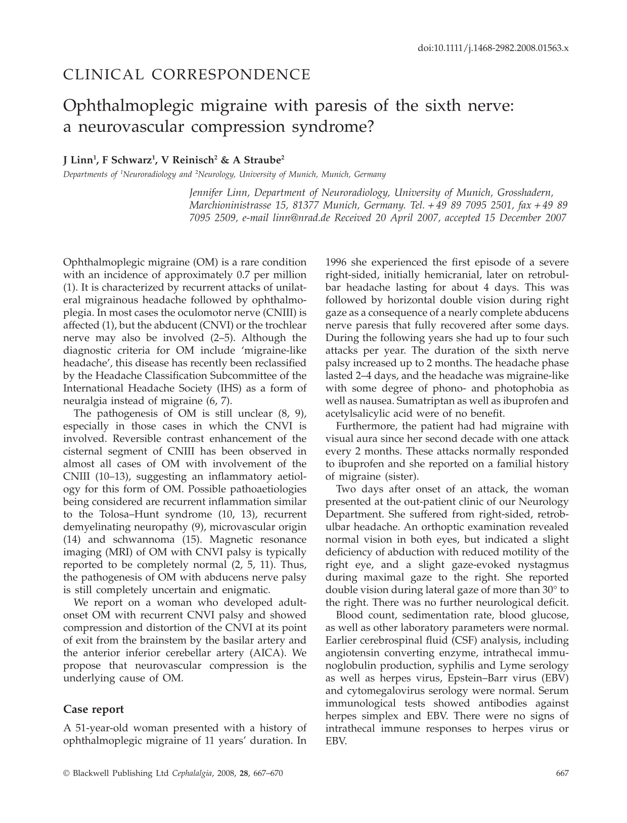
An MRI examination was performed at the day of admission on a 3.0-T scanner (Signa; General Electrics, Fairfield, CT, USA), including a T2-weighted sequence (TR = 5900 ms, TE = 118 ms, slice thickness = 3 mm), a high-resolution 3D-FIESTA- (TR = 4.528 ms, TE = 1.804 ms, slice thickness = 0.6 mm) and a gadolinium-enhanced 3D-FSPGR-sequence (TR = 7.168 ms, TE = 3.15 ms, TI = 500 ms, slice thickness = 1.4 mm), as well as 3D-time of flight MR angiography (TR = 17 ms, TE = 5.7 ms, slice thickness = 0.8 mm). It revealed neurovascular contact between the right CNVI at its point of exit from the brainstem (PE) and the basilar artery as well as the right AICA at the PE of the right AICA from the basilar artery, which caused compression and displacement of the right CNVI to the right side (Fig. 1). Apart from this, the MRI showed no pathological findings, especially no contrast enhancement of the cranial nerves in their cisternal course, no pathology in the cavernous sinus and no aneurysm.

The high-resolution 3D-FIESTA-sequence performed on a 3.0-T scanner demonstrates neurovascular contact between the right CNVI (dotted arrows) at its point of exit from the brainstem and the basilar artery (arrows) as well as the right AICA (arrowheads) at the point of exit of the right AICA from the basilar artery (A–D), which causes compression and displacement of the right CNVI to the right side (C).
Discussion
According to the diagnostic criteria of the IHS (6), our patient had migraine with aura (visual aura) and OM with involvement of the right CNVI. Using high-resolution 3D MRI sequences, we were able to demonstrate a vascular compression of the involved nerve at its PE by the basilar artery and the AICA.
To date, the pathogenesis of OM, especially with involvement of CNVI, is an unsolved problem (2, 8–10). In patients with oculomotor OM, contrast enhancement and thickening of the oculomotor nerve are nearly constant findings (10–13). On the basis of these observations, several pathogenic mechanisms of OM have been proposed, for example, a viral or non-infectious inflammatory disease like the Tolosa–Hunt syndrome (1, 10–12), recurrent demyelinating neuropathy (9) and schwannoma (15). Other groups have found narrowing of the distal portion of the carotid artery in patients with oculomotor OM and have suggested either direct vascular compression or vascular ischaemic neuropathy as the underlying cause (11, 16).
In contrast to the observations in oculomotor OM, cranial MRI in patients with OM and abducens palsy reported so far is typically normal and shows no contrast enhancement of the nerve. Only one case in the literature has reported an enhancing lesion of unknown aetiology in the brainstem in a patient with OM and CNVI palsy (3).
Although extremely rare, vascular compression of the CNVI in its cisternal course can cause intermittent CNVI palsy. Kalidindi et al., for example, have reported a case of persistent trigeminal artery in contact with the CNVI, which resulted in CNVI palsy (17). Nevertheless, neurovascular contact of the CNVI in its cisternal course has not been described before in OM.
To the best of our knowledge, we are the first to apply high-resolution, heavily T2-weighted sequences, such as the 3D-FIESTA sequence, to patients with OM. This sequence offers high-spatial resolution, since it can be performed with thin sections (0.6 mm), and thus is able to depict small structures surrounded by CSF with high contrast. It has been successfully used to visualize various cranial nerves (18).
Compression, displacement or distortion of the cranial nerves by neurovascular contacts is known to cause a wide variety of neurovascular compression syndromes, e.g. trigeminal neuralgia (19, 20), glossopharyngeal neuralgia (21), facial hemispasm (22) and superior oblique myokymia (23).
At present, several hypotheses regarding the exact pathophysiology of neurovascular compression syndromes are under discussion (24). The most widely accepted theory postulates that nerve compression results in reversible demyelination of the respective nerve, a mechanism that is also under consideration in the pathogenesis of ophthalmoplegic migraine with involvement of the third nerve (9).
The attack-like appearance of OM, a typical feature of the neurovascular compression syndromes, the re-classification of this disease by the IHS under the heading of ‘cranial neuralgias and central causes of facial pain’ (6), and the MRI findings in our patient lead us to suggest that OM with abducens nerve involvement might also be a form of neurovascular compression syndrome in some patients. The incidence of neurovascular compression syndromes is known to be higher in elderly patients with elongated intracranial vessels (25). Interestingly, ophthalmoplegic migraine with sixth nerve palsy also typically seems to occur in patients who are older compared with those with ophthalmoplegic migraine with oculomotor nerve palsy (2, 4).
Although discussed for decades now, the underlying mechanisms of several characteristics of neurovascular compression syndromes are still unclear, e.g. regarding the paroxysmal occurrence of the symptoms, as well as the stop mechanism (24, 26). As regards ophthalmoplegic migraine, it remains unclear why the paresis usually starts after a delay of 24–72 h following the onset of the headache attack. To date, cerebral vasodilation is widely seen as one of the predominant causative factors in the development of migraine pain (27). There is transcranial Doppler evidence for vascular dilation in the headache phase, responding to sumatriptan (28). If a neurovascular contact is present, this might be intensified by the dilation of the cerebral vessels, resulting in irritation and distortion of the nerve.
The absence of contrast enhancement of the nerve makes it very unlikely, but not impossible, that there was secondary inflammation or microvascular infarction of the sixth nerve, which finally caused the palsy.
In conclusion, the vascular compression of the affected nerve in a patient with OM with abducens palsy suggests that neurovascular compression is a possible pathogenic factor in OM.