
Abstract

Ophthalmoplegic migraine (OM) has been classified by the Headache Classification Committee of the International Headache Society (IHS) in 2004 under the heading of ‘Cranial neuralgias and central causes of facial pain’ (13.17) (1). OM is defined as consisting of at least two episodes of headache accompanied or followed within 4 days of its onset by paresis of one or more of the third, fourth and/or sixth cranial nerves, with investigations having ruled out parasellar, orbital fissure and posterior fossa lesions. This is a rare condition, of which the vast majority of cases present in childhood. We describe a case of late-onset OM in a 35-year-old female with a previous history of childhood abdominal migraine. We discuss the differential diagnosis and investigations of this condition and review the current literature regarding this disorder.
Case report
A 35-year-old female presented to our service in 1990 with 3-day history of diplopia, which had gradually deteriorated over an initial 24-h period. Her visual complaints had been preceded by a severe headache, which had begun 2 days previously. The headache was in a right frontal area predominantly and was described as a ‘throbbing’ sensation. Our patient had a history of migraine since childhood, which was particularly severe between the ages of 3 and 9 years. During these childhood episodes, abdominal complaints were prominent, with marked abdominal pain, nausea and vomiting associate with her headaches. She described abdominal episodes occurring up to 25 times per year. Since childhood, she had had one to two migraine episodes per year and she described the headache preceding her diplopia as being typical of her migraine. Her usual migraine now consisted of right hemicranial headache without aura, often precipitated by triggers such as chocolate and cheese. She had been a smoker of 10 cigarettes per day for 15 years. There was a family history of migraine affecting at least eight individuals across four generations, with no family history of intracranial haemorrhage.
On examination, our patient was found to have a pupil-involving right third nerve palsy, which manifested as an incomplete ptosis and an inability to elevate and adduct her right eye. Her right pupil was larger than her left, but did react slowly to light and accommodation. There was diplopia in all gaze directions, except for looking laterally to the right. Her other nervous system examination was normal, including corneal reflexes. There were no signs of meningeal irritation.
A computed tomographic (CT) brain scan with contrast was performed, followed by a right carotid angiogram, both of which were normal. Cerebrospinal fluid analysis was normal, including protein, glucose levels, cell counts and negative tests for oligoclonal bands and IgG index. In particular, there was no evidence of xanthochromia. A diagnosis of probable ophthalmoplegic migraine was made and our patient's diplopia resolved over the following 8 weeks.
Since 1990, the patient has had her more typical migraine headaches without ophthalmoplegia, occurring approximately twice per year. She had also had three other episodes of diplopia over the 15-year interval, the most recent in 2005. These episodes all occurred 2–3 days after the onset of her migraine headache, with a documented right third nerve-mediated ophthalmoplegia, which resolved after approximately 3 days. Repeat investigations in 2005 included normal HbA1c, erythrocyte sedimentation rate, vasculitic autoimmune screens, Venereal Disease Research Laboratory and Lyme serology. A 3-D volume-rendering reconstruction contrast CT scan of the circle of Willis was normal (see Fig. 1). Magnetic resonance imaging (MRI) of brain with gadolinium contrast revealed no abnormality (see Fig. 2) and normal venous architecture was demonstrated by a CT maximum intensity projection (MIP). It should be noted that these most recent imaging studies were performed when our patient was asymptomatic, with the last episode of ophthalmoplegia resolving approximately 1 month previously.

Three-dimensional volume-rendering reconstruction contrast computed tomographic scan of the circle of Willis, showing normal architecture. (Right posterior communicating artery arrowed.)
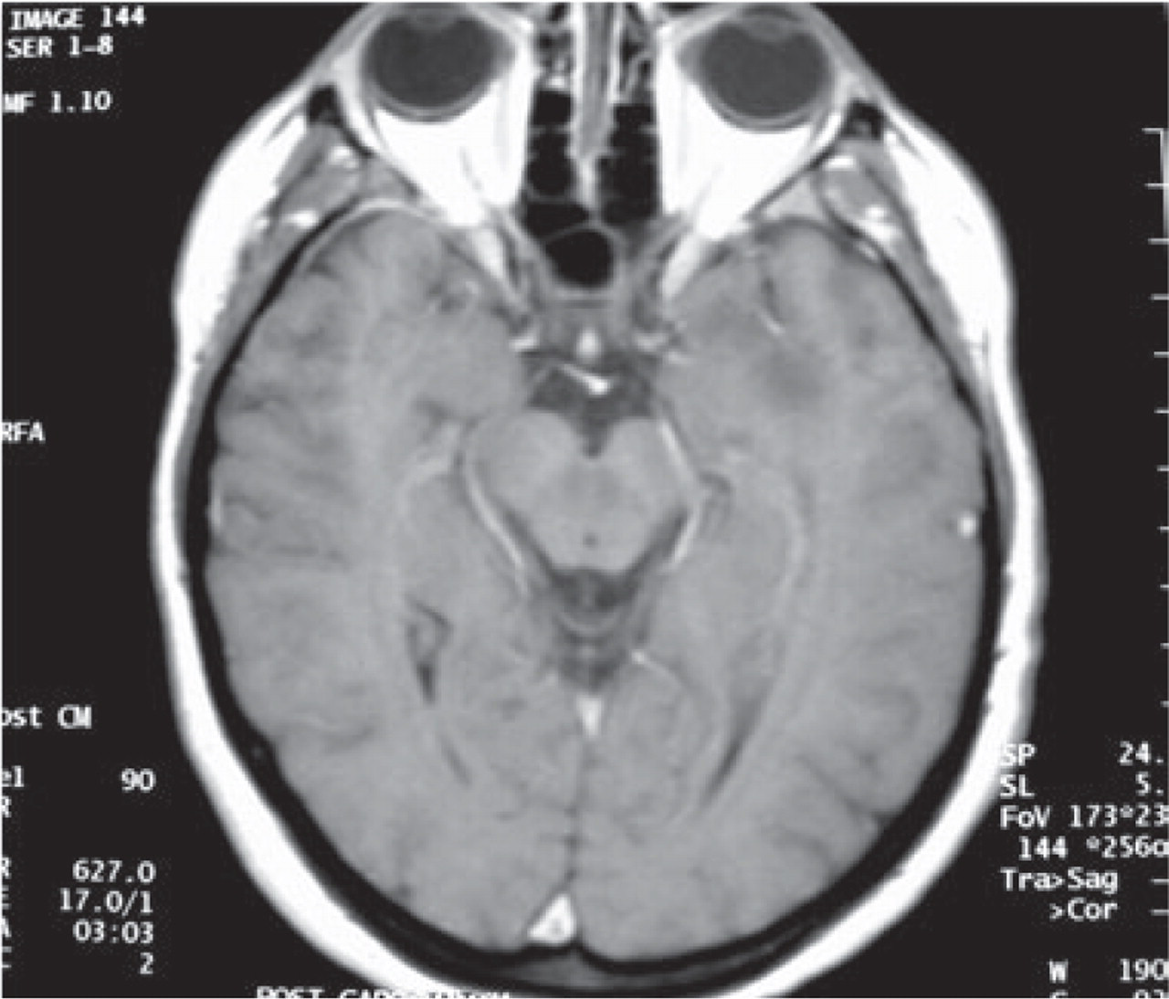
Normal magnetic resonance imaging of brain with gadolinium contrast.
Discussion
We report a case of late-onset ophthalmoplegic migraine in a 35-year-old patient who, in retrospect, was found to have a diagnosis of abdominal migraine in childhood. The prevalence of abdominal migraine has been estimated at 4.1% of children aged 5–15 in population studies (2), of which the majority will develop migraine headaches later in life (1, 3). Despite migraine being a common condition, OM is rare, with a review of 5000 migraine patients by Friedman et al. finding only eight cases of OM (4). A Danish population-based survey by Hansen et al. estimated the annual incidence of OM to be 0.7 per million inhabitants (5), with the majority of these cases presenting before the age of 10. On reviewing the literature, only three cases of OM presented at an age of >35 years, with the oldest documented case being a 50-year-old female (6–8). As in this case, the oculomotor nerve is the most commonly involved, with less than 10% of cases involving abducens, and fourth nerve involvement even less common (9).
OM remains a poorly understood condition and is now considered unlikely to be a migraine variant by the IHS, but rather a possible recurrent inflammatory neuropathy (1). OM still remains a diagnosis of exclusion. Depending on the age of presentation, the main differential diagnoses of ophthalmoplegia to exclude include aneurysm (particularly of the posterior communicating artery in our patient), tumour, diabetes, infiltrative or infective meningitis, Lyme disease, sarcoidosis, myasthenia gravis and sphenoid sinus mucocoele. Tolosa–Hunt syndrome may present with similar symptoms and may be misdiagnosed as OM, especially in studies done before the availability of MRI (3). Troost describes four ‘red flags’ when considering the diagnosis of OM, namely: (i) the absence of a migraine history; (ii) severe persistent headache with total ophthalmoplegia; (iii) onset after the age of 20; and (iv) symptoms and signs of subarachnoid haemorrhage (10).
The role of investigations therefore remains primarily to exclude other potential causes of ophthalmoplegia, of which MRI is generally considered the most important. However, recent reports of MRI findings in OM such as enhancement of the third nerve have shown that MRI is not only useful in excluding other diagnoses, but may also have a role in elucidating the aetiology of this condition. A study by Mark and colleagues on patients with acute OM has demonstrated reversible intraneural enhancement with enlargement of the oculomotor nerve at the point where it emerges from the midbrain and extending along the proximal course of the nerve (11). These findings are considered to be more suggestive of inflammation or demyelination, rather than ischaemic causes of neuropathy. Similarly, a case report by Lee and colleagues showed reversible enhancement of the abducens nerve on MRI in a patient with OM and abducens nerve palsy (8). Repeat MRI imaging performed 2 months after this migraine episode showed a resolution of the enhancement seen previously. Such findings have prompted Lance et al. to suggest that OM may represent a recurrent inflammatory neuropathy and that the pain associated with the ophthalmoplegia may be due to irritation of sensory fibres of the ophthalmic division of the trigeminal nerve, via an association with the oculomotor nerve that is found in other primates (12).
In our patient, we did not demonstrate enhancement of the third cranial nerve on contrast-enhanced MRI. Whether this was due to the interval of approximately 1 month between symptom cessation and imaging, allowing resolution of any possible enhancement, is unknown. Other case reports of OM have also failed to demonstrate MRI enhancement of the involved cranial nerves, although the interval between symptom onset and imaging is unclear (6, 13). An adult OM case described by O'Halloran et al. showed no abnormality on MRI within 24 h of the onset of ptosis. This lack of enhancement was not found to correlate with severity of the OM episode in their patient, who developed a permanent neurological deficit (14). The cause of the cranial neuropathy in these adult cases remains uncertain, with a lack of response to reported migraine treatments supporting the theory that it is unlikely to represent migraine aura (13). It is unclear why some adult patients with OM demonstrate cranial nerve enhancement on MRI and how cranial nerve enhancement correlates with the severity and duration of the OM.
In our patient, we describe the evolution of childhood abdominal migraine into a case of a more typical adult migraine headache without aura, and with subsequent development of OM. To our knowledge, this is the only case demonstrating this pattern of various migraine subtypes in a patient with late-onset OM. OM needs to be considered in the differential diagnosis of ophthalmoplegia in patients presenting at any age, after the exclusion of more sinister conditions.