
Abstract
Introduction
Spontaneous intracranial hypotension is characterized by orthostatic headache (OH), sometimes associated with nausea, neck pain, dizziness, diplopia, hearing disturbance, and visual field defects. CSF can show pleocytosis and increased protein levels (1). Magnetic resonance imaging (MRI) can reveal abnormalities with gadolinium enhancement of the meninges, subdural hygromas, global increase in brain volume, and pseudo Chiari type I malformation (2, 3). Evidence of CSF leakage is generally difficult. In most cases the diagnosis is based on the typical history. The most promising therapy is the lumbar epidural blood patch which closes the CSF leakage by a gelatinous tamponade and later a fibrin deposit. Some authors point out that the patches may not be successful if the leakage is located higher than the lumbar spine (2).
We report on a patient who developed an OH due to a cervical CSF leakage and who had partial relief of his headache after a lumbar patch.
Case report
A 51-year-old physician developed within 5–10 min a severe holocephalic headache (7–8/10 on a visual analogue scale (VAS)) and later nausea. He had neither engaged in physical activity nor experienced any trauma before headache appeared. The headache showed marked postural variation: it began within 2 min when in an upright position and ceased within a few minutes after lying down. The patient did not remember having had a similar headache earlier.
His examination was normal 1 day after the headache had developed. No relevant medical history was apparent. A cranial computed tomography (CT) and the laboratory results were normal. A spontaneous intracranial hypotension was suggested, since the patient's history excluded lumbar or epidural puncture, a traumatic lesion, and recent sexual intercourse. The diagnosis was confirmed by lumbar puncture: the opening pressure was 85 mm H2O in a sitting position and was not measurable in supine position. The CSF showed a slight pleocytosis of 11 cells/ml and protein was elevated to 237 mg/dl.
The patient was treated with a lumbar epidural blood patch at the L3-L4 level. One day later the headache intensity had markedly decreased (2–3/10 VAS), and the patient was able to remain upright for > 30 min.
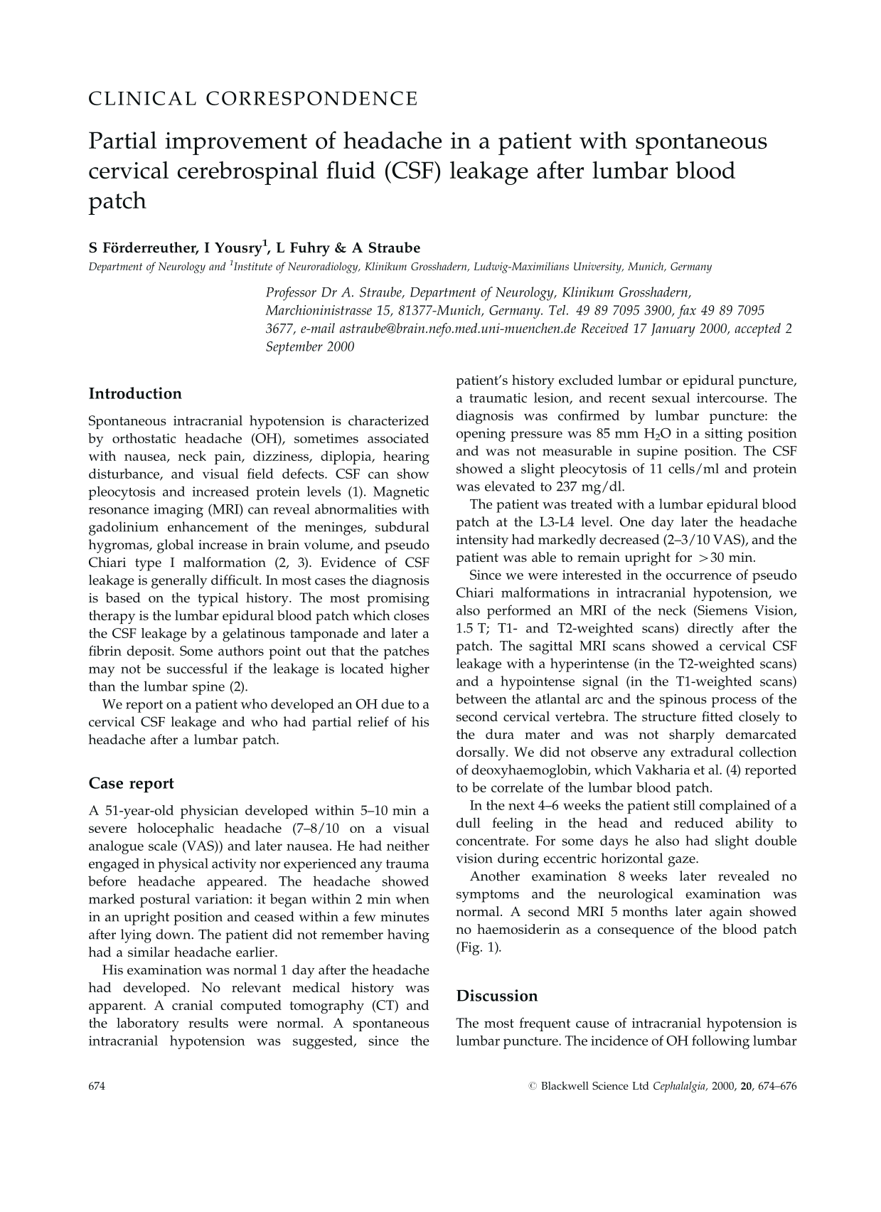
Since we were interested in the occurrence of pseudo Chiari malformations in intracranial hypotension, we also performed an MRI of the neck (Siemens Vision, 1.5 T; T1- and T2-weighted scans) directly after the patch. The sagittal MRI scans showed a cervical CSF leakage with a hyperintense (in the T2-weighted scans) and a hypointense signal (in the T1-weighted scans) between the atlantal arc and the spinous process of the second cervical vertebra. The structure fitted closely to the dura mater and was not sharply demarcated dorsally. We did not observe any extradural collection of deoxyhaemoglobin, which Vakharia et al. (4) reported to be correlate of the lumbar blood patch.
In the next 4–6 weeks the patient still complained of a dull feeling in the head and reduced ability to concentrate. For some days he also had slight double vision during eccentric horizontal gaze.
Another examination 8 weeks later revealed no symptoms and the neurological examination was normal. A second MRI 5 months later again showed no haemosiderin as a consequence of the blood patch (Fig. 1).

T2-weighted magnetic resonance imaging scan (sagittal plane) showing a hyperintense signal between the atlantal arc and the spinous process of the second cervical vertebra. The structure fits closely to the dura mater and is not sharply demarcated dorsally. No signs of the lumbar epidural blood patch can be seen.
Discussion
The most frequent cause of intracranial hypotension is lumbar puncture. The incidence of OH following lumbar puncture is reduced by the use of a non-cutting smaller needle. Vilming & Kloster analysed the time course of the headache after changing the patient's head position and found it to be very stable during the period of symptoms (5). All these findings agree with the view that a leakage of CSF is the primary reason for the headache; it causes intracranial hypotension with the result that in the upright position the brain and spinal cord settle with gravity and cause traction on supporting structures (6). An analogous mechanism has also been proposed to explain spontaneous intracranial hypotension that may occur after sexual activity, small traumas, or without any obvious reason (1–3, 6).
Five findings, however, do not completely agree with this view: (i) orthostatic headaches also occur during normal CSF pressure (2); (ii) patients have been reported to have lowered CSF pressure without positional headache (7, 8). We can confirm this observation. Two years ago we performed a lumbar spinal tap in a patient who did not complain of any headache in order to exclude encephalomyelitis disseminata. The puncture proved difficult, for the CSF did not flow spontaneously and it was necessary to aspirate CSF while the patient remained in a sitting position. Furthermore, Marshall compared the CSF pressure of one lumbar puncture and another lumbar puncture 24 h later in 43 patients and showed that there was no correlation between CSF pressure and the development of OH (8). Not even a decrease of CSF pressure in an individual was correlated to OH (8). This argues against the hypothesis that CSF pressure values may vary significantly in different individuals and that therefore OH can develop after a relative decrease of CSF pressure even when CSF pressure is formally within normal limits; (iii) although the mechanism of effectiveness of caffeine in OH is not entirely clear, it cannot be explained by a simple traction theory (9). It is more likely that caffeine produces intracerebral arterial constriction via blockade of brain adenosine receptors (10); (iv) epidemiological data show that a history of headache or a lower body mass index are risk factors for developing post-lumbar puncture headache (11). This cannot easily be explained by a leakage theory; (v) our observation of a dramatic improvement of the OH after a lumbar blood patch does not agree with the most often cited theory that a patch cures the headache by a tamponade of the leakage. Using MRI examinations, Vakharia et al. recently showed that an epidural patch with 20 ml blood in the mean spread 4.6 intervertebral spaces; they suggested that these findings document that the epidural blood patch has a tamponade effect (4). Our MRI examinations proved that the epidurally injected blood did not reach the cervical spinal level and therefore could not seal the leakage.
These findings suggest that other mechanisms may be important for the development of OH.
Hannerz recently found that the occurrence of post-lumbar puncture headache is more related to a history of chronic tension type than to a history of unilateral headache. He proposed that chronic tension-type headache and post-lumbar puncture headache have certain mechanisms in common (12). One possible link could be that patients with tension-type headache may have a tendency to react with more dilatation of the intracranial vessels. Hannerz showed that in patients with chronic tension-type headache, in contradistinction to controls, nitroglycerin and tilting induced a significant increase of headache intensity (12). Furthermore, there is evidence that the inhibition of nitric oxide synthase has a therapeutic effect on chronic tension-type headache (13). According to this scenario, the rapid decrease in CSF pressure due to the leakage or lumbar puncture would cause a vasodilatation, especially of the veins. This normally causes a compensatory vasoconstriction secondary to the increased cerebral perfusion pressure (14).
On the basis of these findings we would like to discuss a pathophysiological theory for OH which is supported by the fact that patients with OH do not react with such a compensatory vasoconstriction when the perfusion pressure increases. Similar considerations on this subject were recently published by Mokri (15). Thus, the intracranial blood vessels dilate when the CSF pressure decreases and as a consequence the intracranial blood volume increases. This increase causes the headache and/or brain swelling, which is seen in some MRI investigations in patients with OH. The intracranial hypotension is less when the patient is in a supine position, since the orthostatic pressure and therefore the intracranial perfusion pressure decreases, resulting in less intracranial blood volume and headache. The time required for these dynamic procedures may also help explain why the headache regularly takes time to build up when the patient is upright and also to fade when lying down. If the traction on pain-sensitive structures were the cause we would not expect such a delay. In addition, the occasionally positive effects of caffeine on OH could be explained by the vasoconstrictory action of caffeine (10). Finally, the blood patch effect on OH may not only be due to its influence on the leakage but also on the cerebral blood flow, e.g. by inducing an inflammation and production of vasoactive cytokines. This might explain why a patch improved the headache of our patient without stopping the CSF leakage.