
Abstract
Dexmedetomidine is a highly specific α2-adrenergic agonist, which is used clinically as an anesthetic adjuvant and in animal experiments has a neuroprotective effect during ischemia. The current study showed that dexmedetomidine enhances glutamine disposal by oxidative metabolism in astrocytes. This effect occurs at pharmacologically relevant concentrations. It is exerted on α2-adrenergic receptors and not on imidazoline-preferring sites, and it is large enough to reduce the availability of glutamine as a precursor of neurotoxic glutamate.
Dexmedetomidine is a potent and highly specific α2-adrenergic agonist (Virtanen, 1989) used clinically as an anesthetic adjuvant (Aantaa et al., 1997, Venn et al., 1999). Doses of 1–100 μg/kg have a neuroprotective effect during ischemia (Hoffman et al., 1991; Maier et al., 1993; Kuhmonen et al., 1997; Jolkkonen et al., 1999). With a molecular weight of 186 and a volume of distribution of 2–3 liter/kg (Scheinin et al., 1992; Muge et al., 1996), the dose range corresponds to plasma concentrations of 2.5–250 nmol/L.
It was originally assumed that all α2-adrenergic receptors are presynaptic and inhibit noradrenaline release. Although α2-adrenergic agonists have such an effect (Hong et al., 1992; Matsumoto et al., 1993; Gobert et al., 1998), compelling evidence for an additional localization on post-junctional target cells has appeared. The target cells include astrocytes (Ebersolt et al., 1981; Lee et al., 1998a,b). Stimulation of astrocytic α2-adrenergic receptors activates the phoshatidylinositol second messenger system (Enkvist et al., 1996) and increases free cytosolic calcium concentration, [Ca2+]i (Salm and MacCarthy, 1990; Zhao et al., 1992; Muyderman et al., 1997; Chen and Hertz, 1999). Clonidine, a less selective α2-adrenergic agonist, also increases glutamine uptake in astrocytes and its subsequent oxidative degradation (Huang and Hertz, 1994, 1995).
The importance of glutamine for glutamate-induced excitotoxic injury is illustrated by the observation that a decrease of the glutamine concentration in the culturing medium reduces anoxic death of cultured neurons (Goldberg et al., 1988; Huang et al. 1997). Oxidative degradation of glutamine occurs through pyruvate and lactate (Sonnewald et al., 1995; Schousboe et al., 1994). This is probably the main mechanism by which the brain disposes of excess glutamine and glutamate (Hertz et al., 1999, 2000). During anoxia this process is halted, but it can be re-established after reoxygenation (Pascual et al., 1998). In the current study it is shown that dexmedetomidine at pharmacologically relevant concentrations increases glutamine oxidation in astrocytes.
MATERIALS AND METHODS
Chemicals for tissue culture medium were from Sigma Chemical Company (St. Louis, MO, U.S.A.). Dexmedetomidine was donated by Orion Corporation Farmos (Turku, Finland). Idazoxan was bought from Research Biochemicals Inc. (Natick, MA, U.S.A.). L-[U-14C]glutamine was from NEN, Du Pont Canada Inc., Mississauga, Ontario. [1-14C]glutamine was donated by Drs. T. Tildon and H.R. Zielke, University of Maryland, MD, U.S.A.
Cultures of astrocytes were prepared as previously described (Juurlink and Hertz, 1992; Hertz et al., 1998). The neopallia, that is, the parts of the cerebral hemispheres above the lateral ventricles, were dissected out of brains from newborn Swiss mice and mechanically dissociated in a slightly modified Dulbecco's tissue culture medium (Juurlink and Hertz, 1992). The cell suspension was seeded in Falcon Primaria culture dishes and grown under a CO2/air (5%/95%) atmosphere at 37°C. After 2 weeks the culturing was continued in the additional presence of 0.25 mmol/L dibutyryl cyclic adenosine monophosphate, which promotes morphologic and functional differentiation (Meier et al., 1991; Enkvist et al., 1996). The cultures were used after 3 to 6 weeks.
Production of labeled CO2 from 0.2 mmol/L [U-14C]glutamine (0.3 μCi/ml) was measured during 60 minutes of incubation of individual cultures in an air-tight chamber (Yu et al., 1982). The cells were incubated in modified Dulbecco's medium, prepared without glucose to prevent the production of labeled CO2 reflecting an exchange between labeled glutamate and non-labeled α-ketoglutarate formed from glucose. At the end of the experiments the medium was acidified by injection of acetic acid, converting bicarbonate to CO2. During subsequent incubation for 60 minutes on a hotplate (60°C), which created air currents within the chamber, all CO2 was quantitatively trapped into 10% tetramethylammonium hydroxide which had been injected into a presuspended beaker. Accumulated radioactivity was determined in a liquid scintillation counter, and glutamine oxidation rates/mg protein were calculated on the basis of the specific activity of the medium and the protein content in the culture, as measured by the Lowry technique (Lowry et al., 1951).
A few experiments were carried out using [1-14C]glutamine instead of L-[U-14C]glutamine. This provided a quantitatively more correct estimate of glutamine disposal because the carbon in the C-1 position is the first to be oxidized. Under control conditions the rate of 14CO2 production from [1-14C]glutamine was 2.7 ± 0.3 times higher (n = 6) than that from [U-14C] glutamine.
RESULTS
The apparent rate of 14CO2 production under control conditions was 0.24 ± 0.01 nmol/min·mg protein. It increased by ~25% by exposure to either 70 nmol/L or 25 μmol/L dexmedetomidine (Table 1). The apparent control rate and the stimulation of 0.06 nmol/min · mg protein are nominal values that underestimate glutamine oxidation for two reasons: (1) not all 5 carbon atoms of uniformly labeled glutamine are oxidized within the 1-hour incubation period, and (2) the specific activity of intracellular [U-14C]glutamine is lower than that in the medium (Huang et al., 1995). The first of these sources of error can be corrected by aid of the ratio of 2.7 between rate of 14CO2 production from [1-14C]glutamine and [U-14C]glutamine (see Methods). Using this ratio as a correction factor, the dexmedetomidine-induced increase in rate of glutamine disposal can be estimated to ~0.2 nmol/min-mg protein. This value remains an underestimate because extracellular, not intracellular, specific activity was used for the determination (Zielke et al., 1996). However, because of possible intracellular compartmentation of glutamine (Schousboe et al., 1994), no attempt was made to correct this source of error.
Rates of 14CO2 production from 0.2 mmol/L L-[U-14C] glutamine in primary cultures of mouse astrocytes
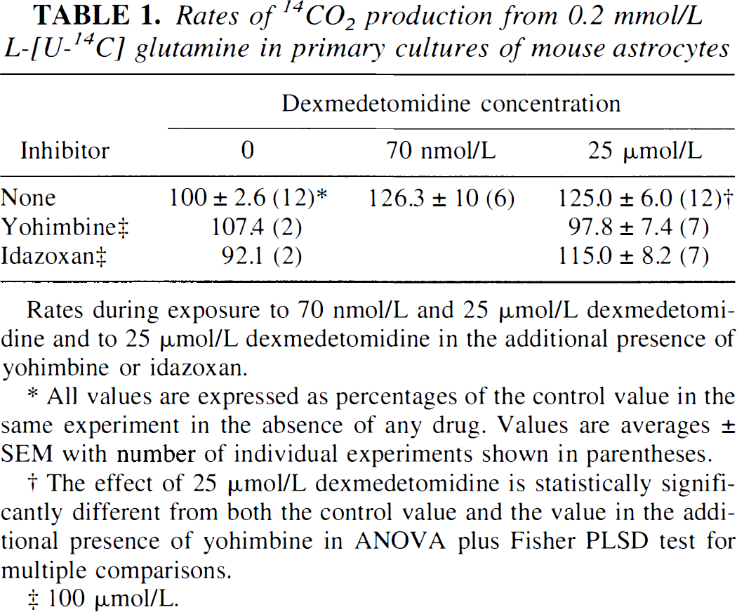
Rates during exposure to 70 nmol/L and 25 μmol/L dexmedetomidine and to 25 μmol/L dexmedetomidine in the additional presence of yohimbine or idazoxan.
All values are expressed as percentages of the control value in the same experiment in the absence of any drug. Values are averages ± SEM with number of individual experiments shown in parentheses.
The effect of 25 μmol/L dexmedetomidine is statistically significantly different from both the control value and the value in the additional presence of yohimbine in ANOVA plus Fisher PLSD test for multiple comparisons.
100 μmol/L.
The dexmedetomidine-induced stimulation of U-[14C]glutamine oxidation was exerted on α2-adrenergic receptors as shown by its inhibition by yohimbine, an α2 antagonist (Table 1), but not by idazoxan, an inhibitor of the imidazoline binding site (Bylund, 1995). The small, insignificant reduction of dexmedetomidine-stimulated 14CO2 by idazoxan reflects that idazoxan, on its own, caused approximately a 10% reduction in the rate of 14CO2 formation.
DISCUSSION
Dexmedetomidine in vivo exerts other actions than inhibiting noradrenaline release as proven by the fact it has a sedative effect and potentiates anesthetics in animals depleted for noradrenaline (Segal et al., 1988; Riekkinen et al., 1993), an effect exerted within the central nervous system (Doze et al., 1989). The current results suggest that some of dexmedetomidine's actions are exerted on astrocytic α2-adrenergic receptors. This is consistent with a dexmedetomidine-induced increase in astrocytic [Ca2+]i (Zhao et al., 1992; Enkvist et al., 1996; Chen et al., 2000).
In heart cells, an increase in [Ca2+]i resulting from adrenergic stimulation leads to uptake of K+ in mitochondria and swelling of the mitochondrial matrix, which in turn stimulates glutaminase activity (Halestrap, 1989). The same mechanisms may operate in astrocytes because they display considerable glutaminase activity (Hogstad et al., 1988; Aoki et al., 1991; Wurdig and Kugler, 1991), which is increased by clonidine (Huang and Hertz, 1994, 1995). Moreover, α2-adrenergic agonists have a direct effect on oxidation of α-ketoglutarate (Subbarao and Hertz, 1991) which, combined with an increased rate of formation of glutamate from glutamine, could explain the increase in glutamine oxidation seen in the current study.
Dexmedetomidine concentration of 70 nmol/L, which stimulated glutamine oxidation by 25%, is within the plasma concentration range expected after administration of dexmedetomidine as a neuroprotectant. The effect is large enough that it may affect glutamine content in the brain, a novel mechanism of action for a neuroprotectant drug. Net synthesis of glutamate and glutamine from glucose depends upon pyruvate carboxylation, which in the brain occurs at a rate 10–15% of total TCA cycle activity (Lapidot and Gopher, 1994; Aureli et al., 1997; Gruetter et al., 1998). Based upon this information and a glucose utilization rate in the mouse brain of 60–70 μmol/min per 100 g (Wree et al., 1989), it can be estimated that the maximum rate of net glutamate/glutamine synthesis from glucose in the mouse brain amounts to 1.2–2.1 nmol/min-mg protein (Hertz et al., 1999). A dexmedetomidine-induced increase of glutamine disposal of at least 0.2 nmol/min-mg protein may cause a substantial change in the balance between formation and disposal of glutamine, thus reducing the availability of glutamine as a glutamate precursor. This concept is consistent with the observation by Talke and Bickler (1996) that glutamate release from hippocampal slices during hypoxia is decreased by dexmedetomidine.