
Abstract


Introduction:
Natural orifice specimen extraction is increasingly utilized in minimally invasive colorectal surgery. This technique is particularly useful in morbidly obese patients, where the usual morbidity associated with abdominal wall extraction sites is avoided. 1 It reduces the postoperative complications of wound infection, ileus, and long-term risk of incisional hernia. 2
This video focuses on the key aspects of performing a transanal natural orifice specimen extraction in anterior resection for oncological purposes. The planning for the rectal and mesorectum division, the specimen extraction technique, the closure of the rectal stump, and the creation of an intra-corporeal purse-string are vital for the success of this technique.
Materials and Methods:
The patient is placed in a lithotomy position with reverse Trendelenburg and left tilt position. A beanbag is used to ensure the patient does not slip down on the table. Bilateral calf compressors are used. An indwelling catheter is used. Standard prophylactic intravenous antibiotics were given.
An on-table colonoscopy is performed to confirm the diagnosis and perform washout with Povidone-Iodine.
Optical entry is performed using a 5 mm port and a 5 mm 30◦ endoscope at the Palmer’s point. Pneumoperitoneum is established. Three further ports are inserted: 5 mm port at the right para-umbilical, a 5 mm port at the right upper quadrant, and a 12 mm port at the right iliac fossa.
Dissection Phase:
The left colon is mobilized in a medial to lateral fashion. The left ureter is identified. A high ligation of the inferior mesenteric artery is performed. The inferior mesenteric vein is ligated high at the inferior border of the pancreas. The splenic flexure is mobilized through a supracolic and transverse mesocolic approach. The upper rectal mesorectum is ligated at the intended site of division.
The upper rectum is stapled off with an endoscopic stapler. The proximal mesocolon is ligated with an energy device. The proximal colonic margin is determined and divided with an endoscopic staple.
Extraction Phase:
The rectal staple line is removed. A small wound retractor is placed through the rectum. The anvil of the circular stapler is introduced through the anus into the peritoneal cavity. The assistant then uses a Rampley’s forceps to extract the specimen. The wound retractor is removed. The rectal stump is closed with an endoscopic stapler.
Reconstructive Phase:
Indocyanine green can be used to assess the perfusion of the colonic conduit and the rectum. A purse-string of the conduit is created intra-corporeally with 3/0 V-Loc. The anvil is inserted and further secured with a PDS endoloop. An end-to-end colorectal anastomosis is created with a circular stapler.
A flexible sigmoidoscopy is performed to assess the colorectal anastomosis and pneumatic test.
Results:
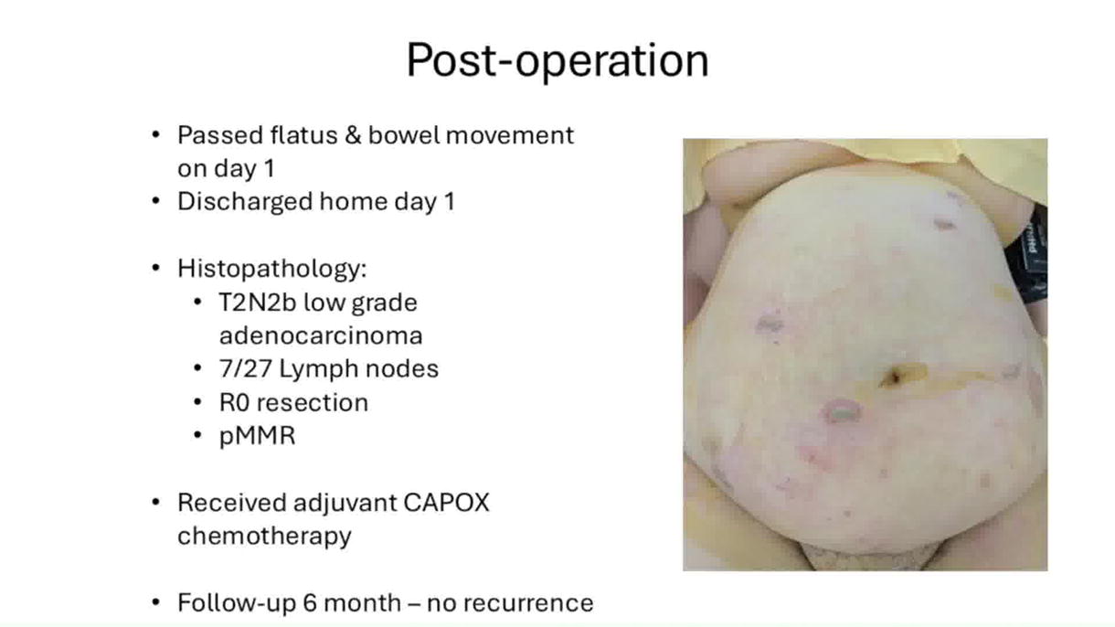
There was no intraoperative complication. The patient had return of gut function on day 1 and was discharged home. Histopathology revealed a T2N2b low-grade adenocarcinoma. There were 7/27 lymph nodes.
Conclusion:
Laparoscopic anterior resection with transanal natural orifice specimen extraction is safe and reproducible through the understanding of the key steps. It avoids morbidity associated with the abdominal wall extraction site, especially in obese patients.
Source of work:
Authors’ own work.
The authors have no commercial associations during the last two years that might create a conflict of interest in connection with the video.
Authors have received and archived patient consent for video recording/publication in advance of video recording of procedure.
The patient has provided written consent for the publication of the article and video.
Runtime of video: 7 mins 15 secs.
Get full access to this article
View all access options for this article.