
Abstract
Introduction:
Aortopexy is a well-established technique for the treatment of severe tracheomalacia that occurs most commonly in the setting of children with esophageal atresia and tracheoesophageal fistula. 1 There is a lack of video evidence of the thoracoscopic technique. We, therefore, present a thoracoscopic aortopexy for severe tracheomalacia.
Materials and Methods:
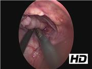
The patient is an 8-month old male who was born with a type A esophageal atresia. He also had a single kidney and cleft lip and palate. A laparoscopic gastrostomy was placed on day 1 of life followed by a thoracoscopic converted to open repair of the esophageal atresia after he was allowed to grow. This was complicated by a small contained leak and prolonged oxygen dependence. He was never able to be weaned off oxygen and did suffer from a respiratory code event at 8 months of life leading to another intubation. A repeat bronchoscopy at that time showed almost complete airway collapse with significant distal secretions. Therefore, aortopexy was indicated. A left thoracoscopic approach was selected because of his previous right-sided operations. A 5 mm port was placed at the anterior axillary line at the nipple level and two 3 mm ports were placed just anterior to the 5 mm port on each side. The left lobe of the thymus was removed bluntly giving good observation of the pericardium and aortic arch. A 3-0 braided polyethylene suture was then passed through the sternum. The first suture was placed through the pericardium overlying the distal aortic arch. This suture was then fed back through the sternum with some effort. The second suture was brought in through the sternum and was placed through the pericardium overlying the proximal aorta. Then an 18-gauge needle with a loop of monofilament suture placed in a loop was passed through the sternum. The loop was placed into the chest and used to lasso the other suture, which was then brought back out of the chest. The third suture was passed through the sternum and was passed through the pericardium at its junction with the aorta. The 18-gauge needle was again used to lasso this suture. Tying the three sutures brought the mediastinal structures into direct apposition with the sternum. Immediate bronchoscopy showed significant distal secretions with some residual airway collapse. A repeat bronchoscopy 1 week later after he was weaned from all oxygen showed the airway was completely open without any residual collapse.
Results:
The patient who tolerated the procedure well was weaned from the ventilator after 1 day and was off all oxygen support after a few days. He was discharged home and continues to be free of respiratory issues during follow-up.
Conclusions:
The thoracoscopic approach for aortopexy is safe, well tolerated, and affords the surgeon excellent observation.
No competing financial interests exist.
Runtime of video: 4 mins 26 secs
Previously presented at IPEG on April 13, 2018.


Get full access to this article
View all access options for this article.