
Abstract
Introduction:
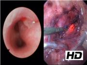
Tracheomalacia refers to collapsibility of the posterior trachea commonly associated with esophageal atresia and tracheoesophageal fistula. Several surgical procedures for the treatment of tracheomalacia have been described. One approach includes aortopexy, which provides anterior displacement of the trachea by attaching the aorta to the back of the sternum. This treatment is appropriate if vascular compression is contributing to the collapsibility of the trachea. Primary posterior tracheopexy is preferred when tracheomalacia is primarily because of posterior tracheal membrane collapse. A thorough preoperative work-up includes bronchoscopy and computed tomography (CT) aid in the determination of the cause of tracheomalacia and appropriate initial treatment. This video (4 minutes 26 seconds) shows the thoracoscopic approach to posterior tracheopexy.
Materials and Methods:
A 3-year-old girl presented with severe tracheomalacia. She underwent extensive evaluation in a multidisciplinary clinic, which included longitudinal flexible and rigid bronchoscopy, swallow study, sleep study, and CT scan. The CT scan demonstrated tracheomalacia in the midtrachea with 53% narrowing of the anterior-posterior diameter of the trachea at the level of the brachiocephalic artery with expiratory imaging. The patient was brought to the operating room, placed under general anesthesia, and positioned for a right thoracoscopic procedure. Three 5 mm step trocars were placed, insufflated to a pressure of 5 mm Hg, and flow was set to 1 L/minute. The esophagus was identified and a 16F orogastric tube was placed by anesthesia. Next, a plane was developed between the esophagus and the trachea. The Vagus nerve was identified and preserved. The anterior spine was dissected free of loose connective tissue. The posterior membranous portion of the trachea was dissected free of connective tissue. With bronchoscopic guidance, three stitches were placed into the posterior membranous trachea using 4-0 prolene on a renal (artery) bypass needle. There was close attention not to go full thickness on the tracheal side. Next, a bite of anterior spinal ligament was then taken and the suture was tightened until there was just enough tension to keep trachea from collapsing. The three stitches were placed between the thoracic inlet and the carina, and hemostasis was ensured. A 12F chest tube was placed under direct observation. The incisions were closed with monocryl and the chest tube was secured with 3-0 silk.
Results and Conclusions:
The duration of surgery was 101 minutes, and the estimated blood loss was 10 mL. The patient did well postoperatively, and was discharged home after 3 days. Since the procedure, the patient initially responded very well with an improved cough and a stable nightly oxygen requirement. Of note, she did require admission to the hospital for a viral upper respiratory infection about 6 months after the surgery. This procedure represents the first thoracoscopic posterior tracheopexy performed by the author. Three previous posterior tracheopexy approaches were performed in an open manner on patients with a history of trachea esophageal fistula. None of these patients required readmission within 30 days of the procedure and all had symptomatic improvement. Thus, posterior tracheopexy is a safe and effective procedure for tracheomalacia, and the thoracoscopic approach may be considered in the appropriate patient population.
No competing financial interests exist.
Runtime of video: 4 mins 26 secs


Get full access to this article
View all access options for this article.