
Abstract
Introduction:
Thymic teratoma is a rare condition in children. 1 –3 We report a clinical case of thymic teratoma submitted to thoracoscopic hemi-thymectomy.
Case History:
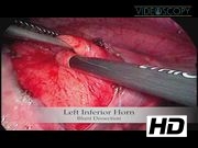
A 14-year-old boy presented with a history of pleuritic chest pain, which worsened with deep breathing. Chest roentgenogram and computed tomography (CT) scan revealed a well-defined cystic thymic mass (32 × 25 mm). Routine blood tests were normal, including tumor markers (alpha-feta protein and beta-human chorionic gonadotropin). A CT scan-guided biopsy was performed (cytology was compatible with epithelial cyst), and simultaneously, the cystic component was aspirated with symptomatic relief. At 1-year follow-up, there was a regrowth of the cyst associated with symptomatic recurrence; therefore, a conservative surgical resection was decided. Using single-lung ventilation, a left side thoracoscopy was achieved with three 5-mm trocars. The phrenic nerve was identified and the pleura was incised anteriorly. Dissection was completed using a combination of blunt dissection and electrocoagulation. A sealing device was used for vessels and thymic parenchyma. After lung expansion, no chest tube was left in place. The postoperative recovery was uneventful and the patient was discharged on the following day. Histologic examination revealed a mature cystic teratoma. During the follow-up, there was no recurrence; the boy is asymptomatic and otherwise healthy.
Conclusions:
Hemi-thymectomy by thoracoscopic approach is feasible and safe for thymic lesions, such as teratoma. This conservative approach avoids any possible immunologic consequences of total thymectomy and does not jeopardize the contralateral phrenic nerve.
No competing financial interests exist.
This video was presented at IPEG Meeting 2015.
Runtime of video: 3 mins 39 secs


Keywords
Get full access to this article
View all access options for this article.
References
Supplementary Material
Please find the following supplemental material available below.
For Open Access articles published under a Creative Commons License, all supplemental material carries the same license as the article it is associated with.
For non-Open Access articles published, all supplemental material carries a non-exclusive license, and permission requests for re-use of supplemental material or any part of supplemental material shall be sent directly to the copyright owner as specified in the copyright notice associated with the article.