
Abstract
Introduction:
Myasthenia gravis (MG) is an autoimmune neuromuscular disease, the effects of which can be improved or alleviated by thymectomy in young patients. However, median sternotomies or thoracotomies have a high degree of morbidity, especially when already weakened from their MG. A thoracoscopic approach allows for a minimally invasive approach, but it can be technically challenging to completely remove all the thymic tissues in the contralateral chest and lower neck, especially in smaller children who typically have larger glands. The video demonstrates the robot-assisted approach for dissection of the thymus gland.
Background:
This patient is a 3-year-old boy who had been suffering from generalized MG. Due to disease progression that was only partially controlled by medications, his neurologist referred him for thymectomy. After a lengthy conversation with his parents, the decision was made to proceed with a robot-assisted left thoracoscopic thymectomy.
Methods:
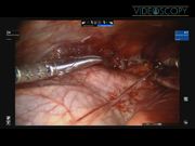
The patient was placed in the supine position on the operating room table. Through a transverse incision in the left axilla, a 5-mm XCEL trocar was placed in the fourth interspace, and pneumothorax was created with a pressure of 4 mm Hg. One additional 5-mm robotic trocar was placed in the left midclavicular line in the sixth interspace, and an additional 8-mm trocar was placed in the sixth interspace in the anterior axillary line. The XCEL trocar was replaced with a 5-mm robotic trocar. At this point, the robot was docked, and the 8-mm camera and 30-degree scope was placed in the central trocar. Hook cautery and a Maryland grasper were used to dissect the gland off of the heart. The left lateral aspect of the thymus was lateral to the left phrenic nerve, which was identified and preserved. Dissection was continued in a caudad direction to free up the entire left lobe of the gland and carried over toward the right chest. A small hole that had been made in the right pleura was widened to prevent a tension pneumothorax from developing. At this point, the dissection was carried around the right lobe of the thymus with care taken to preserve the right phrenic nerve. Dissection was continued to free the gland from the heart as well as both superior horns that extended well into the lower neck. Once the organ was dissected free, it was placed into a 10-mm Endocatch bag that had been placed through the slightly widened 8-mm trocar defect. The lungs were fully inflated before the fascia being closed at all port sites without placing a chest tube. No pneumothorax was seen on the postoperative chest X-ray.
Results:
The patient tolerated the procedure well without any postoperative complications, had minimal blood loss, and was discharged home the following day.
Conclusion:
The use of robotic assistance in thoracoscopic thymectomies with its articulating instruments and 3D visualization has allowed this approach to be offered to younger and smaller patients despite having a larger thymus. This approach allows these patients to benefit from an earlier thymectomy while avoiding the morbidity from a sternotomy or large thoracotomy.
The authors have no relevant financial disclosures.
Runtime of video: 4 mins 13 secs


Keywords
Get full access to this article
View all access options for this article.
Supplementary Material
Please find the following supplemental material available below.
For Open Access articles published under a Creative Commons License, all supplemental material carries the same license as the article it is associated with.
For non-Open Access articles published, all supplemental material carries a non-exclusive license, and permission requests for re-use of supplemental material or any part of supplemental material shall be sent directly to the copyright owner as specified in the copyright notice associated with the article.