
Abstract
Introduction:
Minimally invasive pancreaticoduodenectomy, first described by Gagner and Pomp in 1994, 1 has made a significant progression in the past few years due to the evolution in surgical techniques and advancement in instrumentation. Multiple publications have touted its feasibility and our group has shown its safety in comparison to open pancreaticoduodenectomy. 2 This 15-minute video demonstrates our current technique of laparoscopic pancreaticoduodenectomy with patient and surgeon positioning and trocar placement. Surgeon positioning, camera placement, and trocar use will vary throughout each step of the procedure and these variations are demonstrated.
Materials and Methods:
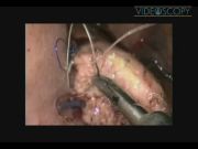
A 61-year-old male presented with painless jaundice and imaging revealed a mass in the head of the pancreas. He was taken for a totally laparoscopic pancreaticoduodenectomy. The patient is positioned in a supine split leg position and six trocars are placed. The operation starts by entering the lesser sac and exposing the head of the pancreas. The transverse and hepatic flexure of the colon is completely mobilized and the duodenum is exposed. The gastrohepatic ligament is opened and the proximal duodenum is transected. The hepatoduodenal ligament is dissected and the common bile duct is divided. The neck of the pancreas is separated from the underlying superior mesenteric vein and the pancreas is transected. Then, an extended Kocher maneuver is performed and the ligament of treitz is opened and the proximal jejunum is reduced underneath the root of the mesentery and divided. The uncinate process and the head of the pancreas are then carefully separated from the lateral border of the mesenteric vessels and the specimen is freed. The specimen is removed through the umbilical site and negative margins are ensured. The reconstruction is begun by performing a single layer hepaticojejunostomy with a 4-0 braided absorbable suture in a running fashion. Proximal to this on the same jejunal limb, a two-layer duct-to-mucosa pancreaticojejunostomy is performed. The outer layers are completed with a running 4-0 monofilament nonabsorbable suture and the inner duct-to-mucosa sutures are placed using an interrupted braided absorbable suture method. Then, the gastrointestinal reconstruction is performed by an antecolic end to side duodenojejunostomy in a two-layer fashion using a running braided absorbable stitch. A single drain is placed in the vicinity of the biliary and pancreatic anastomoses. The total operative time was 431 minutes. The patient recovered very well from surgery, resumed oral intake at 2 days, and was discharged home after 6 days with no complications and is doing very well in follow-up. Final pathology revealed an invasion adenocarcinoma of the distal common bile duct and the patient proceeded to adjuvant therapy.
Results and Conclusions:
Laparoscopic pancreaticoduodenectomy is a very complex procedure and has undergone significant alterations and refinement within our practice over the past few years. The technique demonstrated in this video shows our current method that has proven to be as safe as open pancreaticoduodenectomy. 2 As a large amount of experience is gained and with additional technical advances, we hope to further improve patient safety and outcomes for this procedure.
The authors have no disclosures and no conflicts of interest.
Runtime of video: 15 mins 13 secs


Keywords
Get full access to this article
View all access options for this article.
References
Supplementary Material
Please find the following supplemental material available below.
For Open Access articles published under a Creative Commons License, all supplemental material carries the same license as the article it is associated with.
For non-Open Access articles published, all supplemental material carries a non-exclusive license, and permission requests for re-use of supplemental material or any part of supplemental material shall be sent directly to the copyright owner as specified in the copyright notice associated with the article.