
Abstract
Small renal masses are commonly treated with minimally invasive approaches by nephron-sparing procedures. However, in complex scenarios where the tumor is located in an intra-hilar position, there is a higher likelihood of requiring open conversion or an increased incidence of complications. Here, we demonstrate such a case that was managed using an anterior nephrotomy approach during robotic partial nephrectomy. The procedure had a clamp time of 23 minutes and an estimated blood loss of 100 mL. The surgical margins were negative, and the patient is doing well, with no evidence of recurrence at the 6 months follow-up.
Introduction:
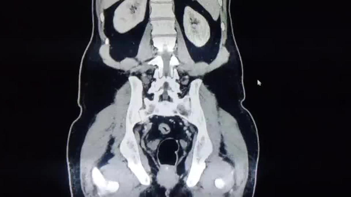
Renal cell carcinoma is relatively common, and the challenges in managing complex intra-hilar tumors have been well documented in the literature. 1,2 We demonstrate one of the techniques for managing a complex intra-hilar tumor through a nephrotomy. This is a case of a 56-year-old man with hypertension, and no other comorbidities who was incidentally found to have a 2.4 cm mass in the left kidney, located in the intra-hilar area, anterior to the pelvicalyceal system. The preoperative plan was either to trim a portion of the renal parenchyma to access the tumor, or to perform intra-hilar dissection to achieve access. As a last resort, an anterior nephrotomy was considered. The procedure was successfully completed using the anterior nephrotomy approach.
Materials and Methods:
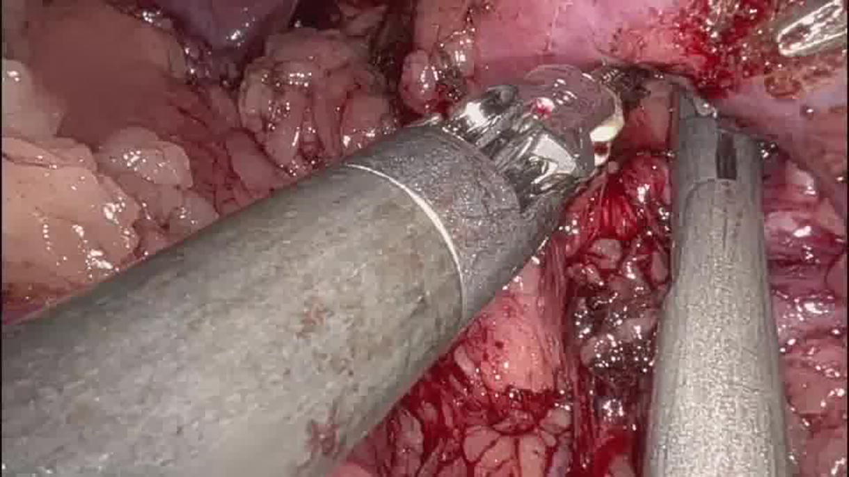
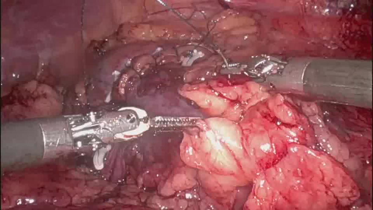
The patient underwent robotic-assisted surgery using the da Vinci Xi system. He was positioned in the right lateral position, and standard ports for a robotic left radical or partial nephrectomy were utilized, including four 8 mm ports (three working and one camera) and two assistant ports (one 12 mm and one 5 mm).
Results:
Key operative metrics included a dock time of 10 minutes, a console time of 180 minutes, and a clamp time of 23 minutes. Intraoperative blood loss was 100 mL. The excised tumor measured 2.6 × 2 × 1.9 cm and was histologically confirmed as a grade II renal cell carcinoma with all margins negative. The patient was discharged on postoperative day 3. The patient is disease-free at 6 months follow-up with preserved serum creatinine, the same as preoperative level (0.8 mg/dL).
Conclusion:
Complex intra-hilar renal tumors can be successfully managed using the nephrotomy approach based on the location of the tumor with respect to the pelvicalyceal system. This patient had an uneventful recovery, demonstrating the effectiveness of this technique in challenging such locations.
The accompanying article provides a structured text breakdown.
The video component details are as below.
Runtime of video: 6 mins 15 secs.
Format: MP4 playable on Windows PC.
We have ensured the video content aligns with the reviewed and accepted article. In addition, all narration is in English, and written informed consent for video publication has been obtained from each patient.
Conflicts of Interest Statement:
No conflict of interest as signed by all authors. Form enclosed as a separate attachment.
Keywords
Get full access to this article
View all access options for this article.