
Abstract
Clinical History:
A 35-year man presented with left flank pain of 6 months and left lower chest pain since 3 months associated with dry cough.
Physical Examination:
On examination there was no palpable mass in the abdomen.
Diagnosis:
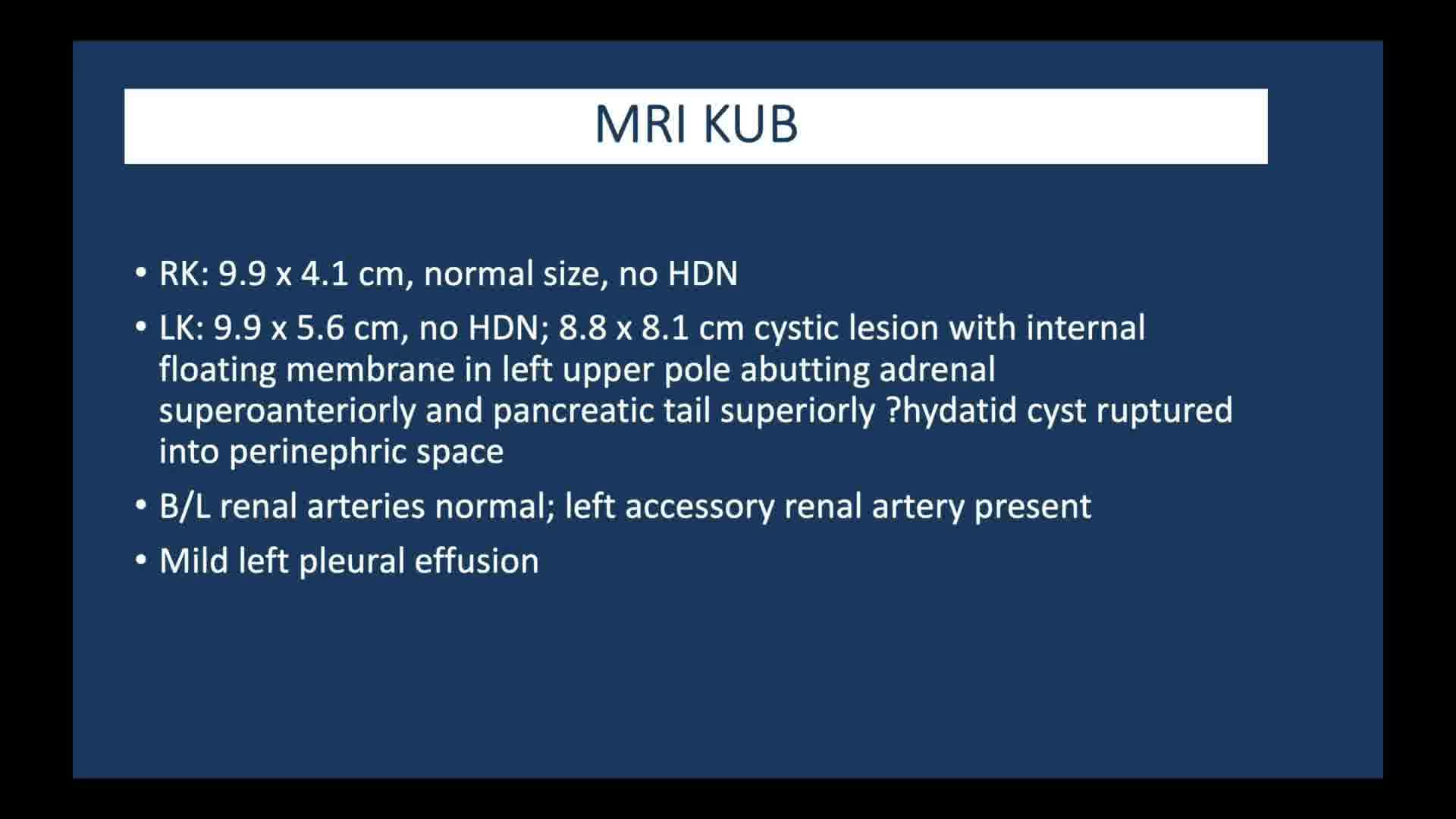
Ultrasonography of the abdomen showed a 9 cm cystic lesion in the upper pole of the left kidney. MRI showed a cystic lesion in the upper pole of the left kidney with internal floating membrane and suspected ruptured hydatid cyst into the perinephric space. Left pleural effusion and perinephric fat stranding were evident on CT of the thorax.
Intervention:
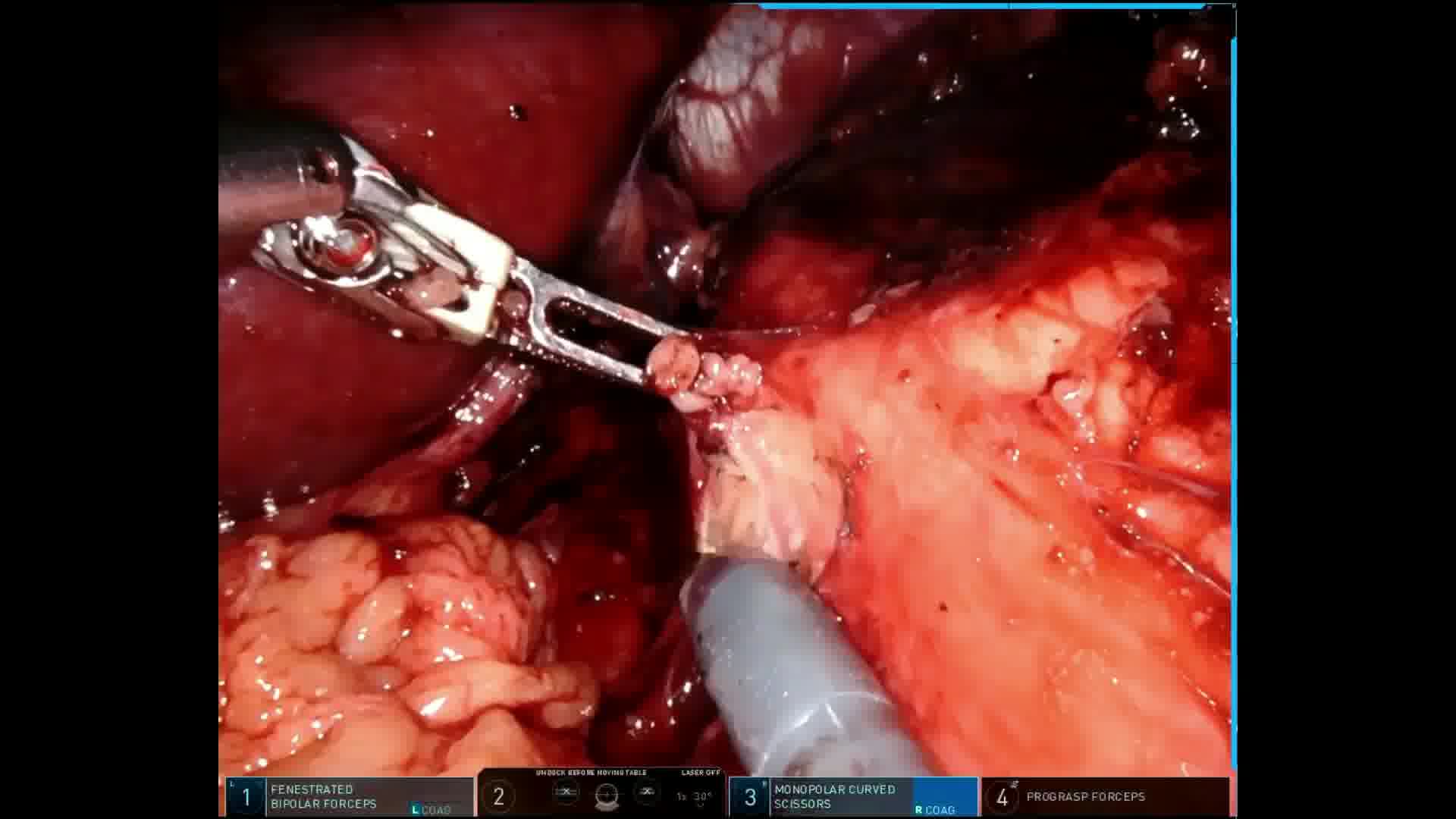
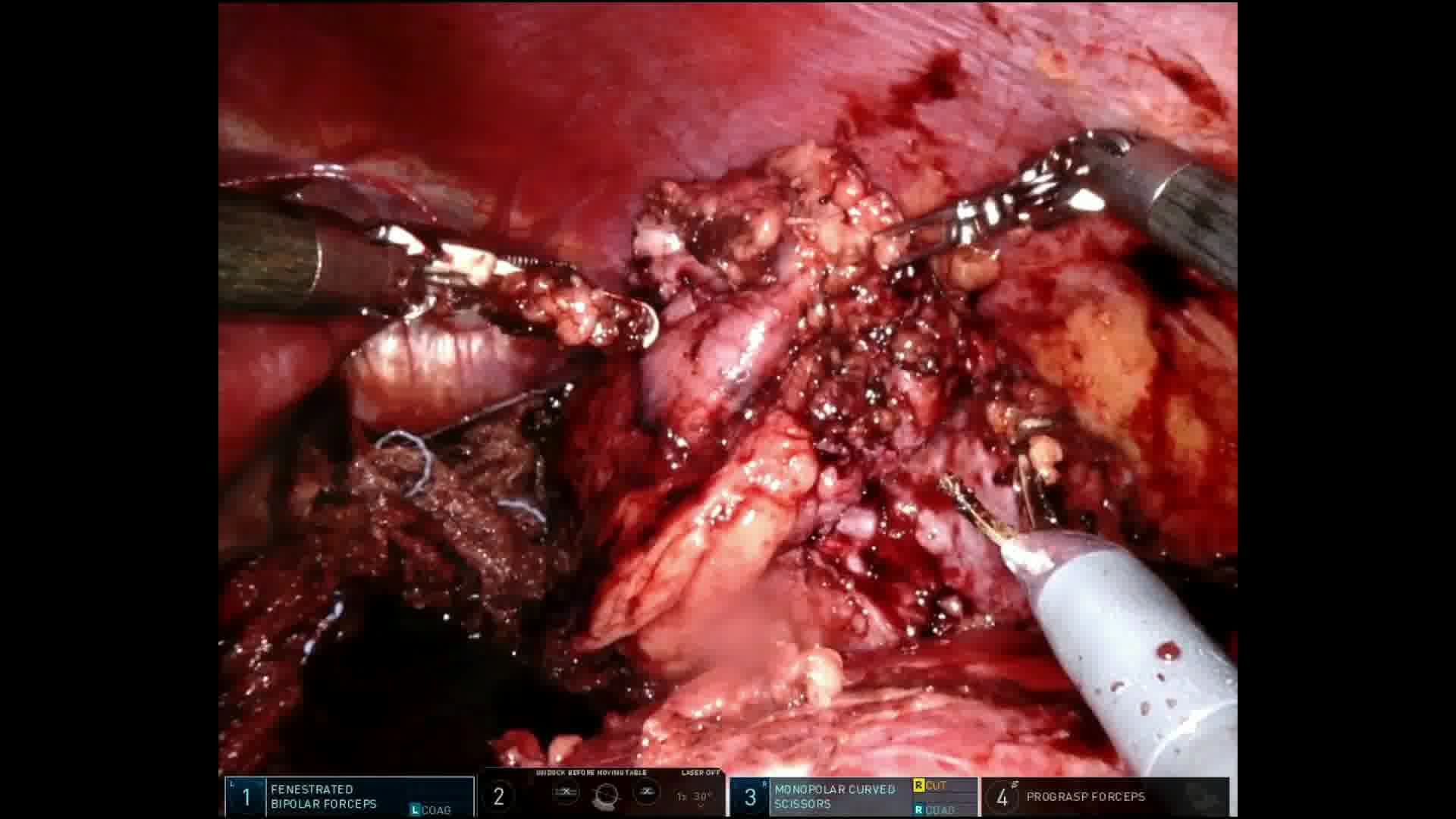
Surgery with da Vinci-Xi was planned. With the patient in right lateral position, pneumoperitoneum was created and 8 mm metallic ports were placed in the paramedian line and 12 mm assistant port was placed near the umbilicus on the right of the midline. Palanivelu hydatid trocar-cannula system was used to suction the cyst contents and irrigate it with scolicidal agents. 1 The cyst was found to be intact and outside the renal capsule. It was carefully excised and extracted using an endobag.
Outcomes:
The docking time was 12 minutes and the console time was 185 minutes. The estimated blood loss was 40 mL. Oral feeds were resumed on postoperative day 1. Pain score was 2 on visual analog scale on day 0. Patient was discharged on day 2 after drain removal. Perinephric hydatid is a rare disease and its management by robot assistance is not published in the literature till date.
Patient Consent Statement:
Authors have received and archived patient consent for video recording/publication in advance of video recording of the procedure.
No competing financial interests exist
.
Runtime of video: 4 mins 53 secs
Get full access to this article
View all access options for this article.