
Abstract
Introduction:
Focal therapy of prostate cancer aims to destroy known cancer while preserving uninvolved prostatic tissue with the goal of preserving genitourinary function. 1,2 Previous methods of focal cryoablation were done using predefined templates, leading to destruction of a greater volume of prostatic tissue than necessary. 2 Multiparametric magnetic resonance imaging (MpMRI) has emerged as the most accurate method to identify and differentiate prostate lesions. 3 MpMRI–ultrasound (US) fusion technique for focal therapy more accurately targets cancerous lesions while minimizing damage to vital structures. In this study we describe the novel technique of MRI–US fusion guided focal cryotherapy using a transperineal fusion platform devised for targeting and treatment of prostate cancer.
Materials and Methods:
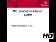
Informed consent is obtained from the patient. The patient in the video had Gleason Grade Group 3 (4 + 3) in a PIRADS4 lesion in the right apex with all systematic cores being negative. The procedure is done in dorsal lithotomy under general anesthesia. The MRI with the target lesion previously outlined by the radiologist is uploaded into the Artemis™ system and a custom cradle for the transperineal targeting is assembled. A biplanar ultrasound probe is inserted into the rectum and a sagittal scan of the prostate is performed. Gland boundary refinement is done in the fusion system followed by three-dimensional reconstruction. Optimal probe placement is then planned using the reconstruction, ensuring lesion ablation with a 5 mm–1 cm margin. Using the fused images as a guide, the length of the isotherm is adjusted on each needle. The cryotherapy needles are then inserted using MRI–US fusion with the transperineal needle guide attachment. Cystourethroscopy is then completed to ensure no urethral damage was done. A urethral warming catheter is then placed. Two freeze–thaw cycles are completed with the ice ball margins extending past the edge of the lesion, ensuring appropriate ablation. Needles are withdrawn and a regular Foley catheter is placed.
Results:
Follow-up prostate specific antigen for the patient at 6 weeks was 0.56 ng/mL, from 3.6 preoperatively. Data from a cohort of 20 men who underwent focal cryoablation showed a median change in Sexual Health Inventory for Men of −1.5 (IQR −6.3 to 0.0) points and International Prostate Symptom Score of 0.0 (IQR −2.3 to 1.0) at 6 months. Repeat MRI and biopsy at 6–9 months showed 76.9% of patients with absence of clinically significant prostate cancer.
Conclusions:
MRI–US fusion technique for focal cryotherapy produces excellent short-term results in both ablation of clinically significant PCa and preservation of genitourinary function. Additional long-term studies are needed for the optimization of this technique.
Authors received and archived patient consent for video recording/publication in advance of video recording.
No competing financial interests exist.
Runtime of video: 6 mins 35 secs

Get full access to this article
View all access options for this article.