
Abstract
Introduction:
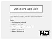
Standard technique in renal pelvis tumor resection involves fluoroscopy-guided access, tract dilatation, tumor resection, and nephrostomy tube placement. A ureteroscope-guided approach offers the advantage of direct visual inspection of the kidney and renal pelvis before access, facilitating optimal placement of the percutaneous access sheath and potentially reducing sheath trauma and bleeding. Incorporation of this ureteroscope-guided access technique ultimately enables more precise resection of the renal pelvis tumor with improved visibility.
Materials and Methods:
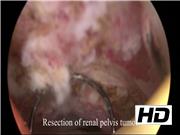
We present a 68-year-old male with a left solitary kidney who underwent management of a left renal pelvis tumor. Prior biopsy revealed low-grade urothelial carcinoma. Renal pelvis washings and voided urine cytology were negative. A computed tomography (CT) urogram showed an enhancing nodular lesion in the left renal pelvis and ureteroscopy showed a low-grade papillary tumor amenable to percutaneous management. Narrow band imaging was used during ureteroscopic evaluation to ensure no additional suspicious areas. Disease was noted to be multifocal with large surface area, so percutaneous resection was selected for management. During the procedure, access to the kidney was obtained under ureteroscopic and fluoroscopic guidance, and a guidewire was passed down the ureter for through-and-through access. The nephrostomy tract was dilated and a percutaneous access sheath was placed under direct vision to ensure optimal depth of sheath placement and minimize trauma. Complete resection of the renal pelvis tumor was performed using bipolar electrocautery and normal saline for irrigation. A nephrostomy tube was placed at procedure completion. A second patient at our institution, a 68-year-old man with congenital absence of left kidney, presented with large right upper pole urothelial low-grade carcinoma. He underwent a similar ureteroscope-guided access and percutaneous resection. Based on the renal anatomy, the upper pole calix—the same location as the tumor—was accessed. This patient had a complete effective resection of the tumor with excellent visibility.
Results:
The first patient was discharged on postoperative day 1 and returned for nephrostomy tube removal 1 week later. Surgical pathology analysis showed noninvasive low-grade papillary urothelial carcinoma. The patient remains recurrence-free 15 months postoperatively on surveillance ureteroscopy. However, follow-up CT imaging and ureteroscopy showed infundibular stenosis of a lower pole infundibulum, requiring laser incision and balloon dilatation. The second patient was also discharged on postoperative day 1. Surgical pathology analysis showed low-grade noninvasive (Ta) urothelial carcinoma. Intracavitary Bacillus Calmette–Guérin or mitomycin was not administered for adjuvant therapy. He remains recurrence free in the kidney 10 months postoperatively, but was noted to have low-grade tumor in the bladder, which was treated.
Conclusions:
Direct ureteroscopic vision to guide puncture location and access sheath depth reduces sheath trauma and bleeding from the tumor and kidney. Fluoroscopy time is also reduced, providing radiation safety benefits. Although spatial anatomy is known preoperatively and assists in access location, direct vision achieves more precise access sheath placement. Both patients were prestented to allow atraumatic placement of a ureteral access sheath. Although perhaps unnecessary for straightforward cases, this technique should be considered in select patients with difficult-to-access tumor locations or tumors in proximity to the planned entry site.
No competing financial interests exist.
Authors have received and archived patient consent for video recording/publication in advance of video recording of procedure.
Music source:
Runtime of video: 6 mins 34 secs

Keywords
Get full access to this article
View all access options for this article.