
Abstract
Introduction:
Standard hydrocelectomy for adult primary vaginal hydrocele may cause postoperative discomfort, limited mobility, and complications, such as hematoma, infection, persistent swelling, chronic pain, and decreased fertility. 1 –3 To decrease the complications, we described a novel endoscopic “Su-Wang technique” for adult primary vaginal hydrocele with the aid of a scrotoscope.
Materials and Methods:
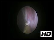
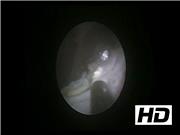
A 56-year-old man with fall bilge feeling of the right scrotum for 12 months was found to have primary vaginal hydrocele by the B-mode ultrasound. First, a transverse incision about 1 cm long is made at the anterior superior part of the right scrotum. Then, we cut the skin, dartos, and superficial fascia of the scrotum layer-by-layer, until the parietal layer of the tunica vaginalis was exposed. We made a length of about 0.5 cm incision, and then a self-made working sheath was passed through the incision into the vaginal cavity, steadily fixed at the parietal layer. Through the self-made working sheath, we inserted the scrotoscope into the vaginal cavity. Using a plasma cylindrical electrode, we burned a circle at the parietal layer of the tunica vaginalis as a marker. The distance between the burned place and the testis or epididymis was 1 to 2 cm. Along the circular mark, the parietal layer of the tunica vaginalis was burned with a plasma cylindrical electrode. The burning depth penetrates only the parietal layer of the tunica vaginalis, but does not reach the deep fascial layer of the scrotal wall. After checking the depth of the burn site, the scrotoscope was pulled out. Then, the parietal layer of the tunica vaginalis was peeled away from the deep fascial layer of the scrotal wall. After that, the parietal layer of the tunica vaginalis could be easily stripped from the scrotum.
Results:
We have completed 15 cases of primary vaginal hydrocele in adults with the “Su-Wang technique,” the average size of hydrocele was 7.8 cm, the average operation time was 43 minutes, the average volume of drainage after operation was 7 mL, the average drain removal time was 2 days postoperatively, and the length of stay was 4 day. Postoperative hematoma or wound infection did not occur. Mild scrotal edema usually dissipated within 2 days after surgery. Patients were able to resume normal daily activity an average of 4 days after surgery (range 2–5). All 15 cases were cured and followed up for 1 year without recurrence.
Conclusions:
Our “Su-Wang technique” allows the surgeon to remove large hydrocele through a 1-cm incision and to almost completely resect the parietal tunica vaginalis, resulting in no recurrence, minimal complications, and early recovery. This procedure may be a viable option for surgical treatment of adult primary vaginal hydrocele.
No competing financial interests exist.
Runtime of video: 7 mins 12 secs

Get full access to this article
View all access options for this article.