
Abstract
Introduction:
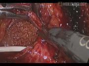
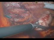
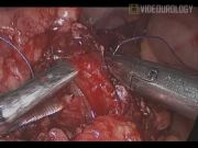
Urolithiasis is a common urological problem with significant morbidity for the patients. Currently, open surgery is indicated for failed percutaneous, ureteroscopic procedure and for calculus in anomalous kidney when other methods are not feasible or not available. In these scenarios, when expertise is available, laparoscopic approach has been proved to be more beneficial with regard to postoperative morbidity, recovery, and clearance of the stone. 1,2 We are presenting a video demonstration of laparoscopic management of urolithiasis in different circumstances. Laparoscopic pyelolithotomy for left renal calculus was performed through transperitoneal approach using four ports in lateral decubitus. Double-J stent was placed in the lithotomy position. After reflecting colon, upper ureter was identified and traced proximally to reach the renal pelvis. Renal pelvis was exposed completely, and inverted V-shaped pyelotomy was performed. Calculus was identified and delivered, followed by pyelotomy incision closure using 3-0 absorbable barbed suture. Laparoscopic ureterolithotomy for upper ureteric large calculus was performed using three ports technique in lateral decubitus. Upper ureter was identified after reflecting colon to identify the stone. Ureterotomy was made on the stone using hook electrocautery. Ureterotomy was closed after delivering the stone. Double-J stent was placed in a retrograde manner in the lithotomy position. In third scenario, laparoscopic pyelolithotomy was performed for left pelvic kidney calculus. Three ports were placed in the supine position with 30° right tilt. Sigmoid colon was reflected to identify the pelvic kidney and renal pelvis. Pyelolithotomy was performed followed by retrograde placement of a Double-J stent. In all patients, drain was placed followed by port closure. Catheter was retained for 24–48 hours. The Double-J stent was removed after 6 weeks.
Materials and Methods:
Case 1: A 72-year-old male patient presented with left loin pain with a history of percutaneous nephrolithotomy twice in the recent past. Detailed evaluation revealed dense left renal calculus measuring 4×3.8 cm with normal renal parameters. Laparoscopic pyelolithotomy was contemplated. Case 2: A 59-year-old male patient presented with left upper ureteric calculus with longitudinal dimension of 3.8 cm. He had a history of cystolithotripsy done for bladder calculus in the recent past. Laparoscopic ureterolithotomy was performed. Case 3: A 32-year-old male patient presented with a 3-cm calculus in the left pelvic kidney. Laparoscopic pyelolithotomy was performed. All patients with urolithiasis who were treated with laparoscopic approach were analyzed to assess the operative and postoperative outcomes.
Results:
In total, 40 patients were treated with laparoscopic approach for urolithiasis at our center. Three patients developed a urine leak during the postoperative period, which subsided with conservative treatment. There were no other operative and postoperative complications.
Conclusion:
Laparoscopic ureterolithotomy should be considered the first option for large ureteric calculus not amenable for endoscopic approach. Laparoscopic pyelolithotomy is a viable alternative to laparoscopic-assisted percutaneous nephrolithotomy (PCNL) for pelvic kidney calculus. However, laparoscopic pyelolithotomy for calculus in normally located kidney is an alternative to open approach when a percutaneous procedure has been failed or not feasible.
No competing financial interests exist.
Runtime of video: 6 mins
Keywords
Get full access to this article
View all access options for this article.