
Abstract
Introduction:
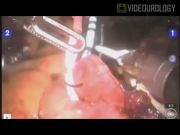
We present a 10-minute video of robot-assisted laparoscopic Studer pouch formation (RASP) after robot-assisted laparoscopic radical cystoprostatectomy.
Materials and Methods:
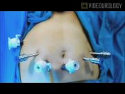
Between December 2009 and April 2010, we performed 12 RASP procedures. 1,2
Urethroileal anastomosis and segregation of the ileal segment:
The patient is placed in a slight (5°) Trendelenburg position, and a 0° lens is used. The most distal 20-cm segment of the terminal ileum is marked with the Cadier forceps. Most dependent part of the ileal segment is determined for uretral anastomosis. A 1-cm incision on the antimesenteric border, 15–20-cm proximal to the spared terminal ileal segment, is made for urethral anastomosis, and the posterior wall is finished first. An indwelling 20–22F urethral catheter is passed through the urethra into the intestinal lumen, and anastomosis is completed. Another 15–20 cm of the ileal segment left to the urethroileal anastomosis is assigned for Studer pouch formation. The third segment of 10-cm proximal to this is assigned for an afferent loop. Two intestinal endoGIA staplers are introduced from a 15-mm port on the left side and placed at the junction of the terminal ileum and pouch segment and proximal ileum and the afferent loop segment.
Ileoileal anastomosis:
A triangular shaped intestinal wall segment is removed just proximal to the staple line, and openings snug to fit for endoGIA blades. A second endoGIA stapler is introduced into the ileal ends and fired after antimesenteric borders are brought close proximity. The remaining intestinal opening is closed with the use of third endoGIA blades.
Formation of Studer pouch:
Optic is changed to 30° lens. Sparing the afferent loop, rest of the segregated ileal segment to be used for the formation of the Studer pouch is opened at the antimesenteric border by monopolar scissors. Adjacent walls of the intestinal walls on each side of the urethroileal anastomosis are sutured by using 3/0 monocryl sutures. The posterior wall is then completed. Thereafter, the anterior wall is formed asymmetrically.
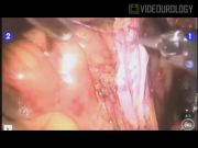
Uretero–ureteral and ureteroileal anastomosis:
In the right gutter, a Wallace type anastomosis is done. Ureteral catheters are passed through the abdominal wall into the pouch and then into ureters. The remaining anterior wall of the proximal intestinal segment and the medial edge of the ureteral plate are completed. Then, posterior plate anastomosis is completed. The cut edges of ureters are sent for frozen. After retroperitonealization of the ureteroileal anastomosis, the opening on the anterior pouch wall is closed.
Results and Conclusions:
Postoperative outcomes could be accessed in Reference 2. At a mean follow-up of 7.1±2.3 months, 3 patients died due to metastatic disease. Of the available 7 patients, 6 were fully continent, and 1 had mild day-time incontinence. RASP is a safe procedure that can be learned and performed with acceptable short-term surgical and functional outcomes.
No competing financial interests exist.
Runtime of video: 10 mins
Presented as a video during the 28th World Congress of Endourology & SWL, September 1–4, 2010, Chicago, IIlinois.
Keywords
Get full access to this article
View all access options for this article.