
Abstract
For certain select cases, laparoscopic pyelolithotomy is a practical and efficacious method to treat renal stone disease. One such case is that of an ectopically located pelvic kidney with a large stone burden. Herein, we describe our technique and provide what we feel is a trick to performing a key part of this procedure—flexible nephroscopy while using CO2 gas to inflate the collecting system.
Problem:
Most patients who suffer from renal calculus disease undergo one or a combination of shockwave, ureteroscopic, or percutaneous treatments. In certain select cases, alternative therapies may be necessary. One of those circumstances is that of an ectopically located kidney with a very large stone burden. Recently, El-Kappany et al 1 have described their techniques and results for both laparoscopy-guided percutaneous nephrostolithotomy and for laparoscopic pyelolithotomy in patients with pelvic kidneys with large stone burdens. Their stone-free rate was 91% with a zero complication rate. Kramer et al 2 have also described the technique of a transperitoneal laparoscopic pyelolithotomy for treatment of renal stones in anomalous kidneys—including horseshoe and ectopic pelvic kidneys. Via a transperitoneal approach, large renal stones may be easily grasped with laparoscopic instruments after the renal pelvis has been incised. However, using rigid laparoscopic instruments to grasp caliceal stones may be challenging and potentially traumatic. In contrast, flexible nephroscopy is practical for a full inspection of the collecting system and for grasping or treating additional stones. However, using saline or water as an irrigation fluid during nephroscopy has certain pitfalls. One problem encountered is that the open renal pelvis will leak fluid to the peritoneal cavity. In addition, the interface of a liquid and the CO2 gas used to establish pneumoperitoneum alters and hinders one's visualization. Herein we offer our approach of using CO2 gas to insufflate the collecting system to facilitate nephroscopy.
Technique:
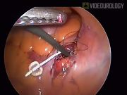
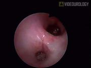
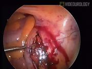
The companion video demonstrates the technique as performed in a patient who had a preoperative computerized tomography, which revealed four stones within the renal pelvis of an ectopically located, pelvic kidney. Each stone was roughly 1000 Hounsfield units and 10 to 12 mm in diameter. The patient was placed in low lithotomy position under general anesthesia, and prepared for both cystoscopy and standard laparoscopy. Via a retrograde approach, an occlusion balloon catheter was advanced to the renal pelvis over a guidewire placed via the cystoscope. A laparoscopic port was placed at the umbilicus, as well as along each midclavicular line at points several centimeters below the umbilicus. Manipulation of the occlusion balloon, including gentle traction and retrograde irrigation with saline to fill the pelvis, allowed for easy identification of the renal pelvis through the posterior peritoneum. The pyelotomy was approximately 2 cm in length. Once incised, the renal pelvis was identified, mobilized, and opened between stay sutures. Stones in the renal pelvis were removed with grasping forceps. To remove the last of the four stones, which was not visible using rigid laparoscopy, flexible nephroscopy was performed. A flexible nephroscope was guided through a 10-mm port and into the renal pelvis. The CO2 gas line, which had been initially connected to the laparoscopic port, was then connected to the irrigation port of the flexible nephroscope. Nephroscopy was performed in a dry but open space in which there was excellent observation. Periodic venting of the pneuperitoneum via a laparoscopic port allowed for fresh CO2 gas to flow into the collecting system. The fresh CO2 gas inflated the collecting system relative to the lower pressure of the periodically vented peritoneal cavity, facilitating localization of the stones within the calices. With appropriate observation of the collecting system, a 0 tip basket (Boston Scientific, Natick, MA) was used to remove any remaining stones from the kidney. Although we did not use a pneumatic lithotripter or holmium laser in this case, both of those technologies could be utilized in a CO2 gas medium, as has been previously reported. 3,4 After removal of the stones, a guidewire was placed through the balloon occlusion catheter, which was then removed, and an internal Double-J stent was placed in a retrograde fashion via a cystoscope. The open renal pelvis and the overlying posterior peritoneum were closed with intracorporeal sutures in separate layers using 4-0 Vicryl. A Jackson Pratt drain was placed via a laparoscopic port to rest adjacent to the peritoneal closure. All laparoscopic port sites were then closed in standard fashion. In this case, the patient was rendered completely stone free based upon postoperative computerized tomography.
No competing financial interests exist.
Runtime of video: 3 mins
Get full access to this article
View all access options for this article.