
Abstract
Abstract
Introduction:
To minimize risk and improve outcomes after parathyroidectomy, a number of tools are used to distinguish the diseased parathyroid gland from its surrounding structures.1–3 Examples include preoperative sestamibi scanning, intraoperative nerve monitors, and intraoperative quick parathyroid hormone (PTH) assays. Indocyanine green (ICG) fluorescence imaging, new applications of which continue to be described, has recently been used in dogs to identify the parathyroid gland.4–6 In this video, we demonstrate a parathyroidectomy with the use of intravenous ICG fluorescence and a near-infrared camera.
Case Presentation:
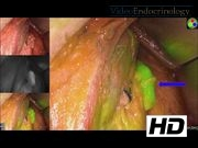
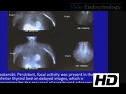
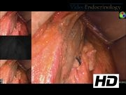
A 50-year-old woman presented with symptomatic primary hyperparathyroidism. She described difficulty concentrating, fatigue, nausea, vomiting, and abdominal pain. Laboratory results confirmed primary hyperparathyroidism, and sestamibi showed a focal activity present in the left inferior thyroid bed on delayed imaging. Parathyroidectomy was recommended. Intubation was done using the NIM® EMG endotracheal tube. An incision was made two fingerbreadths above the sternal notch. Subplatysmal flaps were raised. The strap muscles were divided and on the left were retracted laterally. The thyroid was mobilized away from its lateral attachments revealing a fatty-appearing tissue. We utilized the PINPOINT® fluorescence laparoscope after administration of 3 mL of ICG (2.5 mg/mL). The tissue displayed some fluorescence, but no more than background thyroid gland. It was removed, sent to pathology, and the frozen section returned with a benign fibroadipose tissue. Further mobilization and exploration revealed an enlarged left upper parathyroid gland. A nerve monitor was used to identify the recurrent laryngeal nerve. Three milliliters of ICG was again administered; this time the parathyroid gland fluoresced bright green on the PINPOINT® laparoscope, allowing careful identification of the parathyroid adenoma and ligation of its vascular stalk. The parathyroid was easily dissected and the specimen was sent to pathology. A postoperative PTH was measured showing an appropriate response after removal of what the frozen section returned as hypercellular parathyroid tissue.
Conclusion:
ICG fluorescence imaging continues to find new applications in medicine and surgery. ICG imaging can be used in the operating room to identify the parathyroid gland during parathyroidectomy. Further study is needed to determine the clinical significance of using this technology routinely.
No competing financial interests exist.
Runtime of video: 3 mins 30 secs
Keywords
Get full access to this article
View all access options for this article.
References
Supplementary Material
Please find the following supplemental material available below.
For Open Access articles published under a Creative Commons License, all supplemental material carries the same license as the article it is associated with.
For non-Open Access articles published, all supplemental material carries a non-exclusive license, and permission requests for re-use of supplemental material or any part of supplemental material shall be sent directly to the copyright owner as specified in the copyright notice associated with the article.