
Abstract
Abstract
Introduction:
Ectopic parathyroid adenomas can be located within the residual thymic tissue, in the aortopulmonary window (APW), in the posterior mediastinum, or within the parenchyma of the thyroid gland. Only 2% of them are not accessible through standard cervical incision and require a thoracic approach. 1 Recently, a number of minimally invasive strategies for their management have emerged, including video-assisted thoracoscopic surgery (VATS) and video-assisted mediastinoscopy (VAM). We report the case of an ectopic parathyroid adenoma located in the APW, which was successfully managed by VAM under continuous intraoperative neuromonitoring to ascertain the function of the left recurrent laryngeal nerve (LRLN).
Materials and Methods:
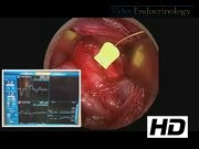
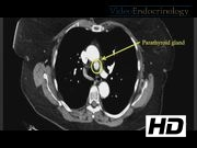
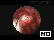
A 75-year-old woman with a history of primary hyperparathyroidism and of Graves' disease previously underwent a total thyroidectomy and bilateral cervical exploration without any preoperative localization imaging. Two hyperplastic parathyroid glands were removed and a third one left in place; the left superior one was not found. Unfortunately, the biological markers of primary hyperparathyroidism did not improve. An ectopic parathyroid adenoma could then be shown by a Technetium-99m-labeled sestamibi scintigraphy and a chest CT scan to sit in the APW. Its size was 14 × 14 × 12 mm. Among the multiple surgical options, we chose an approach by VAM. The low cervical transverse incision was partially reopened and dissection carried down in the pretracheal space with the video-mediastinoscope (Richard Wolf). Because of the position of the ectopic adenoma, close to the LRLN, a vagal probe for continuous intraoperative stimulation and neuromonitoring (Automatic Periodic Stimulation electrode with Medtronic NIM®Response 3.0 nerve monitoring system) was placed around the left vagus nerve after its exposure through a lateral approach. The carina, the pulmonary artery, and the aorta as well as the LRLN were identified. The parathyroid adenoma was found, as expected, in the middle of these structures and completely excised. The functional integrity of the LRLN was once again confirmed by direct and vagal stimulation at the end of the procedure. The postoperative course was uneventful. Histological analysis confirmed parathyroid tissue and there was a normalization of serum parathormone and calcium levels at the first postoperative day.
Results:
In our patient, we chose to perform the second surgery by VAM, which permitted to avoid the single-lung ventilation and the need of a chest tube, which are mandatory with VATS, and additional scars. One of the most common complications during mediastinoscopy is damage to the LRLN,2,3 with an incidence of up to 6.5%. 4 In our patient, the risk was higher because of the scar tissue and the location of the adenoma close to the LRLN. Intraoperative identifying and monitoring of the LRLN is feasible and safe and helps to ascertain its functional integrity. 5 For these reasons, we performed the surgery under continuous intraoperative neuromonitoring.
Conclusions:
VAM is a good surgical option to manage ectopic parathyroid adenomas located within the reach of the video-mediastinoscope, and the continuous intraoperative neuromonitoring is a simple and safe technique to identify and test the functional integrity of the LRLN.
No competing financial interests exist.
Runtime of video: 5 mins 38 secs
Keywords
Get full access to this article
View all access options for this article.
References
Supplementary Material
Please find the following supplemental material available below.
For Open Access articles published under a Creative Commons License, all supplemental material carries the same license as the article it is associated with.
For non-Open Access articles published, all supplemental material carries a non-exclusive license, and permission requests for re-use of supplemental material or any part of supplemental material shall be sent directly to the copyright owner as specified in the copyright notice associated with the article.