
Abstract
Introduction:
Electronic cigarette (e-cigarette) aerosol is understood to provide reduced exposure to harmful toxicants compared with tobacco cigarette smoke, as it delivers nicotine and flavors without the use of tobacco. Published studies have shown that e-cigarette aerosol is chemically simple compared with tobacco smoke and corresponding reductions in toxicity in vitro have been demonstrated. However, comprehensive analytical and in vitro assessments of many widely available and currently marketed products, including pod-based systems, are limited.
Materials and Methods:
Here we report comparative data for aerosol emissions and in vitro toxicity, using the neutral red uptake, the bacterial reverse mutation, and in vitro micronucleus assays, for a pod system e-cigarette compared with 3R4F reference cigarette smoke.
Results and Discussion:
Many of the harmful and potentially harmful constituents found in cigarette smoke were not detected in e-cigarette aerosol. Using established in vitro biological tests, e-cigarette aerosol did not display any mutagenic or genotoxic activity under the conditions of test. By contrast, 3R4F cigarette smoke displayed mutagenic and genotoxic activity. E-cigarette aerosol was also found to be ∼300-fold less cytotoxic than cigarette smoke in the neutral red uptake assay.
Conclusion:
Data presented here show clear differences between a tobacco cigarette reference product and a commercially available nontobacco containing e-cigarette product in terms of emissions and in vitro toxicity profile. Our results demonstrate that high-quality e-cigarettes and e-liquids may offer the potential for substantially reduced exposure to cigarette toxicants in adult smokers who use such products as alternatives to cigarettes.
Introduction
Electronic cigarettes (E-cigarettes) have been characterized by Public Health England as being ∼95% less harmful than conventional (traditional tobacco) cigarettes 1 with research showing that these devices can assist smokers in replacing conventional cigarettes and reducing their number of cigarettes per day consumption.2,3 E-cigarettes are battery-powered devices that have prefilled cartridges/pods or refillable tanks containing a liquid mixture composed primarily of propylene glycol and/or glycerol, nicotine, and flavoring. 4 Typically for pod-based e-cigarettes, drawing breath activates a pressure-sensitive circuit that heats the atomizer and turns the liquid into an aerosol (popularly referred to as “vapor”) that is inhaled by the user through the mouthpiece.
For decades, scientists have worked to characterize the toxicants in cigarette smoke 5 and several regulatory authorities have mandated the reporting of constituents in smoke emissions from cigarettes.6–8 Given the rise in popularity of e-cigarettes worldwide as an alternative to conventional cigarettes by adult smokers, there is increasing public health and regulatory interest in toxicant emissions from e-cigarettes. On May 10, 2016, the U.S. Food and Drug Administration (FDA) published the final rule to deem e-cigarettes to be subject to the Federal Food, Drug, and Cosmetic Act, providing the FDA authority to regulate e-cigarettes and e-liquids, and published industry guidance on premarket tobacco product applications for e-cigarettes in June 2019. 9 The guidance provided a list of harmful or potential harmful constituents (HPHCs), which includes certain analytes contained in the abbreviated HPHC list for conventional cigarette smoke. 8 During the development and implementation of the European Union Tobacco Products Directive (2014/40/EU), which also encompasses e-cigarettes, the European Commission issued a data dictionary that includes a recommended list of emissions for product notification purposes across EU member states. 10 Although there are standardized analytical procedures for the measurement of toxicants in conventional cigarette smoke, currently there are few standardized test methods and no reference products for e-cigarettes.
The HPHCs in conventional cigarette smoke are well documented and have been linked to a number of negative health outcomes, including cancer, emphysema, and cardiovascular disease. 4 Research has indicated e-cigarettes can provide reduced exposure to cigarette smoke constituents because they deliver flavor and nicotine through aerosolization of a liquid rather than by burning tobacco. 11 The majority of studies in the literature performed on older generation e-cigarette devices, typically using cartomizers, have demonstrated that the limited number of constituents in e-cigarette aerosols are tens to thousands of times lower on a per-puff basis than in conventional cigarette smoke.12,13 Many of the toxicants in tobacco smoke are simply not present in e-cigarette aerosols at detectable levels when assessed using machine-based aerosol generation or are at levels equivalent to the tolerances allowed in medicinal products.11–19
A recent review of chemical, toxicological, and clinical studies for both e-cigarette liquids and aerosols indicated that they contain reduced levels of harmful chemicals and emissions, induced significantly less cytotoxicity, and resulted in fewer cardiovascular and respiratory functional effects when compared with reported data on tobacco cigarettes. 1 Romagna et al. reported e-cigarette aerosol to be significantly less cytotoxic than tobacco smoke in fibroblasts, 20 and Farsalinos et al. concluded the same findings in myocardial cells. 21 Scheffler et al. found cell viability was lower in primary human bronchial cells exposed to tobacco smoke than in e-cigarette aerosol. 22 Husari et al. found e-cigarette aerosols exhibited significantly less toxic effects on lungs of experimental animals and on A549 cell cultures than smoke from tobacco products. 23 Wieczorek et al. (in press) compared two e-cigarette aerosols from blu GO™ disposable and blu PLUS+™ rechargeable cartridge-based devices with the smoke from a reference cigarette (3R4F) in an in vitro battery of established assay: neutral red uptake (NRU) for cytotoxicity, in vitro micronucleus (IVM) for genotoxicity, and the bacterial reverse mutation (Ames) assay for mutagenicity. Results from this study showed that the e-cigarette fresh whole aerosol resulted in a significant 250–1000-fold reduction in in vitro cytotoxic response in the BEAS-2B cell line compared with cigarette smoke and displayed no mutagenetic response in TA100 or TA98 or genotoxicity in V79 cells. In addition, Wieczorek et al. (in press) showed device type could impact the cytotoxicity of the aerosol. Aerosol generated from blu GO was significantly more active than blu PLUS+ aerosol in the NRU assay, although these responses were substantially less cytotoxic than cigarette smoke exposure. 24 The blu GO device operates at a much higher power level than blu PLUS+; although this may generate larger puff volumes and deliver higher doses per puff to the user than the blu PLUS+ product, the differences seen is this study may be due to changes in the chemical nature of the aerosol. Published clinical research has shown that adult smokers who switch to e-cigarettes have significantly lower exposure to carcinogens and toxicants found in cigarette smoke, with reductions largely indistinguishable from complete smoking cessation or use of licensed nicotine replacement products.25–27
In summary, it has been demonstrated that older generation e-cigarette aerosols are chemically simple when compared with cigarette smoke. The inhalation of e-cigarette aerosol, compared with cigarette smoke, has the potential to induce significantly less adverse toxicological effects and reduce various negative health effects when used by adult smokers who would otherwise continue to smoke. However, comprehensive analytical and toxicological assessments of many widely available and currently marketed e-cigarette products, including pod-based products, are limited.
This study aimed to characterize the aerosol generated by a commercially available e-liquid in the myblu e-cigarette pod-system device and compare the emissions and in vitro toxicity with the reference cigarette smoke. The e-cigarette aerosol and tobacco smoke were characterized for 44 analytes. These analytes included carbonyls, phenolics, tobacco-specific nitrosamines, polyaromatic amines, and polycyclic aromatic hydrocarbons. Many of these compounds are included in guidance issued by the FDA, 8 which includes reporting obligations for 20 HPHCs in cigarette smoke that the FDA considers cause or could cause harm to smokers. In addition, established in vitro toxicological assays were used to examine the cytotoxicity (NRU), mutagenicity (Ames test), and genotoxicity (IVM test) of fresh cigarette smoke and myblu aerosol.
Materials and Methods
Reagents
All reagents and equipment were purchased from Sigma-Aldrich (St. Louis, MO) unless stated otherwise. Aroclor 1254-induced rat liver microsomal fraction S9 (Lot Number: MolTox S93604) was stored at −70°C until use.
Cultures of BEAS-2B (human bronchial epithelial) (ECACC 95102433) and V79 (Hamster Chinese lung) cells were obtained from the European Collection of Authenticated Cell Cultures (ECACC). All cell stocks were stored frozen in liquid nitrogen before use. Each batch was checked for the presence of mycoplasma contamination using a standard polymerase chain reaction mycoplasma test kit. Salmonella typhimurium strains, TA98 and TA100, were obtained from Trinova Biochem GmbH (Giessen, Germany) and stored frozen (−70°C) in aliquots before use.
Test articles
The test articles were the 3R4F Kentucky Reference Cigarette and a commercially available pod-based e-cigarette.
The 3R4F reference cigarettes (lot number V351X61B5) were obtained from the University of Kentucky, Center for Tobacco Reference Products (Lexington, KY). Before analysis, 3R4F sticks were conditioned at 22°C ± 2°C and 60% ± 5% relative humidity for a minimum 48 hours (but no more than 10 days), according to International Organization for Standardization (ISO) method 3402. 28
E-cigarettes from brand myblu™ contained tobacco flavored e-liquid with 1.6% (w/w) nicotine and were purchased from UK retailers. The myblu liquid is formulated using pharmaceutical and food-grade ingredients. myblu (see Appendix 1) is a rechargeable closed pod-system e-cigarette, consisting of two segments: a rechargeable battery section (battery capacity, 350 mAh) and a replaceable e-liquid containing pod (volume, 1.5 mL; coil resistance, 1.3 Ω). E-liquids and devices were stored at room temperature until use.
Smoke and aerosol generation
All smoking machines are validated for the specific tests, as described hereunder.
For the characterization of analytes, mainstream smoke and aerosol were generated on a linear smoking machine LMC4 (Borgwaldt, Germany) and a rotary smoking machine RM20D (Borgwaldt, Germany).
For mutagenicity assessment, aerosol from e-cigarettes and smoke from 3R4F were generated using a three-port adapter RM158 connected with a single-port smoking machine RM1 (Burghart Instruments, Wedel, Germany).
For the NRU and IVM assays, fresh aerosol/whole smoke was generated using a bespoke smoking machine “Smoke Aerosol Exposure In Vitro System” (SAEIVS) (Burghart Tabaktechnik, Wedel, Germany) (Fig. 1). The SAEIVS is a five-port smoking machine directly connected with the exposure device and equipped with smoke “distributors” for 24 and 96 multiwell plates. A smoke distribution device disperses the smoke/aerosol across the multiwell plate. All wells of the plate are provided with separate smoke/aerosol inlet and suction ducts. The computer-controlled smoke dilution system allows precise and rapid dilution of freshly generated cigarette smoke if necessary (e.g., high cytotoxicity) in <10 seconds to prevent sample aging. The rapid mixing and dilution process uses an exact predefined volume of humidified and filtered air and is performed in a closed system (using impingers). The two exposure chambers have separate independent dilution systems to allow parallel exposure to the same smoke/aerosol and their gas vapor phase at different dilution levels. Smoke/aerosol is rapidly delivered to the cells (∼10 seconds). All wells of each plate are provided with an individual smoke inlet and outlet ducts for exposure and extraction at the end of each puff. The use of a blanking plate in each exposure chamber enables puff by puff determination for dose–response analyses. Furthermore, the separate chambers enable testing of the same product in two independent in vitro assays and in different multiwell plates at the same time.
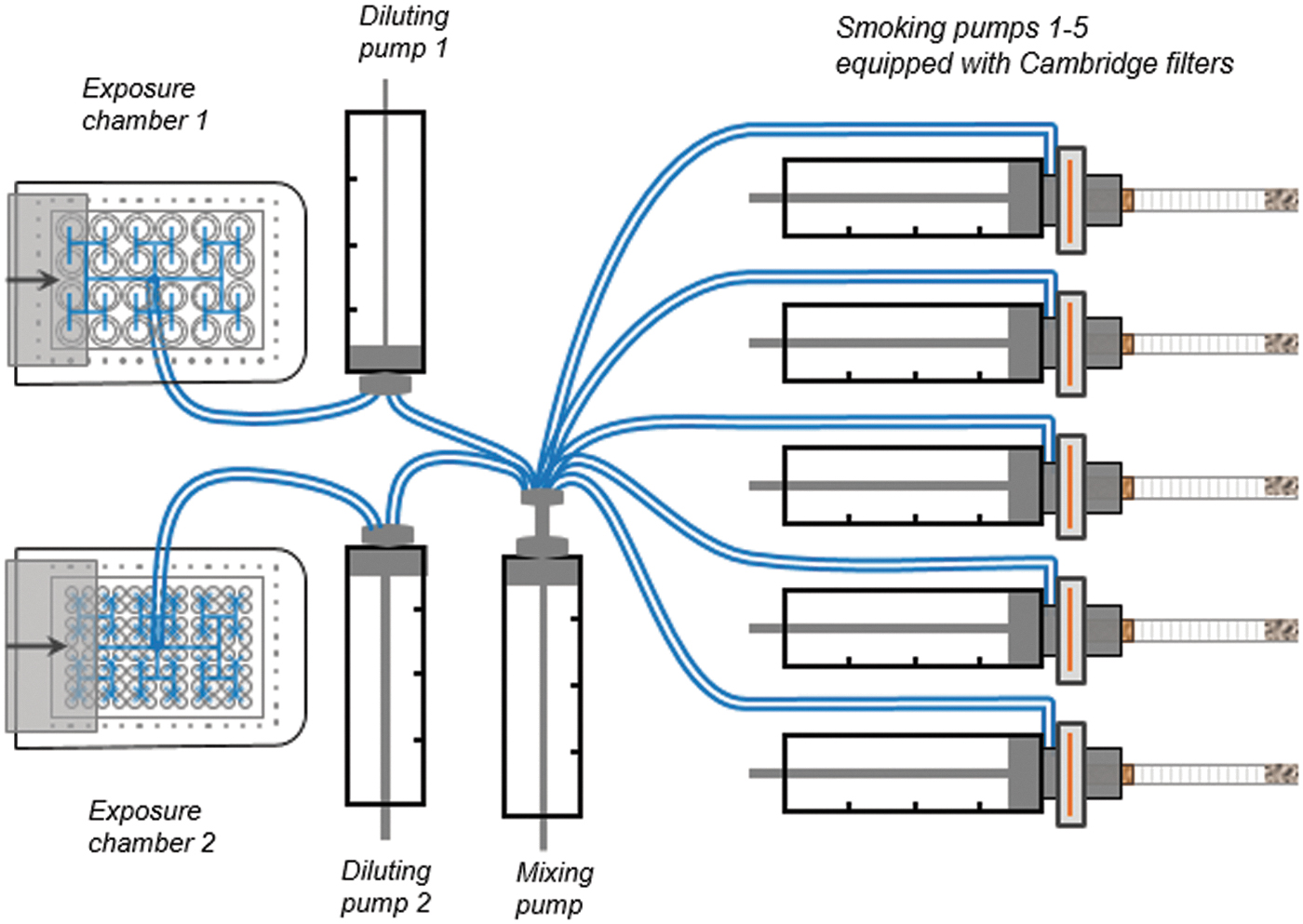
Diagram of the SAEIVS smoking machine. SAEIVS, smoke aerosol exposure in vitro system.
The SAEIVS system has been validated internally regarding delivery of smoke/aerosol and the biological effects induced by the gaseous components by using appropriate positive controls and puffing parameters described herein. E-cigarette aerosol is delivered undiluted and 3R4F smoke diluted 1:6–1:16 dependant on the assay. After 3 seconds exposure, the aerosols or smoke is removed by vacuum.
Cigarettes were machine smoked according to Health Canada Intense smoking regime (55 mL puff volume, 2 seconds puff duration, 30 seconds puff frequency; bell-wave profile), with 100% ventilation block. 29 For emissions testing, the smoke was collected from three replicates.
The e-cigarettes were machine puffed according to the CORESTA (Cooperation Centre for Scientific Research Relative to Tobacco) Recommended Method No. 81 puffing regime (55 mL puff volume, 3 seconds puff duration, 30 seconds puff frequency; square-wave profile). 30
For emissions testing, the aerosols were collected from three separate 50-puff blocks with three replicates measured. Each test product was weighed before and after aerosol collection to verify that product mass changes and filter pad mass changes were comparable. For the determination of ammonia, a bell profile rather than square was used for e-cigarette aerosol collection due to methodological limitations.
Blanks were prepared by puffing ambient air (50 puffs) through an empty smoking machine port to the appropriate trapping system for the analysis method. These air blank samples were prepared and analyzed in the same manner and at the same time as the smoke and aerosol samples. Blanks were included where appropriate to exclude environmental contamination, if any, from the data assessment.
Characterization of smoke and aerosol
All methods used in this study were established and validated for factory made cigarette (FMC) ISO smoking regime (35 mL puff volume, 2 seconds puff duration, 60 seconds puff frequency; bell-wave profile).
All analyses measured and analytical methods used to quantify the smoke and aerosol constituents are detailed in Appendix 2.
In vitro biological test methods
Cytotoxicity: NRU assay
Cytotoxicity of whole fresh e-cigarette aerosol and tobacco smoke was measured in BEAS-2B using the NRU method of Borenfreund and Puerner, 1985. 31 Diluted 3R4F smoke was used as a positive control. BEAS-2B cells were routinely taken from a preprepared stock and incubated with bronchial epithelial growth medium (BEGM) (BEGM supplemented with Lonza Bullet Kit, CC-3170).
A total of 100 μl of BEAS-2B cells (0.5 × 10⁴/mL), in serum-free medium, were seeded into each of the inner 60 wells of a 96-well tissue culture plate coated with Collagen I solution (20%, PureCol® EZ Gel; 2%, 1 M HEPES buffer; and 78% BEGM) and preincubated at 37°C in a humidified incubator with 95% air and 5% CO2 for 20 ± 3 hours. Directly before exposure, the medium was removed by suction and reverse plate centrifuged (10 g for 10 seconds). The plates were then placed in the SAEIVS chamber and the cells were exposed to the e-cigarette aerosol (0 to 140 puffs, given the maximum exposure time of 1 hour in this assay) or to diluted whole smoke from the 3R4F cigarette (0–10 puffs at 1:14–1:16 dilutions) at the air–liquid interface (ALI).
After ALI exposure, 200 μL of fresh medium was added to each well and incubated for 65 ± 2 hours. After incubation, the medium was replaced by neutral red staining solution in culture medium (supplemented with 20 mM HEPES and 10% fetal bovine serum) and further incubated at 37°C, 5% CO2 for 3 hours to allow dye uptake by the viable cells. After staining, the cells were washed with 150 μL of 1.34% calcium chloride and lysed with 100 μL of ethanol/acetic acid solution (1% glacial acetic acid and 50% in water) for 30 minutes at room temperature and pressure, with agitation. The absorption was measured at 400 nm on a microplate reader (Tecan Sunrise).
All exposures were conducted in triplicate with two independent experiments.
Mutagenicity: Ames screen
The in vitro mutagenicity of fresh 3R4F smoke and myblu was determined using the in vitro Ames test. The Ames screen was employed using S. typhimurium strains TA98 and TA100 (Trinova Biochem GmbH) +S9 treatment, conducted in accordance with OECD (Organisation for Economic Co-operation and Development) test guideline 471. 32
2-Aminoanthracene (1 μL/plate) was used as a positive control for both S. typhimurium stains TA98 and TA100. Each concentration of test vapor or smoke and positive controls were testing in triplicate. Six replicate readings were conducted for spontaneous revertants (vehicle).
An appropriate number of 16-hour Nutrient Broth No. 2 (OXOID) cultures of the TA98 and TA100 strains were prepared by inoculating 30 mL of medium with 0.5 mL of a 6-hour preculture in a 100 mL Erlenmeyer flask with one bacterium-coated CRYO-glass bead followed by incubation overnight at 37°C while shaking at 120 rpm.
After overnight incubation, the bacterial suspensions were prepared by centrifugation of 120 mL culture (four flasks of 30 mL) at 1800 g for 15 minutes and the pellet was resuspended in 12 mL of Ca2+, Mg2+-free Dulbecco's phosphate buffered saline (PBS).
The bacterial suspensions were exposed to test aerosol at room temperature under protection from direct light. In total, 10 ml of PBS bacteria suspension in a glass tube was placed in an impinger and bubbled with freshly generated smoke (1–5 cigarettes) and aerosol (up to 300 puffs) from the RM1 smoking machine (Burghart Instruments).
After each puff, a flushing step with fresh charcoal filtered ambient air was applied. After each exposure, 200 μL of bacteria suspension was taken from the tube and immediately used for the Ames screen.
The S9 mix, bubbled bacteria suspension, and Top-Agar were added to sterile 15 mL test tubes in the following order: 50 μL culture of the bacteria suspension (TA98 or TA100), 0.5 mL of S9 mix, then 2 mL of Top-Agar (45°C).
The solution was thoroughly mixed and then poured on top of a Vogel-Bonner agar plate. The plate was rotated and tilted to distribute the top agar evenly. When the top agar was solidified, the plates were inverted and placed in an incubator at 37°C. After 48 hours of incubation, the number of revertant colonies growing on the plates was counted.
The used bacteria were diluted to 1 × 10⁻⁶ with saline buffer. Then, 100 μl of the bacteria suspension was mixed with low melting top agar and poured on top of a Nutrient Broth-Plate (three plates per test item, per test day).
The total number of colonies growing on the plates was determined using the Synbiosis ProtoCOL SR—Automatic Colony Counter (Meintrup-DWS) and recorded.
Genotoxicity: IVM assay
The IVM assay was performed in concordance with OECD test guideline no. 487, 33 but with metabolic activation only.
Genotoxicity of whole fresh e-cigarette aerosol and tobacco smoke was measured in the Hamster lung V79 cell line (ECACC) with metabolic activation. The plates were placed in the exposure chamber and the cells were exposed to the e-cigarette aerosol or to diluted (1:6) whole smoke from the 3R4F cigarette at the ALI. Positive controls of fresh whole smoke for 3R4F reference cigarettes (1:4 and 1:5 dilutions with filtered air) were used to show the responsiveness of the test system and cyclophosphamide A (CAS 6055-19-2) to show the metabolic activity of the S9 fraction used.
Only the inner wells of each 24 multiwell plate were filled with 250 μL/well of Dulbecco's modified Eagles medium and supplemented with 10% fetal calf serum. Inserts with 0.4 μm membrane (Nunc; #140620) were inserted into the well and filled with 10 × 104 V79 cells/mL. Preincubation time was 20 ± 2 hours at 37°C and 5% CO2. Directly before the aerosol or smoke treatment, the medium was removed and the inserts were transferred into wells of a fresh multiwell plate filled with 250 μL HEPES buffer (20 mM final concentration) supplemented medium. The 24 multiwell plates were fixed in the exposure chamber and the cells at the ALI were exposed to the undiluted vapor from myblu or diluted (1:6) whole smoke from the 3R4F reference cigarette.
After ALI exposure, the cell-containing inserts were transferred to a new fresh serum-containing medium plate. Immediately 300 μL of S9 mix was added to each insert and the cells were incubated for 3 hours at 37°C. After incubation, the apical S9 medium was removed, and the cells were covered with serum-containing medium. For the expression of micronuclei, the inserts were incubated for another 20 ± 2 hours to allow for at least one cell division cycle. Smoke/aerosol was tested in three replicates.
After recovery, the cells were counted using a handheld cell counter (Scepter™ Cell Counter, Millipore). The cells were then fixed to slides and DNA-containing structures were stained with DAPI (1 μg/mL) in mounting medium (Vectashield, H-1000). After 20 ± 2 hours recovery, the cells were harvested and number of cells of the treatment groups were determined in a Vi-cell™XR cell counter (Beckman Coulter). Relative cell count (RCC) was the cytotoxicity measure used for the assessment.
The prepared slides were fully evaluated microscopically using the Metafer imaging system coupled to a fully automated microscope (Imager, Z2; Zeiss) in >1000 cells per slide of two parallel replicate cultures (slides).
Statistical analysis
In all cases, analysis was performed using the statistical software GraphPad Prism version 8.0. A p < 0.05 was considered significant.
Cytotoxicity
A calculation of relative cell viability expressed as relative NRU absorbance was made for each concentration of the test sample by using the mean NRU of the valid replicate values. This value was compared with the mean of the control. Relative cell viability is expressed as percentage of absorbance of untreated control. The EC50 is defined as the concentration that causes a response halfway between minimum and maximum responses. The following approach is used to determine the EC50 values:
To fit a sigmoidal dose–response curve and determine the best fit values for the logEC50, the Hill slope, and the bottom and top plateaus, of a four parameter, the nonlinear regression model was applied.
Concentrations associated with 50% viability using the Hill slope and EC50 from the Hill function analysis were also determined. The Hill function analysis was performed using the statistical software GraphPad PRISM® version 6.07. The cytotoxicity was deemed as significant, if confirmed in all three replicates over EC20 (p < 0.05 greater than the corresponding unexposed control). If the EC20 is not reached, the test item is not considered cytotoxic. Significant differences (p < 0.05) in cytotoxicity between myblu aerosol and 3R4F smoke were determined using a one-way analysis of variance (ANOVA) with a Dunnett's post hoc comparison test.
Mutagenicity
The Ames assay acceptance criteria must be met, including the mean negative control colony falling within the normal historical range, the positive control inducing a clear increase in revertant numbers, confirmation of an active S9 preparation, and no more than 5% of the plates were lost through contamination or other unforeseen event. For consideration of a positive mutagenic result for the test article, the following should be met: (1) it produces a twofold increase in the number of induced revertants, compared with negative control (ambient air), (2) revertant number of three of more test substance concentrations are significantly higher than the negative control, (3) a positive linear dose–response is observed, and (4) the positive responses were repeatable.
Mutagenic activity was calculated from the linear slope of the dose–response curve (nonthreshold model) using the statistical software GraphPad PRISM version 8.0. In the case of results with a positive slope in the nonthreshold model and Dunnett's test (p < 0.05 greater than the corresponding unexposed control), the tests were repeated. A test substance was deemed as mutagenic if the effect was confirmed in three replicates.
Genotoxicity
The IVM acceptance criteria must be met, including the negative control micronucleus frequencies should be in the range of historical data and positive controls should induce a statistically significant increase in micronucleus frequencies; at least one of the conventional smoke dilutions applied should induce a statistically significant increase in micronucleus frequencies and the population doubling of the solvent/medium control cultures should match a range between 1.0 and 2.5.
For consideration as a positive IVM response, there needs to be (1) a reproducible dose-dependent increase in micronucleus frequency and (2) the increased frequency, at any dose, must be significantly different to that of the negative control. The differences between median values were statistically analyzed using the chi-square test (p < 0.05 greater than the corresponding unexposed control). Duplicate cytotoxicity and micronucleus frequency determinations were made for each dose of test article and control. For each test article, three independent test days were conducted.
Results
Chemical characterization of smoke and aerosol
The aerosol from a myblu tobacco flavor e-cigarette was compared with 3R4F cigarette smoke for toxicants of public health interest, when generated under comparable smoking regimens.
A standard of 150 puffs (in 3 blocks of 50 puffs) was adopted for all e-cigarette analyses, which provides a similar collected mass per filter pad between the e-cigarette samples (aerosol collected mass [ACM]) and the conventional cigarette testing (total particulate matter [TPM]). This represents ∼15 times more puffs than typically observed for conventional cigarette smoke chemistry studies; the 3R4F cigarette averaged ∼10 puffs per cigarette when machine smoked. The device mass loss for the myblu in this study was ∼7.7 mg per puff, which is consistent with the ACM amounts.
Major constituents of 3R4F smoke and myblu aerosol are given in Table 1. The percentage composition of nicotine in the ACM from the tested e-cigarettes is around a third of the nicotine in the TPM from the 3R4F cigarette. This is reflected in the relative nicotine yields per puff from the e-cigarettes. The nicotine yield for myblu was 85 μg per puff, correspondingly this was 51% less than the 176 μg per puff nicotine yield for the 3R4F cigarette.
Summary of Major Constituents of Cigarette Smoke and myblu E-Cigarette Aerosol (Milligrams per Total Puffs Collected)
Three replicates for each 50-puff block; values represent the average of 3 replicates of 50-puff blocks.
“<” indicates some or all of the values were below the LOD or LOQ; where below the LOD or LOQ, the LOD or LOQ value is used in calculation (see Appendices 3 and 4).
ACM, aerosol collected mass (relevant for e-cigarettes); e-cigarette, electronic cigarette; LOD, limit of detection; LOQ, limit of quantification; TPM, total particulate matter (relevant for cigarettes).
Aerosol emissions testing
A total of 44 analytes were quantified in the e-cigarette aerosols and cigarette smoke. A comparison of the analytical results for the various classes of analytes is reported in Table 2 on a “total puff basis” (i.e., yields from the single 3R4F cigarette of ∼10 puffs were compared with yields from 150 puffs on the e-cigarette product). The limit of detection (LOD) and limit of quantification (LOQ) levels differ between test articles due to the amount of used sticks for level determination. LOQ and LOD values were derived from analytical methods used for FMC products, which are calculated on a per stick basis. The FMC values were adapted to 50-puff blocks used in e-cigarettes aerosol testing. The LOQ based on the concentration of aerosol or smoke is equal. Individual replicates are shown in Appendices 3–5.
Emissions Analysis of Cigarette Smoke and myblu E-Cigarette Aerosol (per Total Puffs Collected)
The mean of each analyte is presented, from 1 3R4F cigarette stick and 150 puffs from myblu. Mean of three independent replicates. myblu aerosol was collected and analyzed in 50-puff blocks. The measured analyte was below LOD or LOQ, the LOD or LOQ value is given.
LOQs and LODs are calculated from analytical methods used for FMC to adjust for puff count differences.
Analytes incorporated in the FDA abbreviated list of 20 HPHCs for cigarette smoke. 8
FDA, Food and Drug Administration; FMC, factory made cigarette; HPHCs, harmful and potentially harmful constituents; SD, standard deviation; TSNA, tobacco-specific nitrosamine.
Of the 44 analytes investigated in the tested e-cigarette aerosol, all were below the level of quantification or level of detection, except for ACM, nicotine, and water.
The analysis of 20 HPHCs of smoke specified by the FDA Tobacco Products Scientific Advisory Committee 8 demonstrated >99% reduction of e-cigarette aerosol compared with cigarette smoke, on a per puff basis.
In vitro biological tests
All raw data for the NRU assay, Ames screen, and IVM assay can be found in Appendices 6–8.
Cytotoxicity
The in vitro cytotoxicity of fresh smoke from the reference cigarette and whole aerosol from the myblu e-cigarette were determined using the in vitro NRU assay in BEAS-2B.
Cytotoxicity was assessed on the basis of the concentration of aerosol or smoke that resulted in a 50% inhibition of cell viability (EC50), shown in Figure 2. EC50 values are reported in Table 3. Compared with the negative control cultures, the e-cigarette showed weak, but statistically significant, cytotoxicity in the in vitro NRU assay. The smoke of the reference cigarette 3R4F presented >300 times higher cytotoxicity than the e-cigarette.
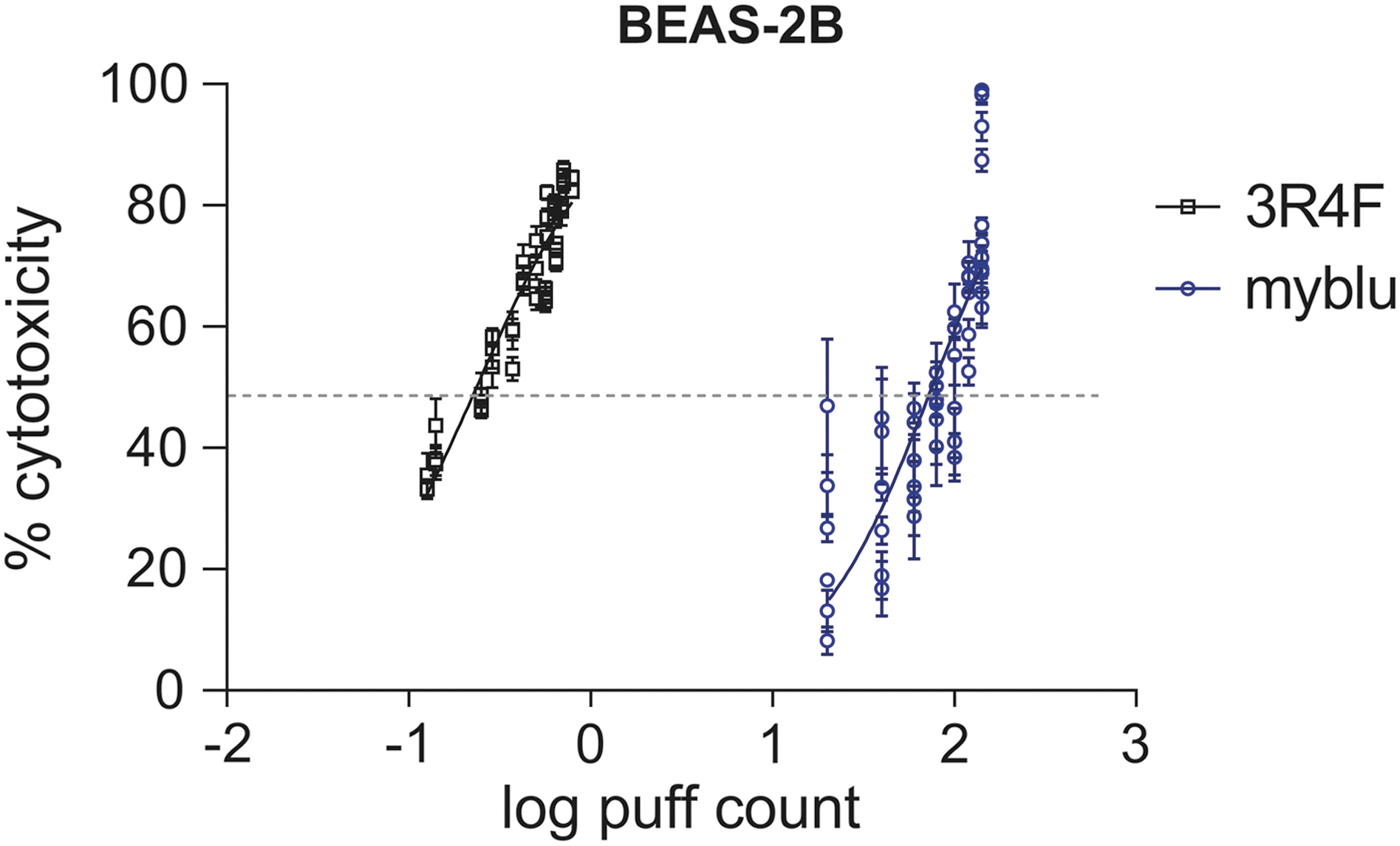
The puff-specific cytotoxicity of the myblu e-cigarette (blue line) aerosol and smoke from 3R4F cigarette (black line) in the NRU assay with the BEAS-2B cell line. Three independent experiments per test article were performed with six replicate measurements per dose level. Single data points represent the average of replicates ± the standard error of the mean. A nonlinear regression curve fit was applied to illustrate the dose–response behavior. The dotted gray line represents the EC50 value. e-cigarette, electronic cigarette; NRU, neutral red uptake.
Puff-Specific Cytotoxicity in BEAS-2B Cells Expressed as EC50
Mutagenicity
All positive controls significantly increased the revertant number. The mutagenic activity of whole smoke/aerosol from the myblu e-cigarette and 3R4F cigarette product is shown in Figures 3 and 4.
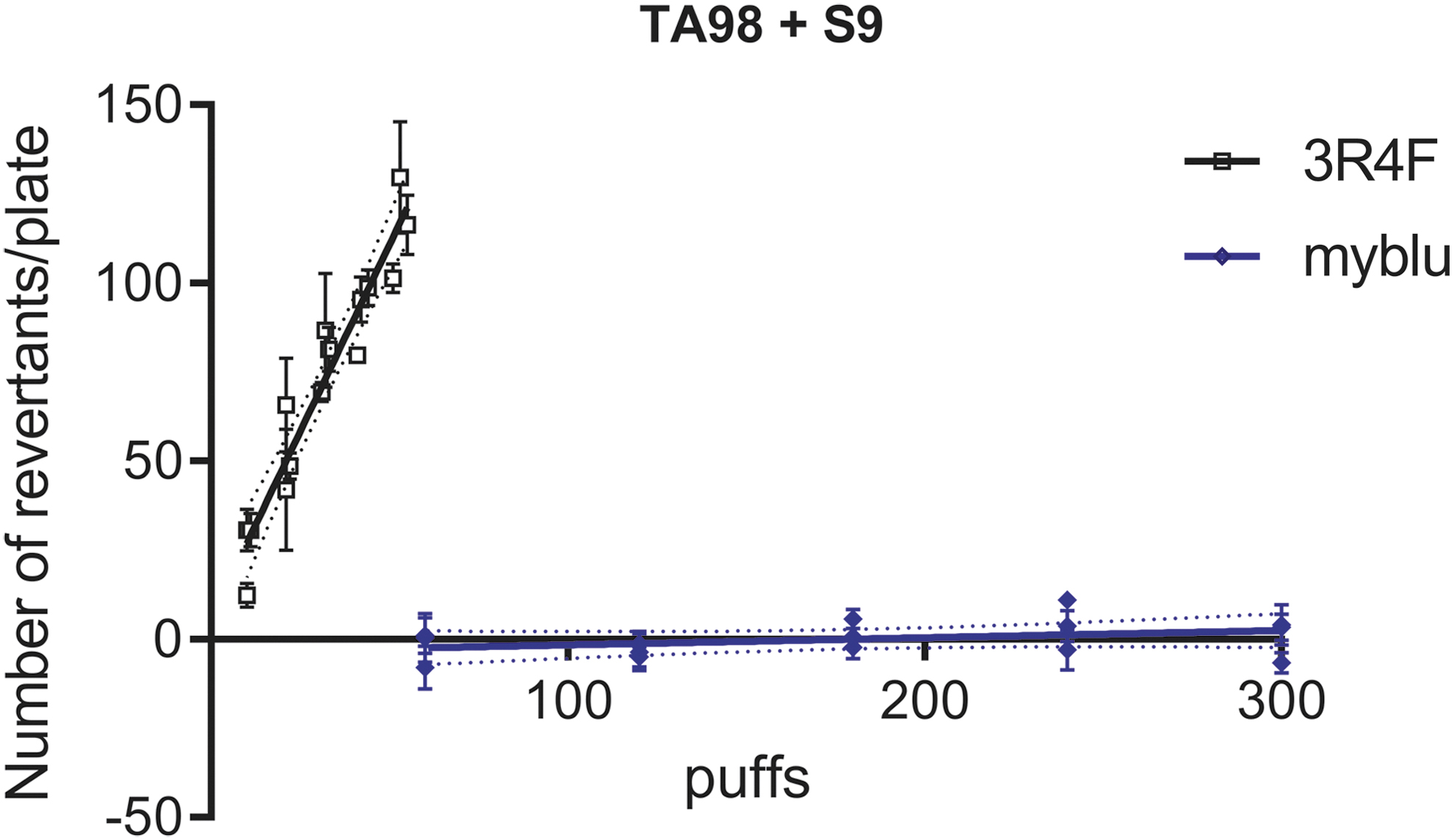
Puff-specific mutagenicity in Salmonella typhimurium TA98 with S9 metabolic activation after exposure to aerosol from myblu e-cigarette (blue line) or smoke from 3R4F cigarette (black line) cigarette using the in vitro bacterial reverse mutation test (Ames test). At least two independent experiments per test article were performed with two to three different dose ranges each. Each data point in Figure 3 represents the average of three replicate agar plates ± standard error of the mean (Dotted line represents the standard error of the mean of linear regression).
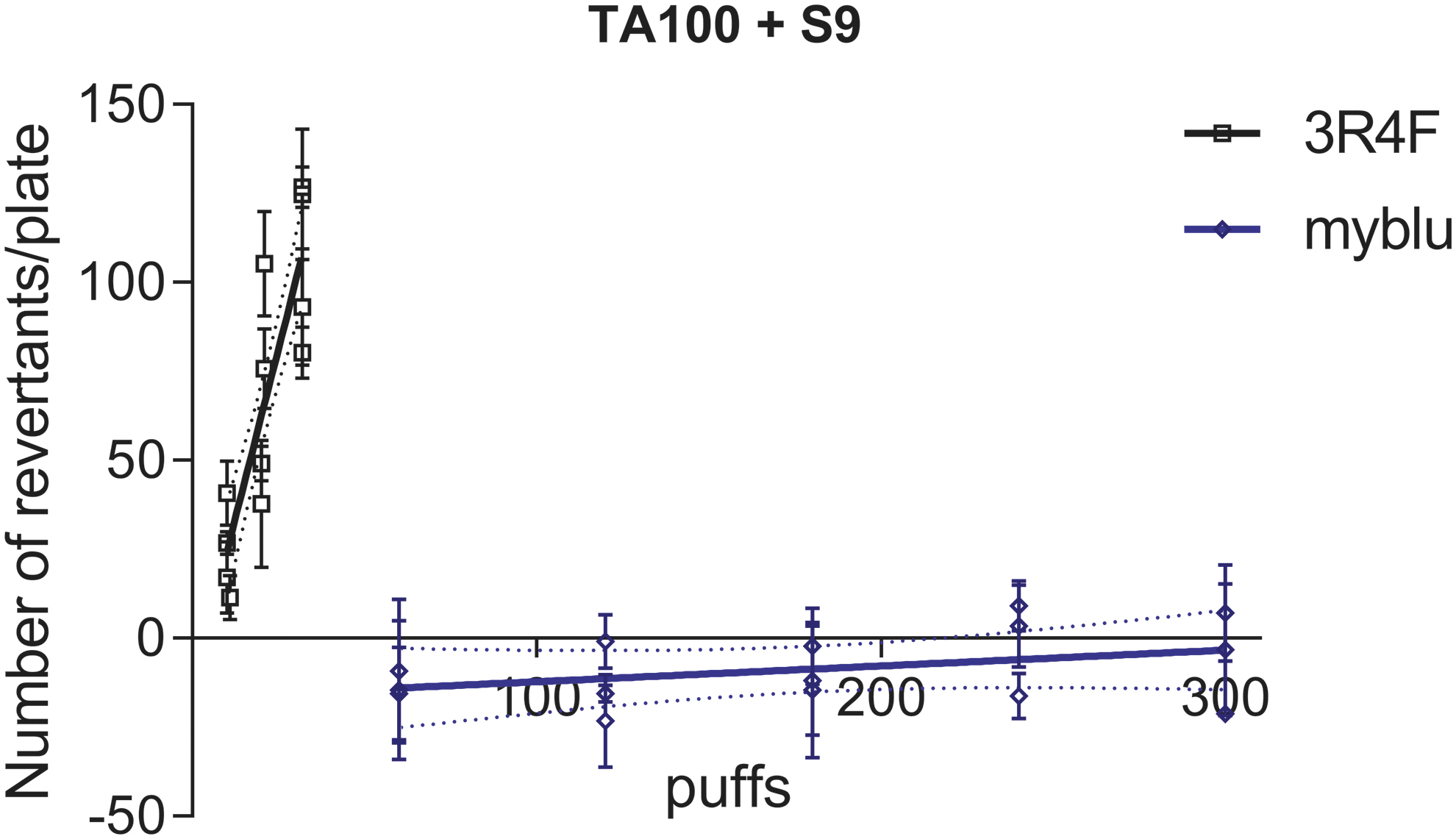
Puff-specific mutagenicity in S. typhimurium TA100 with S9 metabolic activation after exposure to aerosol from myblu e-cigarette (blue line) or smoke from 3R4F cigarette (black line) cigarette using the in vitro Ames test. At least two independent experiments per test article were performed with two to three different dose ranges each. Each data point in Figure 4 represents the average of three replicate agar plates ± standard error of the mean (Dotted line represents the standard error of the mean of linear regression).
For each strain, mutagenic activity was calculated from the linear slope of the dose–response curve (nonthreshold model) with differences in the number of revertants on the treated plates and the untreated controls tested for significance (Tables 4 and 5).
Puff-Specific Mutagenicity in Salmonella typhimurium TA98 with S9 Metabolic Activation: Analysis of the Slope of the Dose–Response Curve (Fig. 3)
Puff-Specific Mutagenicity in S. typhimurium TA100 with S9 Metabolic Activation: Analysis of the Slope of the Dose–Response Curve (Fig. 4)
A statistically significant (p < 0.05) dose-dependent increase in revertant number was observed in both TA98 and TA100 with S9 metabolic activation after 3R4F cigarette smoke exposure. In contrast, the e-cigarette aerosol did not induce any statistically significant increase in number of revertants compared with the negative control in either stain (up to 300 puffs).
Genotoxicity
In all studies, the positive control induced a statistically significant increase in cytotoxicity and micronucleus frequency when compared with the vehicle control. This positive response in micronucleus frequency indicates a responsive assay, regardless of the exposure matrix. No precipitate or significant morphological changes were observed for ambient air control. For e-cigarette aerosol and diluted 3R4F smoke, the average maximum cytotoxicity (RCC) ranged between 47% and 57%.
The activity in the IVM assay is shown in Figure 5. The micronucleus values induced by the different treatment groups were compared pairwise with those from the corresponding negative controls using chi-square analysis per test day. Diluted smoke from the 3R4F cigarette induced reproducible and significantly increased micronucleus frequencies compared with the negative control cultures (p < 0.05), indicative of significant genotoxic potential in this assay. Conversely, the e-cigarette aerosol did not induce any significant increase in micronucleus formation in V79 cells over 100 puffs on any test day.
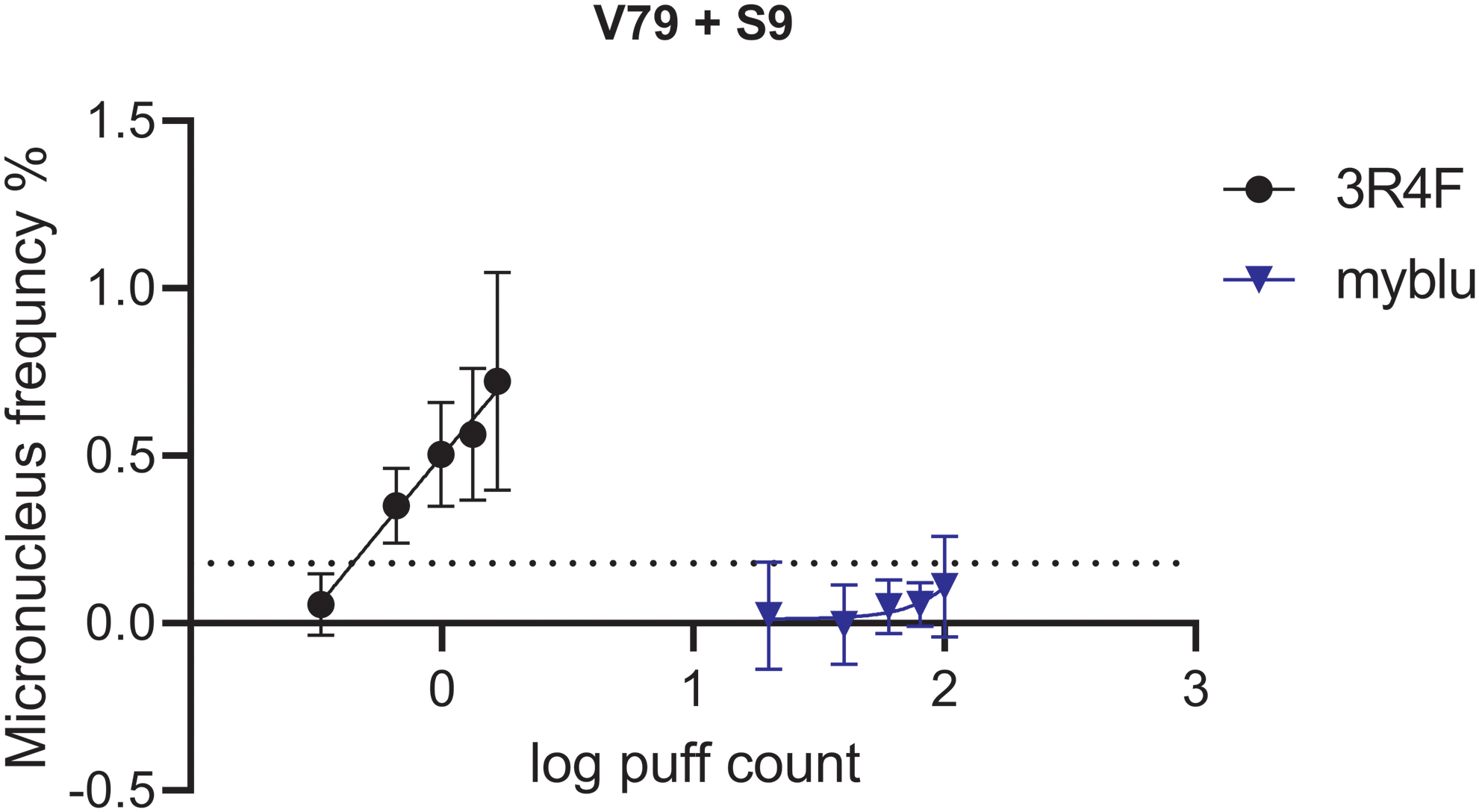
Puff-specific genotoxicity in Chinese hamster lung V79 cells in the in vitro micronucleus assay. Percentage micronucleus dose–response curve after exposure to aerosol from myblu e-cigarette (blue line) or 3R4F smoke (black line). Three independent experiments per test article were performed. Single data points represent the average of replicates ± the standard error of the mean. The dotted line represents the corrected threefold background value of micronuclei frequency for 3R4F (0.18%). Values were subtracted by average background MN frequencies (background for 3R4F = 0.09%; for myblu experiments = 0.18%) of the replicate experiments to focus on the smoke/aerosol-induced genotoxicity. Only non zero dose level micronuclei values are shown.
In addition to the standard OECD guidance for genotoxicity determination, for product comparison, the dose levels needed to induce the threefold increase in micronucleus formation over background (EC-MN3) was calculated (dotted gray line in Fig. 5).34,35 For example for 3R4F smoke, the values from the three test days were averaged and the effective concentration to achieve EC-MN3 was calculated using nonlinear regression (Table 6). No statistical comparison could be modeled for the myblu aerosol because it did not induce any significant increase in micronuclei frequencies in V79 cells (Table 6).
Determination of Genotoxic Activity in Chinese Hamster Lung V79 Cells
Discussion
This study was designed to compare the emissions and in vitro toxicity of the myblu closed pod-system e-cigarette with those of the 3R4F reference cigarette. Clear and substantial differences between the e-cigarette aerosol and tobacco smoke have been demonstrated.
Levels of chemicals from the myblu e-cigarette aerosol were found to be substantially lower than those from cigarette smoke. Analytical results indicate the relative chemical simplicity of the e-cigarette aerosols with no detectable levels of the toxicants analyzed for, compared with cigarette smoke. Substantially more ACM was generated from the e-cigarette aerosol than 3R4F TPM; however, the composition between ACM and TPM is considerably different. ACM is predominantly formed of liquid droplets containing nicotine, propylene glycol, and glycerol, recognized impurities in Pharmacopoeia-quality nicotine, and eight thermal decomposition products of propylene glycol or glycerol, whereas TPM contains solid particles, formed from tobacco combustion, with the majority of these particles identified as carbon.15,36 The data are consistent with other studies in older generation e-cigarette products that have found no quantifiable levels of tested toxicants, including HPHCs, or extremely low levels of measurable constituents relative to cigarette smoke in older generation products.11,15,18,37 Although this study included a wide range of potential toxicants, heavy metals were not analyzed. The literature suggests that although e-cigarettes are less likely to increase exposure to cadmium, as is associated with the use of combustible cigarettes, exposure to other metals, including chromium and nickel, may still increase with the use of e-cigarettes compared with nonusers. Several studies suggest that exposure to metals associated with the use of e-cigarettes is likely associated with the device itself as opposed to the e-liquid.38–42 Extractable and leachable studies conducted on the e-cigarette device should address this risk for new products before market launch.
Established in vitro toxicological studies, the NRU assay to assess product cytotoxicity, 31 the IVM assay for mammalian genotoxicity (OECD, 2016. Test No. 487), 33 and the Ames screening assay to determine mutagenicity (OECD 1997, Test No. 471) 32 in TA98 and TA100 were adopted to assess the toxicity of e-cigarette aerosol to cigarette smoke. Under the test conditions, e-cigarette aerosol demonstrated significantly less toxicity than 3R4F smoke.
The smoke from the 3R4F cigarette was highly cytotoxic to cells in the NRU assay, presenting >300 times higher cytotoxicity than the e-cigarette aerosol. The cytotoxicity findings from this study are consistent with those from a number of other in vitro studies in different cell lines. Misra et al. 43 showed that older closed system e-cigarette products displayed no cytotoxic effects in human alveolar basal epithelial cells. Romagna et al. 20 reported e-cigarette aerosol to be significantly less cytotoxic than tobacco smoke in fibroblasts, and Farsalinos et al. 21 concluded the same in myocardial cells. In addition, Scheffler et al. 22 demonstrated that exposure of primary human bronchial cells to tobacco smoke significantly lowered cell viability compared with e-cigarette aerosol.
The OECD guideline 471 for the Ames test recommends at least five bacterial strains to detect point mutations by base substitutions or frameshifts, incorporating four S. typhimurium strains (TA98, TA100, TA1535, and TA1537) and the TA102 strain. 32 Two strains, TA98 and TA100, are of particular interest for tobacco products because they have been shown to be sensitive to combustion products, notably nitroarenes and aromatic amines. 44 TA98 is sensitive to basic and neutral fractions, such as the heterocyclic amines and aromatic amines that are one of the primary sources of mutagenicity in TPM and smoke extracts and TA100 because of its added sensitivities to carbonyl compounds in the gas vapor phase 45 compared with TA98 and ability to distinguish between tobacco products.46,47 There are no test guidelines currently available for testing of e-cigarette aerosols; therefore, TA98 and TA100 were selected as the most appropriate and responsive strains for this study. In this study, neither TA98 nor TA100 demonstrated a mutagenic response after e-cigarette exposure, whereas clear mutagenicity was observed for cigarette smoke, in line with the results reported by Wieczorek et al. (in press). To fully determine the mutagenicity of e-cigarette aerosol, the full set of Ames strains should be incorporated according to the OECD guideline 471. 32 The use of strains including TA104 may bring value to a more extensive testing strategy beyond mutagenicity screening, due to its known sensitivity to carbonyl compounds.45,48 It should, however, be noted that in this study that all carbonyls measured were below the LOD for the myblu aerosol.
Use of the Ames assay to evaluate the mutagenicity of e-cigarette aerosol has been reported in multiple studies, with results in line with what has been reported in this study. Misra et al. showed that ACM from older closed system e-cigarette products displayed no mutagenic activity in the Ames assay. 43 Thorne et al. used two exposure methods (ACM and aerosol) for Ames testing of e-cigarettes compared with a combustible reference cigarette (3R4F). 49 Both e-cigarette ACM and whole aerosol were found to be negative for mutagenicity in TA98 and TA100. In addition, Thorne et al. evaluated the mutagenic potential of direct e-cigarette aerosol exposure in S. typhimurium (TA98, TA100, TA97, and TA104) and E. coli WP2 uvrA pKM101. 50 This exposure paradigm revealed no statistically significant increase in mutagenicity for any e-cigarette aerosol up to and including the maximum 900-puff exposure, in any strain, both with and without metabolic activation.
The myblu e-cigarette aerosol did not display genotoxic effects in the IVM assay; by contrast, the smoke from the 3R4F cigarette was found to exhibit genotoxicity. The IVM assay used V79 cells, as recommended by the OECD guideline 487. 33 V79 cells show a good responsiveness to cigarette smoke extracts and that the use of V79 cells results in robust reproducible genotoxicity results.35,51,52 Within this study, OECD guidelines were followed, although e-cigarette aerosol and cigarette smoke were tested with metabolic activation only (no −S9 condition within the study); although this is a potential study limitation, the treatment V79 cells do not express P450 enzymes and the inclusion of metabolic activation increases the human relevance of the assay. However, Thorne et al. showed that V79 cells were most responsive to cigarette smoke constituents after an extended recovery/expression period without S9. 52 Therefore, future studies should consider following the OECD test guideline for both short- and long-term treatment. Two different cigarette smoke dilutions (1:4 and 1:5 with filtered air) are included as positive controls within the assay, due to the known genotoxicity of cigarette TPM or whole smoke without metabolic activation.52–54 This is to ensure a minimum of one concentration will induce micronucleus generation and confirm efficient smoke delivery to the cellular system. Future studies should validate the cigarette smoke as a positive control in the IVM assay against OECD recommended positive controls. Published studies using the IVMassay for e-cigarette genotoxicity testing are limited. Misra et al. found that direct e-liquid or ACM from e-cigarettes did not affect micronucleus induction, whereas combustible cigarettes caused a dose-dependent induction of micronucleus formation. 43
A further limitation of this research is that no smoking regimen accurately reflects actual user puff topographies, thus the actual dose of compounds an e-cigarette user inhales may be different to that measured in the study. To address this, further research on pod-based e-cigarette systems would be informative, particularly, topography, clinical biomarker, and behavioral and population studies.
The myblu e-cigarette device and liquids undergo stringent toxicological and product stewardship assessment before launch, including the exclusion of ingredients with carcinogenic, mutagenic, or reproductive toxicity properties. Although e-cigarettes, including myblu, are not risk free, they are a potentially less harmful alternative to cigarettes for the adult smoker. 1 Within this study, results have shown that the myblu e-cigarette aerosol displayed a reduced hazard profile compared with the 3R4F reference cigarette. To assess the biological effect of e-cigarettes, these results should be incorporated into a larger assessment framework using a weight-of-evidence approach. This can include other in vitro human-based assays such as 3D lung models and high content screening.55,56
Conclusions
The in vitrotoxicity data show that the e-cigarette has a low toxicity profile compared with the reference cigarette under the conditions applied. This is perhaps unsurprising, given the demonstrated relative simplicity of the heated e-cigarette aerosol compared with the combusted cigarette smoke, including the absence of many of the analytes tested for. The results obtained in the aforementioned studies and in this study demonstrate that high-quality e-cigarettes and e-liquids offer the potential for substantially reduced exposure to cigarette toxicants in adult smokers who use such products as an alternative to conventional cigarettes. Further studies, including biomarkers of exposure studies in adult smokers, are required to validate the findings in the presented study and to establish the reduced toxicant exposure for the myblu e-cigarette. The findings of this study are an encouraging starting point for future research and development.
Footnotes
Authors' Contributions
The article was written through contributions of all authors. All authors have given approval to the final version of the article.
Acknowledgments
The authors thank Sarah Weaver, Paul Hardman, Rana Tayyarah, and Liliana Ferreira Chaves for critical review of the article.
Author Disclosure Statement
All authors were employees of Imperial Brands PLC or subsidiaries at the time of this study. Imperial Brands PLC is the sole source of funding and sponsor of this project. Fontem Ventures B.V., the manufacturer of the e-cigarettes used in this study, is a wholly owned subsidiary of Imperial Brands PLC.
Funding Information
Imperial Brands PLC is the sole source of funding of this project. No external sources of funding were involved in this project.
Appendix
Appendix 2
ISO 10315:2000 - Cigarettes – Determination of nicotine in smoke condensates: gas chromatographic method
ISO 10362-1:1999-12 Amd.1:2011-07) Cigarettes – Determination of water in smoke condensates – part1: gas chromatographic method
ISO 8454:2007-06/Amd.1:2009-10) Cigarettes – Determination of Carbon Monoxide in vapour phase of cigarette smoke NDIR method
Appendix 3 – 3R4F raw data
| 3R4F | ||||||||||
|---|---|---|---|---|---|---|---|---|---|---|
| Analyte | Unit | LOQ | LOD | Rep 1 | Rep 2 | Rep 3 | mean | sd | COV | |
| TNCO |
|
9.97 | 10.25 | 10.49 |
|
0.26 | 2.5% | |||
|
|
|
35.83 | 37.5 | 36.43 |
|
0.85 | 2.3% | |||
|
|
|
9.51 | 10.64 | 10.17 |
|
0.57 | 5.6% | |||
|
|
|
1.77 | 1.81 | 1.81 |
|
0.02 | 1.3% | |||
|
|
|
4.44 | 4.87 | 4.59 |
|
0.22 | 4.7% | |||
|
|
|
27.768 | 31.313 | 30.204 |
|
1.81 | 6.1% | |||
| Tobacco-specific N-nitrosamines |
|
10.6 | 10.38 | 10.36 |
|
0.13 | 1.3% | |||
|
|
|
|
|
372.09 | 364.58 | 355.23 |
|
8.45 | 2.3% | |
|
|
|
|
|
326.44 | 317.82 | 321.52 |
|
4.33 | 1.3% | |
|
|
|
|
|
35.50 | 34.78 | 40.68 |
|
3.22 | 8.7% | |
|
|
|
|
|
232.71 | 249.15 | 231.74 |
|
9.78 | 4.1% | |
|
|
|
966.75 | 966.33 | 949.17 |
|
10.03 | 1.0% | |||
|
|
|
|
|
115.4 | 109.4 | 107.5 |
|
4.14 | 3.7% | |
|
|
|
|
|
0.4 | 0.3 | 0.3 |
|
0.05 | 16.5% | |
|
|
|
|
|
100.4 | 93.7 | 93.3 |
|
4.02 | 4.2% | |
|
|
|
|
|
19.9 | 18.8 | 18.4 |
|
0.79 | 4.2% | |
|
|
|
|
|
5.2 | 4.8 | 4.4 |
|
0.38 | 7.9% | |
|
|
|
|
|
6.4 | 6.4 | 6.1 |
|
0.18 | 2.9% | |
|
|
|
|
|
8.1 | 8.1 | 7.7 |
|
0.20 | 2.5% | |
| Polyaromatichydrocarbons |
|
10.1 | 10.1 | 9.8 |
|
0.17 | 1.7% | |||
|
|
|
|
|
13.57 | 14.01 | 13.09 |
|
0.46 | 3.4% | |
| Carbonyls |
|
11.68 | 11.76 | 11.8 |
|
0.06 | 0.5% | |||
|
|
|
|
|
91.5 | 92.8 | 97.7 |
|
3.28 | 3.5% | |
|
|
|
|
|
1572.0 | 1588.9 | 1685.8 |
|
61.38 | 3.8% | |
|
|
|
|
|
573.0 | 568.6 | 605.5 |
|
20.15 | 3.5% | |
|
|
|
|
|
161.6 | 161.9 | 174.8 |
|
7.56 | 4.5% | |
|
|
|
|
|
128.6 | 128.6 | 137.6 |
|
5.22 | 4.0% | |
|
|
|
|
|
51.5 | 51.2 | 55.4 |
|
2.33 | 4.4% | |
|
|
|
|
|
170.2 | 163.6 | 166.2 |
|
3.33 | 2.0% | |
|
|
|
|
|
83.6 | 83.9 | 89.0 |
|
3.04 | 3.6% | |
| Aromatic Amines |
|
10.02 | 10.08 | 9.92 |
|
0.08 | 0.8% | |||
|
|
|
|
|
29.7 | 28.6 | 28.3 |
|
0.70 | 2.4% | |
|
|
|
|
|
18.6 | 18.1 | 18.2 |
|
0.23 | 1.2% | |
|
|
|
|
|
4.6 | 4.4 | 4.4 |
|
0.12 | 2.8% | |
|
|
|
|
|
3.8 | 3.7 | 3.7 |
|
0.08 | 2.2% | |
| Gas phase compounds |
|
10.6 | 10.38 | 10.36 |
|
0.13 | 1.3% | |||
|
|
|
|
|
<LOQ | <LOQ | <LOQ | ||||
|
|
|
|
|
107.6 | 102.8 | 101.8 |
|
3.09 | 3.0% | |
|
|
|
|
|
379.9 | 354.9 | 332.4 |
|
23.78 | 6.7% | |
|
|
|
|
|
1533.6 | 1516.3 | 1484.1 |
|
25.11 | 1.7% | |
|
|
|
|
|
447.5 | 437.8 | 442.3 |
|
4.87 | 1.1% | |
|
|
|
|
|
36.8 | 38.0 | 39.2 |
|
1.22 | 3.2% | |
|
|
|
|
|
56.3 | 53.2 | 53.8 |
|
1.66 | 3.0% | |
|
|
|
|
|
632.2 | 557.0 | 564.5 |
|
41.43 | 7.1% | |
|
|
|
|
|
2.5 | 2.4 | 2.3 |
|
0.09 | 3.8% | |
|
|
|
|
|
182.4 | 174.5 | 170.2 |
|
6.17 | 3.5% | |
|
|
|
|
|
740.1 | 739.4 | 725.7 |
|
8.09 | 1.1% | |
|
|
|
|
|
340.3 | 339.0 | 327.8 |
|
6.91 | 2.1% | |
|
|
|
|
|
29.3 | 28.0 | 27.4 |
|
0.99 | 3.5% | |
|
|
|
|
|
0.8 | 0.9 | 0.8 |
|
0.02 | 2.0% | |
|
|
|
|
|
1.6 | 1.3 | 1.2 |
|
0.17 | 12.6% | |
|
|
|
|
|
103.3 | 103.1 | 99.8 |
|
1.99 | 1.9% | |
|
|
|
|
|
1.9 | 1.9 | 1.8 |
|
0.06 | 3.4% | |
|
|
|
|
|
197.9 | 198.9 | 196.3 |
|
1.33 | 0.7% | |
|
|
|
|
|
22.0 | 22.3 | 22.1 |
|
0.16 | 0.7% | |
|
|
|
|
|
14.9 | 15.3 | 15.3 |
|
0.23 | 1.5% | |
|
|
|
4831.0 | 4687.0 | 4608.9 |
|
112.63 | 2.4% | |||
Appendix 4
| myblu | myblu | |||||||||||||||||||||||||||
|---|---|---|---|---|---|---|---|---|---|---|---|---|---|---|---|---|---|---|---|---|---|---|---|---|---|---|---|---|
| Rep 1 | Rep 2 | Rep3 | Average Replicates | |||||||||||||||||||||||||
| Analyte | block 1 | block 2 | block 3 | total | mean | sd | COV | block 1 | block 2 | block 3 | total | mean | sd | COV | block 1 | block 2 | block 3 | total | mean | sd | COV | mean/replicate | sd | COV | ||||
| Unit x | LOQ | LOD | 50 puffs | 50 puffs | 50 puffs | 150 puffs | 50 puffs | 50 puffs | 50 puffs | 50 puffs | 150 puffs | 50 puffs | 50 puffs | 50 puffs | 50 puffs | 150 puffs | 50 puffs | 150 puffs | ||||||||||
| Weight loss |
|
|
398.69 | 389.95 | 356.47 |
|
|
22.29 | 5.8% | 424.13 | 373.18 | 346.09 | 1143.40 |
|
|
10.4% | 412.03 | 405.57 | 365.16 |
|
|
25.40 | 6.4% |
|
|
1.9% | ||
| TNCO, Menthol |
|
|
294.3 | 301 | 310.5 |
|
|
8.14 | 2.7% | 272.8 | 267.2 | 284.8 | 824.80 |
|
|
3.3% | 258.5 | 250.3 | 261.2 |
|
|
5.68 | 2.2% |
|
|
8% | ||
|
|
|
19.53 | 20.14 | 19.81 |
|
|
0.31 | 1.5% | 16.58 | 15.95 | 16.85 | 49.38 |
|
|
2.8% | 14.85 | 15.27 | 15.47 |
|
|
0.31 | 2.1% |
|
|
14.0% | |||
|
|
|
4.45 | 4.59 | 4.69 |
|
|
0.12 | 2.7% | 4.16 | 4.18 | 4.38 | 12.73 |
|
|
2.9% | 4.00 | 3.94 | 4.01 |
|
|
0.04 | 1.0% |
|
|
7.0% | |||
|
|
|
<0.01 | <0.01 | <0.01 |
|
|
0.00 | #DIV/0! | <0.01 | <0.01 | <0.01 | <0.01 |
|
|
#DIV/0! | <0.01 | <0.01 | <0.01 |
|
|
0.00 | #DIV/0! |
|
|
#DIV/0! | |||
|
|
|
<0.01 | <0.01 | <0.01 |
|
|
0.00 | #DIV/0! | <0.01 | <0.01 | <0.01 | <0.01 |
|
|
#DIV/0! | <0.01 | <0.01 | <0.01 |
|
|
0.00 | #DIV/0! |
|
|
#DIV/0! | |||
| Tobacco- specific N-nitrosamines |
|
|
|
|
<LOD | <LOD | <LOD | <LOD | <LOD | <LOD | <LOD | <LOD | <LOD | |||||||||||||||
|
|
|
|
|
<LOD | <LOD | <LOD | <LOD | <LOD | <LOD | <LOD | <LOD | <LOD | ||||||||||||||||
|
|
|
|
|
<LOD | <LOD | <LOD | <LOD | <LOD | <LOD | <LOD | <LOD | <LOD | ||||||||||||||||
|
|
|
|
|
<LOD | <LOD | <LOD | <LOD | <LOD | <LOD | <LOD | <LOD | <LOD | ||||||||||||||||
|
|
|
|||||||||||||||||||||||||||
| Phenolic compounds |
|
|
|
|
<LOD | <LOD | <LOD | <LOD | <LOD | <LOD | <LOD | <LOD | <LOD | |||||||||||||||
|
|
|
|
|
<LOD | <LOD | <LOD | <LOD | <LOD | <LOD | <LOD | <LOD | <LOD | ||||||||||||||||
|
|
|
|
|
<LOD | <LOD | <LOD | <LOD | <LOD | <LOD | <LOD | <LOD | <LOD | ||||||||||||||||
|
|
|
|
|
<LOD | <LOD | <LOD | <LOD | <LOD | <LOD | <LOD | <LOD | <LOD | ||||||||||||||||
|
|
|
|
|
<LOD | <LOD | <LOD | <LOD | <LOD | <LOD | <LOD | <LOD | <LOD | ||||||||||||||||
|
|
|
|
|
<LOD | <LOD | <LOD | <LOD | <LOD | <LOD | <LOD | <LOD | <LOD | ||||||||||||||||
|
|
|
|
|
<LOD | <LOD | <LOD | <LOD | <LOD | <LOD | <LOD | <LOD | <LOD | ||||||||||||||||
| Polyaromatichydrocarbons |
|
|
|
|
<LOD | <LOD | <LOD | <LOD | <LOD | <LOD | <LOD | <LOD | <LOD | |||||||||||||||
| Carbonyls |
|
|
|
|
<LOD | <LOD | <LOD | <LOD | <LOD |
|
<LOD | <LOD | <LOD | |||||||||||||||
|
|
|
|
|
<LOD | <LOD | <LOD | <LOD | <LOD | <LOD | <LOD | <LOD | <LOD | ||||||||||||||||
|
|
|
|
|
<LOD | <LOD | <LOD | <LOD | <LOD | <LOD | <LOD | <LOD | <LOD | ||||||||||||||||
|
|
|
|
|
<LOD | <LOD | <LOD | <LOD | <LOD | <LOD | <LOD | <LOD | <LOD | ||||||||||||||||
|
|
|
|
|
<LOD | <LOD | <LOD | <LOD | <LOD | <LOD | <LOD | <LOD | <LOD | ||||||||||||||||
|
|
|
|
|
<LOD | <LOD | <LOD | <LOD | <LOD | <LOD | <LOD | <LOD | <LOD | ||||||||||||||||
|
|
|
|
|
<LOD | <LOD | <LOD | <LOD | <LOD | <LOD | <LOD | <LOD | <LOD | ||||||||||||||||
|
|
|
|
|
<LOD | <LOD | <LOD | <LOD | <LOD | <LOD | <LOD | <LOD | <LOD | ||||||||||||||||
| Aromatic Amines |
|
|
|
|
<LOD | <LOD | <LOD | <LOD | <LOD | <LOD | <LOD | <LOD | <LOD | |||||||||||||||
|
|
|
|
|
<LOD | <LOD | <LOD | <LOD | <LOD | <LOD | <LOD | <LOD | <LOD | ||||||||||||||||
|
|
|
|
|
<LOD | <LOD | <LOD | <LOD | <LOD | <LOD | <LOD | <LOD | <LOD | ||||||||||||||||
|
|
|
|
|
<LOD | <LOD | <LOD | <LOD | <LOD | <LOD | <LOD | <LOD | <LOD | ||||||||||||||||
| Gas phase compounds |
|
|
|
|
< LOQ | < LOQ | < LOQ | < LOQ | < LOQ | < LOQ | < LOQ | < LOQ | < LOQ | |||||||||||||||
|
|
|
|
|
< LOQ | < LOQ | < LOQ | < LOQ | < LOQ | < LOQ | < LOQ | < LOQ | < LOQ | ||||||||||||||||
|
|
|
|
|
< LOQ | < LOQ | < LOQ | < LOQ | < LOQ | < LOQ | < LOQ | < LOQ | < LOQ | ||||||||||||||||
|
|
|
|
|
< LOQ | < LOQ | < LOQ | < LOQ | < LOQ | < LOQ | < LOQ | < LOQ | < LOQ | ||||||||||||||||
|
|
|
|
|
< LOQ | < LOQ | < LOQ | < LOQ | < LOQ | < LOQ | < LOQ | < LOQ | < LOQ | ||||||||||||||||
|
|
|
|
|
< LOQ | < LOQ | < LOQ | < LOQ | < LOQ | < LOQ | < LOQ | < LOQ | < LOQ | ||||||||||||||||
|
|
|
|
|
< LOQ | < LOQ | < LOQ | < LOQ | < LOQ | < LOQ | < LOQ | < LOQ | < LOQ | ||||||||||||||||
|
|
|
|
|
< LOQ | < LOQ | < LOQ | < LOQ | < LOQ | < LOQ | < LOQ | < LOQ | < LOQ | ||||||||||||||||
|
|
|
|
|
< LOQ | < LOQ | < LOQ | < LOQ | < LOQ | < LOQ | < LOQ | < LOQ | < LOQ | ||||||||||||||||
|
|
|
|
|
< LOQ | < LOQ | < LOQ | < LOQ | < LOQ | < LOQ | < LOQ | < LOQ | < LOQ | ||||||||||||||||
|
|
|
|
|
< LOQ | < LOQ | < LOQ | < LOQ | < LOQ | < LOQ | < LOQ | < LOQ | < LOQ | ||||||||||||||||
|
|
|
|
|
< LOQ | < LOQ | < LOQ | < LOQ | < LOQ | < LOQ | < LOQ | < LOQ | < LOQ | ||||||||||||||||
|
|
|
|
|
< LOQ | < LOQ | < LOQ | < LOQ | < LOQ | < LOQ | < LOQ | < LOQ | < LOQ | ||||||||||||||||
|
|
|
|
|
< LOQ | < LOQ | < LOQ | < LOQ | < LOQ | < LOQ | < LOQ | < LOQ | < LOQ | ||||||||||||||||
|
|
|
|
|
< LOQ | < LOQ | < LOQ | < LOQ | < LOQ | < LOQ | < LOQ | < LOQ | < LOQ | ||||||||||||||||
|
|
|
|
|
< LOQ | < LOQ | < LOQ | < LOQ | < LOQ | < LOQ | < LOQ | < LOQ | < LOQ | ||||||||||||||||
|
|
|
|
|
< LOQ | < LOQ | < LOQ | < LOQ | < LOQ | < LOQ | < LOQ | < LOQ | < LOQ | ||||||||||||||||
|
|
|
|
|
< LOQ | < LOQ | < LOQ | < LOQ | < LOQ | < LOQ | < LOQ | < LOQ | < LOQ | ||||||||||||||||
|
|
|
|
|
< LOQ | < LOQ | < LOQ | < LOQ | < LOQ | < LOQ | < LOQ | < LOQ | < LOQ | ||||||||||||||||
|
|
|
|
|
< LOQ | < LOQ | < LOQ | < LOQ | < LOQ | < LOQ | < LOQ | < LOQ | < LOQ | ||||||||||||||||
|
|
|
0 | 0 | 0 |
|
|
0.00 | #DIV/0! | 0 | 0 | 0 |
|
|
0.00 | #DIV/0! | 0 | 0 | 0 |
|
|
0.00 | #DIV/0! | ||||||
Appendix 5 – Ammonia testing
| Unit | LOQ | LOD | Rep 1 | Rep 2 | Rep 3 | Mean | sd | COV (%) | |
|---|---|---|---|---|---|---|---|---|---|
| 3R4F | μg/stick | 3.88 | 1.29 | 35.9 | 37.3 | 37.0 | 37.2 | 0.7 | 1.98 |
| myblu | μg/150 puffs | 15.5 | 5.17 | <LOQ | <LOQ | - | - | - | - |
Appendix 6 – Raw Data Neutral Red Assay
Appendix 7 – Raw Ames Screen data
Appendix 8 – Raw IVM data
IVM test – 1st Assay with whole smoke from the 3R4F reference cigarette
IVM test – 2nd Assay with whole smoke from the 3R4F reference cigarette
IVM test – 3rd Assay with whole smoke from the 3R4F reference cigarette
IVM test – 1st Assay with e-vapour from myblu eliquid (1.6% nicotine) generated in myblu device
IVM test – 2nd Assay with e-vapour from myblu eliquid (1.6% nicotine) generated in myblu device
IVM test – 3rd Assay with e-vapour from myblu eliquid (1.6% nicotine) generated in myblu device