
Abstract
Proximal femoral focal deficiency (PFFD) is a rare congenital anomaly of the femur. Rotationplasty has been described as a reconstructive procedure in the management of PFFD.
A 68-year-old woman with PFFD of the right leg and rotationplasty at the age of 12 years had prosthetic fitting problems after a fall. The authors describe the analysis of the prosthetic fitting problems and the considerations made in prosthetic management. Following a fall, 56 years after rotationplasty, this woman has a good prosthetic fitting and a satisfying level of functioning.
Introduction
Congenital defects of the femur are most often described as a proximal femoral focal deficiency (PFFD). It is a rare congenital malformation of the femur due to hypoplasia or partial aplasia of the femur. The reported incidence is 1 out of 50,000 children. The exact etiology of PFFD is unknown (Meihuizen-de Regt et al. 2003; Goncalves et al. 1996).
PFFD is often associated with hip instability, malrotation of the hip, insufficient proximal musculature, unequal limb length and contractures or instability of the knee (Fowler et al. 1999). The reported incidence of paraxial fibular hemimelia in the affected limb is 69% (Kritter 1977).
Aitken's classification is a classification system used to describe the severity of PFFD based on radiographic findings using the femoral shaft length as a criterion. In class A, B and C the acetabulum and femoral head are progressively dysplastic and the shaft length progressively shorter. In class D the acetabulum and femoral head are not present (Aitken 1969).
Because there is such diversity in deformities caused by PFFD, there is no standard treatment. Van Nes rotationplasty combined with an arthrodesis of the knee is a frequently used reconstructive procedure in the treatment of PFFD. Sometimes this procedure is followed by a Syme amputation in a later stage. The purpose of the rotationplasty is to provide a full-weight bearing leg with preservation of the ankle joint to function as a knee joint and the abililty to fit a prosthesis (Alman et al. 1995; Fowler et al. 1999). The duration of follow-up after rotationplasty varied in several articles from 2 – 14 years (Alman et al. 1995; Friscia et al. 1989; Kostuik et al. 1975; Kristen et al. 1978; Kritter 1977; Murat et al. 1967).
The authors present the case of a 68-year-old woman who underwent a rotationplasty when she was 12 years old. She came to the clinic with a complex prosthetic problem. To the best of the authors' knowledge no such case, 56 years after rotationplasty, has been described in literature.
Case description
A 68-year-old woman was born with a deficiency of her right leg. The right foot was located at the level of the opposite knee. There was possibly a foot deformity with one missing ray. Documentation of the exact deficiencies was not available to the authors. Most likely there was a PFFD of the right leg. Until the age of 12 years she had walked with her right leg supported by a wooden stelt (a peg or a piece of wood to lean on). At the age of 12 years she underwent a rotationplasty. Documentation of this surgical procedure and the immediate follow-up was destroyed by the hospital. She received a prosthesis and had an active, pain-free life.
In 1988 she had a minor stroke with coordination problems. She visited the authors' outpatient clinic for several months. Because of wear of the old prosthesis a new prosthesis was provided in this period. She was able to walk without walking-aids and do her household activities.
In April 2005 she fell at home and was hospitalized with complaints of severe pain in her right hip and not being able to walk with the prosthesis. Radiographs of the pelvis and right hip showed no fractures. In June 2005 she was admitted to the rehabilitation clinic to evaluate her situation. She was unable to walk properly and she had a fear of falling.
On physical examination we saw a 68-year-old woman, slightly obese and quite nervous. She had a normal left leg and no congenital deformities of the upper limbs. The right leg showed a rotationplasty with the ankle at knee level (Figure 1). Proximal hip- and pelvic musculature were atrophic. The range of motion of the right hip was flexion/extension 40/20/0°, abduction/adduction 45/5/0°, internal rotation/external rotation 30/0/0°. The resting position of the leg was in flexion-adduction-internal rotation.
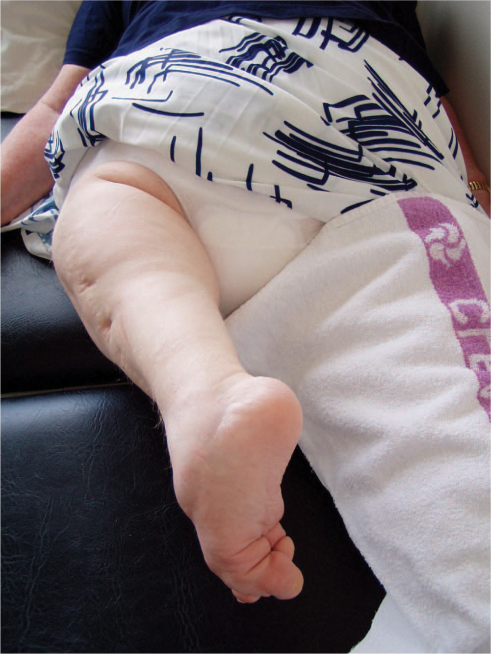
Rotationplasty. (Please see colour online)
The muscle strength of the proximal hip musculature was MRC 3 (MRC scale 0 being no strength and 5 being maximal strength), the hip abductors were MRC 2. She experienced no pain on passive or active movement of the hip.
Inspection of the foot showed multiple deformities: absence of one ray, and equinovarus. The range of motion of the ankle was dorsal flexion (knee-flexion)/plantar flexion(knee-extension) 90/20/0°.
Radiographs of the pelvis showed a dysplastic right hip, with varus position, severe deformity of the femoral head, a short femoral neck and absence of the trochanter minor and a deformity of the trochanter major. There was severe osteoarthritis of the right hip joint (Figure 2). Radiographs of the right leg showed an intramedullary pin and a complete arthrodesis of the knee joint. The patella was present and the fibula absent.
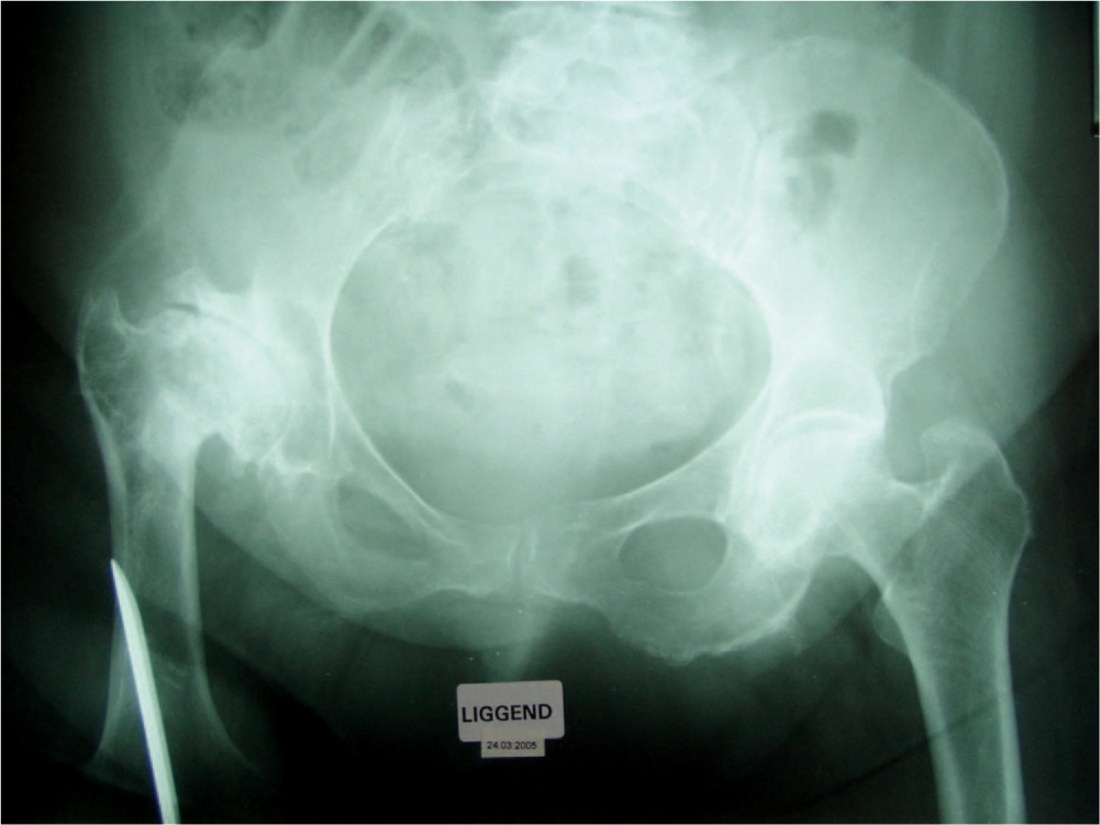
Radiograph of the pelvis: Severe deformity and osteoarthritis of the right hip.
Radiographs of the right foot and ankle showed only four rays, a talocalcaneal coalition and osteoarthritis. An internal rotation of the foot in the ankle-fork was also present (Figure 3).
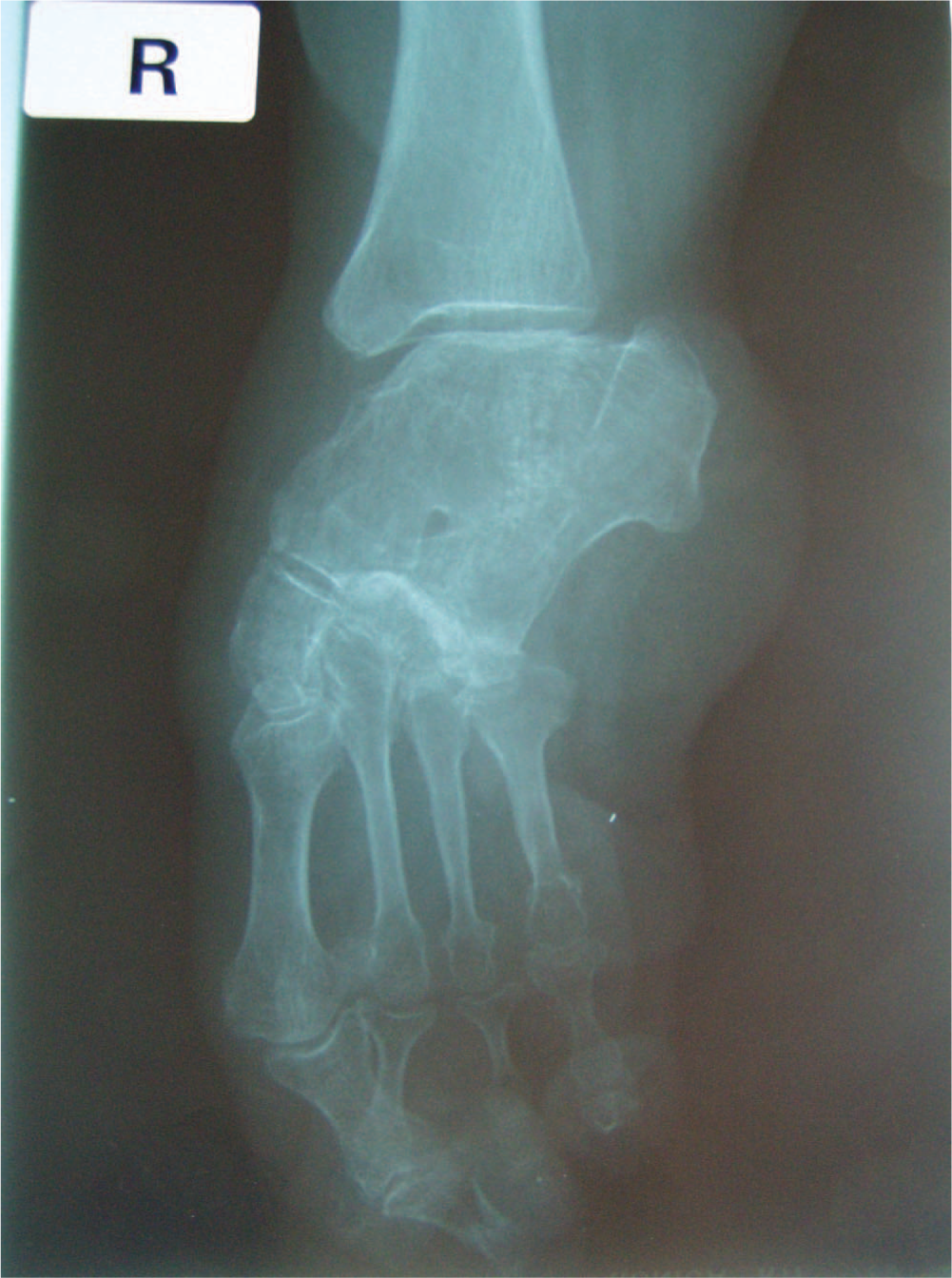
Radiograph of the right ankle: Anteroposterior view.
She was transferred to the authors' rehabilitation centre to evaluate her situation. Four questions were to be answered: (i) what was causing the prosthesis fitting problems; (ii) was adaptation of the prosthesis possible; (iii) would the patient be able to walk with the prosthesis after adaptation; and (iv) would total hip arthroplasty be an option for this patient?
Prosthetic details
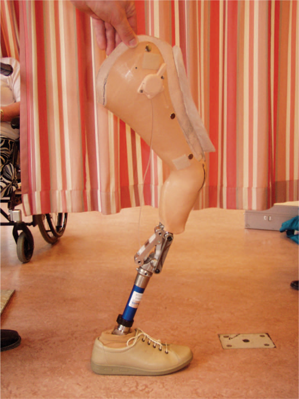
Figure 4 shows the prosthesis she had been wearing since 1988. The leg was fitted in a circumferential leather socket, closed by a lace on the anterior side of the leg. The tuber ischiadicus rested on an ischial shelf and was the primary weight bearing area. Knee flexion and extension were supported by external hinges. The foot was fitted in a foot socket made of thermoplastic. A SACH-foot completed the prosthesis. The leather socket was positioned in the natural position of the leg: Flexion-adduction-internal rotation.
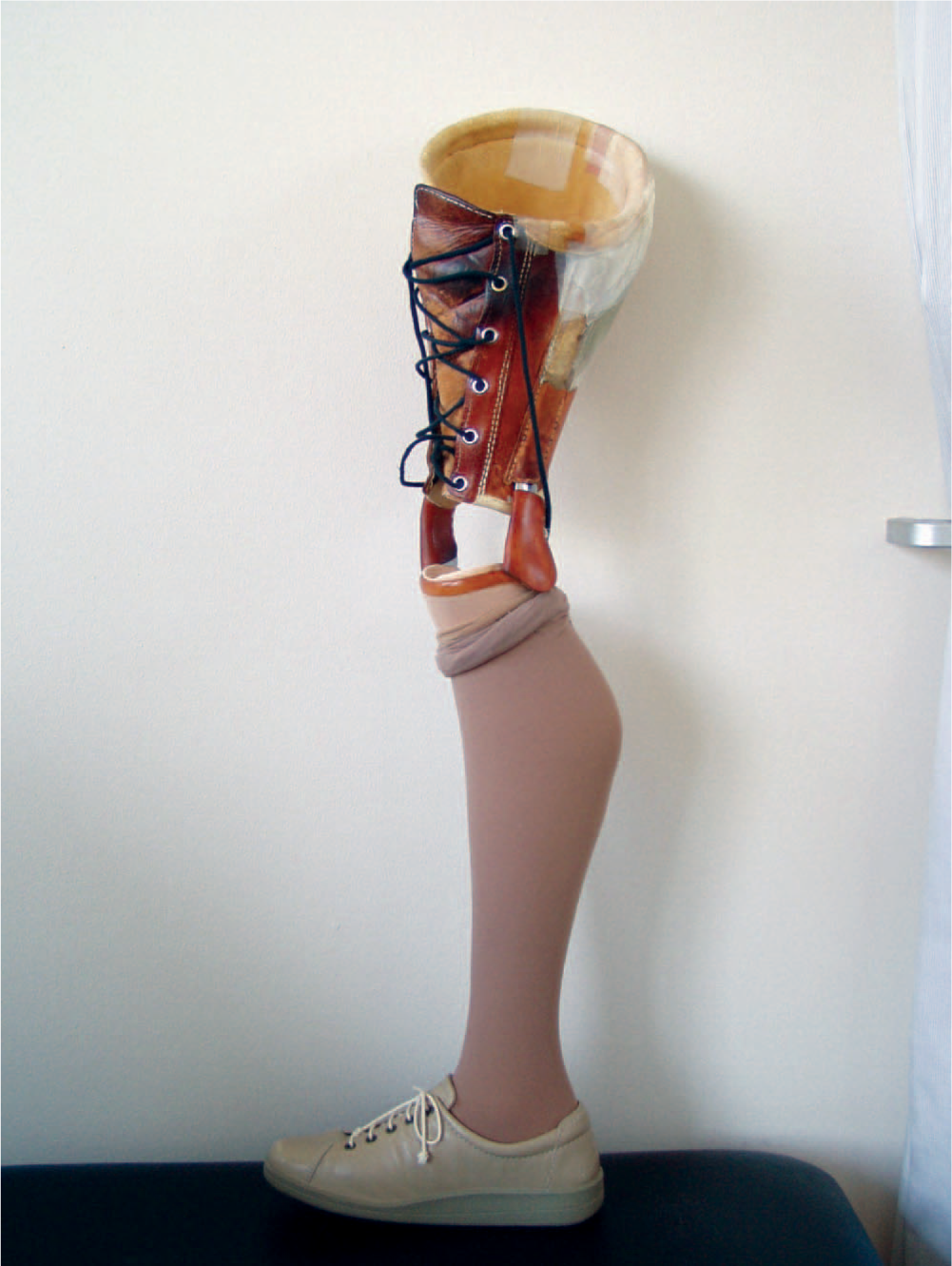
Prosthesis with external hinges and leather socket in internal rotation. (Please see colour online)
The first question about the causes of the prosthetic fitting problems can be answered. Due to the internal rotation of the foot in the ankle-fork the foot socket did not fit anymore. Wearing the prosthesis after the fall caused pain in the foot and leg and made walking with the prosthesis very difficult. Considering the changed anatomical situation, it was impossible to adapt the prosthesis.
A new prosthesis was provided (Figure 5). The primary weight-bearing area remained the ischial tuberosity, resting on an ischial shelf. The prosthesis consisted of a rigid upper leg-ankle-foot socket and a modular component on the lower part. The foot was protected by a Pelite inner socket, which provided an optimal pressure distribution for the bony protrusions of the foot. A rigid socket was made to prevent the foot from further internal rotation in the ankle fork and to prevent the osteoarthritis from worsening, causing more pain. To acquire the desired amount of weight bearing, the inner socket from the heel to the groin was made from leather that could be tightened in the front with a lace. The whole of the foot and the back of the upper leg were covered with a carbon fibre reinforced laminated outer socket. At the distal end of this socket a connector for the modular system was built in, taking into account the flexion-adduction contracture of the hip joint and the desired position of the foot underneath the rigid socket.
Prosthesis with carbon fibre rigid socket and disarticulation knee. (Please see colour online)
The prosthetic knee was a 3R32 disarticulation knee from Otto Bock that remains locked during gait, but which can be unlocked when sitting down. The prosthetic foot was a 1H38 single axis foot from Otto Bock that allows plantar flexion at heel strike to ensure the swift forming of a steady floor contact. Considering the extreme amount of ankle dorsal flexion needed, a tube clamp with a 10° inclination was used. A foam cover finally covered the modular system of the lower part, to give the prosthesis a more aesthetic appearance; however, in the case of this patient function prevailed above cosmetics.
The prosthesis had a good fitting and caused no pain during wearing. The prosthesis was aligned after which the patient was able to walk between the parallel bars. After two weeks of exercising she was able to walk with a rolling walker. When walking longer distances she still experienced pain in her right hip, caused by the degenerative joint disease. A total hip arthroplasty could increase the range of motion of the right hip and possibly attaining a more optimal result regarding the prosthesis. Psychological factors (fear, inability to cope with stress) and social factors (son getting married) made surgical interventions impossible at the time of rehabilitation but will be offered to her in a later stage, when her personal life is more stable. Despite this discomfort she was satisfied with her new prosthesis and level of activity.
Discussion
Considering the patient several remarks can be made. As far as could be reconstructed from her medical history she was born with, according to the Aitken classification, a PFFD type A of the right leg. In this sub-type a bony connection is present between the shaft of femur and the head, neck and trochanteric component at skeletal maturity (Aitken 1969). However difficult to reconstruct we believe our patient has a true PFFD and not a congenital short femur (Gillespie and Torrode 1983). Many authors proceeded to establish accurate classifications of PFFD based on anatomic and radiological criteria, but establishing the most appropriate classification for our patient was not the goal of this report.
The unilateral PFFD was combined with a longitudinal defect of the right leg: absence of the fibula, talocalcaneal coalition and absence of one ray of the right foot. The longitudinal defects are a relative contraindication for rotationplasty (Kostuik et al. 1975; Kritter 1977), but this was possibly not known in 1949, the time the rotationplasty of the patient was performed.
In the past, internal rotation of the ankle has been reported as a complication of rotationplasty (Murat et al. 1967). It is believed that the patient had an internal rotation of the ankle. She did not experience problems with the ankle/foot until the day she fell. This is quite remarkable since the patient also had the talocalcaneal coalition, causing a rigid valgus deformity of the ankle (Grogan et al. 1994). It is not sure whether her fall enhanced instability of the ligaments around the ankle joint. There were no fractures visible on the X-rays.
However, the anatomical changes in the ankle/foot region made walking and wearing the prosthesis more difficult. The prosthesis made in 1988 already showed an internally rotated ankle/foot and until 2005 the fitting was sufficient.
The rotationplasty was done at the age of 12 years, an appropriate age, as suggested in the literature (Murat et al. 1967; Kritter 1977, Kostuik et al. 1975; Friscia et al. 1989). It is amazing that the patient experienced no major complications after the rotationplasty for at least 38 years, but more likely 52 years, and was able to live an active life with a prosthesis. To the best of the authors' knowledge there is no report in the literature concerning follow-up after rotationplasty after so many years. There are no reports concerning hip function in patients with PFFD after a long period of time of patients. However Köse et al. (1998) described a case report of a 40-year-old woman with an isolated unilateral femoral focal deficiency in which a total hip arthroplasty was performed due to decreased activities of daily life caused by pain. A total hip arthroplasty remains an option for the present patient, when pain and discomfort in the right hip increase. Degenerative joint disease has not been reported in association with a type A PFFD and little is known about degenerative processes in such cases.
Conclusion
This case report shows that complex prosthetic fitting problems many years after rotationplasty in PFFD can be managed adequately and lead to a satisfactory situation for the patient. A good evaluation of the problem was the basis for good prosthetic management.
Footnotes
Acknowledgements
The authors would like to thank Alwin Huber (Livit Orthopedie) for his help in providing the prosthetic details.