
Abstract
The use of arm prosthesis in trans-humeral amputees is limited; due to the cone form of the amputation stump. A Humerus-T-Prosthesis was implanted in three patients to create artificial humerus condyles. Two of the patients were successfully rehabilitated with the application of a new type trans-humeral arm prosthesis. This arm prosthesis had a socket which is suspended and stabilized by the humerus and implant only. Traction and rotational stability were secured by adjustable pressure adaptation around the artificial condyles. The third patient developed a pressure wound over the lateral part of the artificial condyle that later healed. He also was subject to a new trauma with a fracture of the ipsilateral scapula and until now has had limited the use of his new arm prosthesis. It was concluded that this new concept for prosthesis fitting of trans-humeral amputees looks promising, but alternative designs of the implant should be tested.
Introduction
Approximately 50% of trans-humeral amputees do not use arm prosthesis (Heger et al. 1985; Stürup et al. 1988; Wright et al. 1995). This is partly explained by major limitations of the conventional arm prosthesis used by trans-humeral amputees. Due to the cone form of the amputation stump, the prosthesis has a shoulder harness with straps around the contralateral shoulder. Thus, the arm prosthesis has a limited range of motion and stability. Furthermore, the patients complain of pain from the neck and the contralateral shoulder. The use of roll-on liners to suspend a trans-tibial prosthesis reduces or eliminates the need for straps. However, these patients still need a harness around the shoulder to assist in rotational stability. Furthermore, to put on a roll-on liner with just one arm can be difficult. Marquardt and Neff (1974) published a study on angulation osteotomy of the humerus as an attempt to improve the mechanical coupling between the humerus and the arm prosthesis (Figure 1). Straightening of the angulation osteotomy has been a problem when this technique has been applied to children (Neusel et al. 1997). Due to the configuration of the humeral condyles, patients with an elbow disarticulation have an amputation stump that aids prosthetic suspension. However, when supplying elbow disarticulated patients with arm prosthesis, the cosmetic result is poor. Therefore, a shortening osteotomy of the humerus has been performed in a patient with elbow disarticulation (De Luccia and Marino 2000). In an experimental clinical study on three patients, the authors surgically modified the trans-humeral amputation stump by use of a cemented Humerus-T-Prosthesis (HTP). The functional results were studied after employing a new type of trans-humeral prosthesis suspended and stabilized by the humerus and implant only.
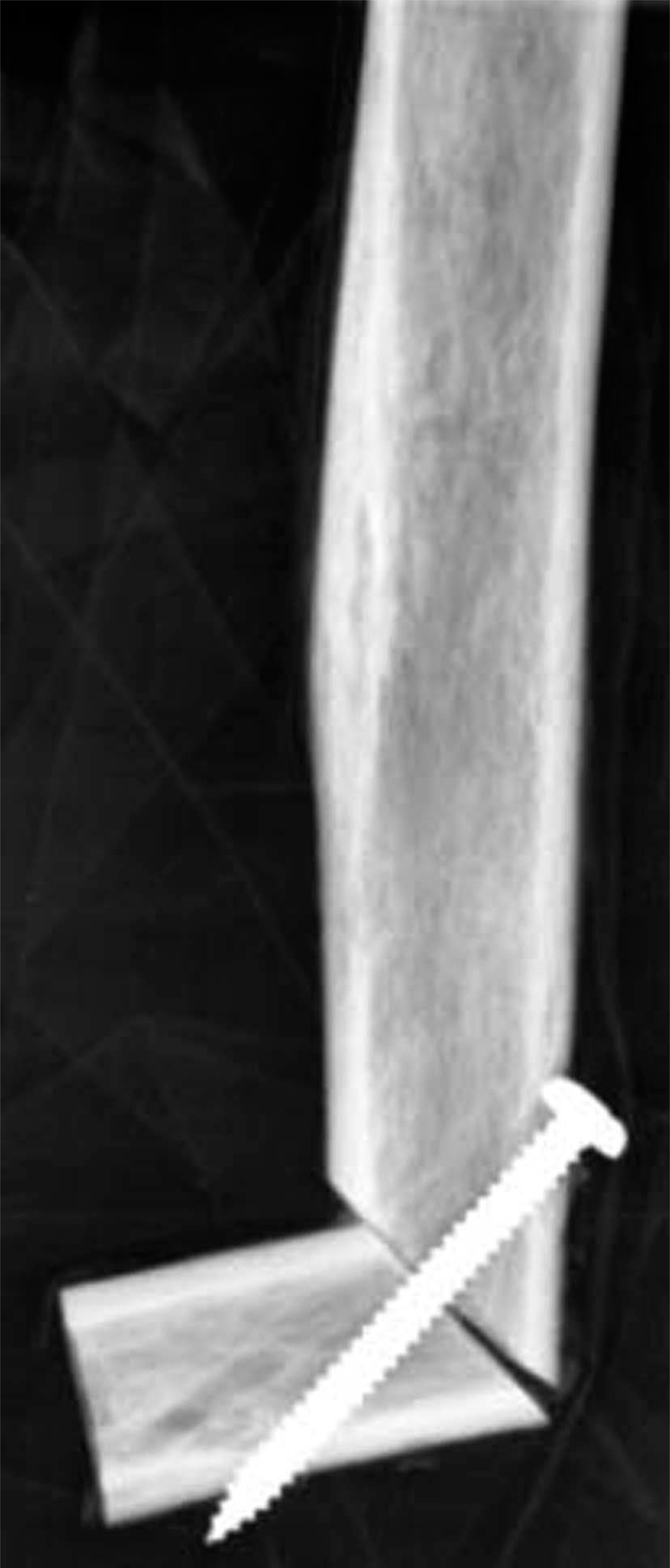
Marquardt's angulation osteotomy.
Material and methods
Design of the implant
The overall idea was to create artificial humeral condyles for better prosthetic fitting. Therefore the authors decided to use a T-shaped implant, where the intercondylar diameter would simulate the normal anatomic humeral intercondylar diameter, i.e., 5 – 7 cm. In most trans-humeral amputees, the intramedullary cavity of the humerus has an increasing diameter from distal to proximal. Therefore, it was decided to use an implant for cemented fixation (Figure 2).
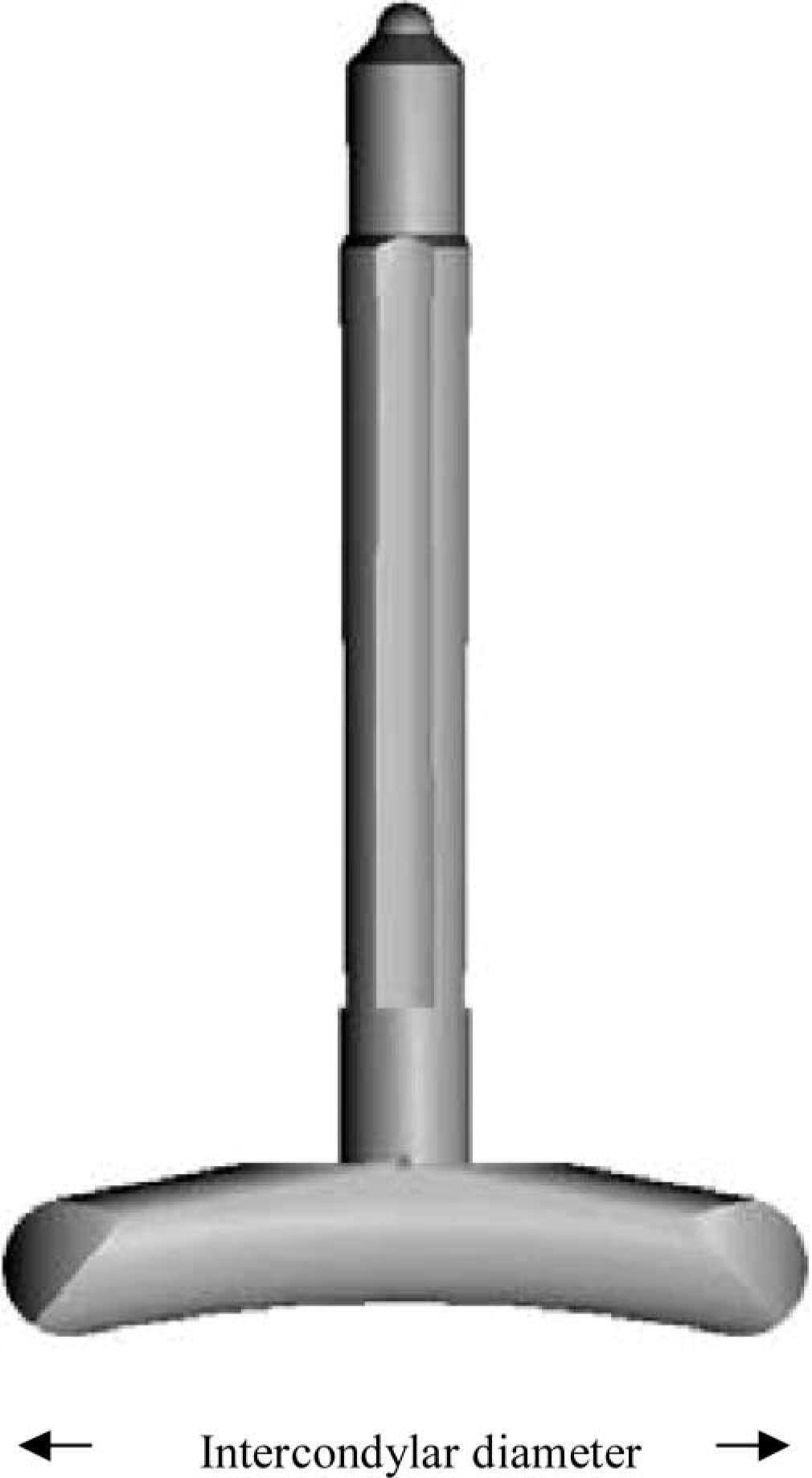
The Humerus-T-Prosthesis (HTP) with stem and condyle. The implant was made of titanium alloy. The condyle surface was polished (Ra-value 0.12 μm). The stem surface was grit blasted (Ra-value 4.5 μm). Four longitudinal flutes with a depth of 0.75 mm were machined into the stem.
In vitro mechanical testing
A Humerus-T-Prosthesis (HTP) was cemented into the distal end of a human cadaver humerus using Palacos® R-40 cum gentamicin bone cement (Schering-Plough Europe, Brussels, Belgium). The bone specimen with the prosthesis was mounted on a jig in a MTS Bionix servo-hydraulic testing machine (MTS, Minneapolis, USA) and subjected to alternating axial (traction) and torsional loading until failure of the fixation of the stem.
Clinical study
The Regional Ethical Committee approved the study, which included three patients. Each patient signed a confirmation of consent.
Patient I
Male, age 67 years, right side amputated through the distal half of the humerus epiphysis 15 years prior to the study. No shortening or elongation of the amputation stump was planned in this patient.
Patient II
Male, age 58 years, left side amputated through the distal half of the humerus epiphysis 33 years prior to the study. In this patient, the amputation stump was so long that the arm prosthesis he had used prior to the study was cosmetically unacceptable. Hence, the patient had been considered for reamputation with shortening of the amputation stump. A shortening of the amputation stump of approximately 5 cm was planned in this patient.
Patient III
Male, age 22 years, left side amputated through the proximal half of the humerus 3 years prior to the study. Due to a short amputation stump this patient had problems using an arm prosthesis. An elongation of the amputation stump was planned in this patient.
At inclusion the patients used a myoelectric arm prosthesis with a harness and straps around the contralateral shoulder. Their main complaints were pain from the neck and discomfort from the contralateral shoulder while using the prosthesis.
In all three patients the soft tissue over the amputation stump was of good quality. None of the patients had any amputation neuroma. Prior to the operation the clinical status and prosthetic function of the patients were evaluated according to a standardized formula.
A CT scan was taken of the humerus and a customized Humerus-T-Prosthesis was made. Several models were made for each patient, with varying degrees of intercondylar diameter. The diameter of the stem was to allow a cement mantel of no less than 3 mm. The condyle of the implant was to be placed under the deep fascia. The patients were operated on in a supine position under general anaesthesia. A fish-mouth incision was made with sharp dissection down to the bone. An osteotomy was made to open the intramedullary cavity. Standard high-pressure cementing technique was used with Palacos® R-40 cum gentamicin bone cement (Schering-Plough Europe, Brussels, Belgium). The patients received systemic antibiotic prophylaxis with cefalotin (Keflin®, Lilly, Eli Lilly & Co., Indianapolis, USA), 2 g preoperatively and 1 g 8 and 16 h post-operatively. After three weeks the skin sutures were removed, and anti-oedema treatment was started. The patients were followed up at 3, 6, 9 and 12 months after the operation, and thereafter at yearly intervals.
Results
In vitro mechanical testing
The mechanical testing showed that loosening of the humeral stem occurred at axial loading of 2700 N. When a pull of 2700 N was applied to the prosthesis, a permanent displacement of 2.5 mm was measured. However, the prosthesis did not appear to be grossly loose. Also, an incremental torsional load up to 52.5 Nm was applied to the prosthesis. Even at this load the implant did not migrate, but a rotation of 5° was observed. This rotation, however, fully recovered when the implant was unloaded.
Clinical study
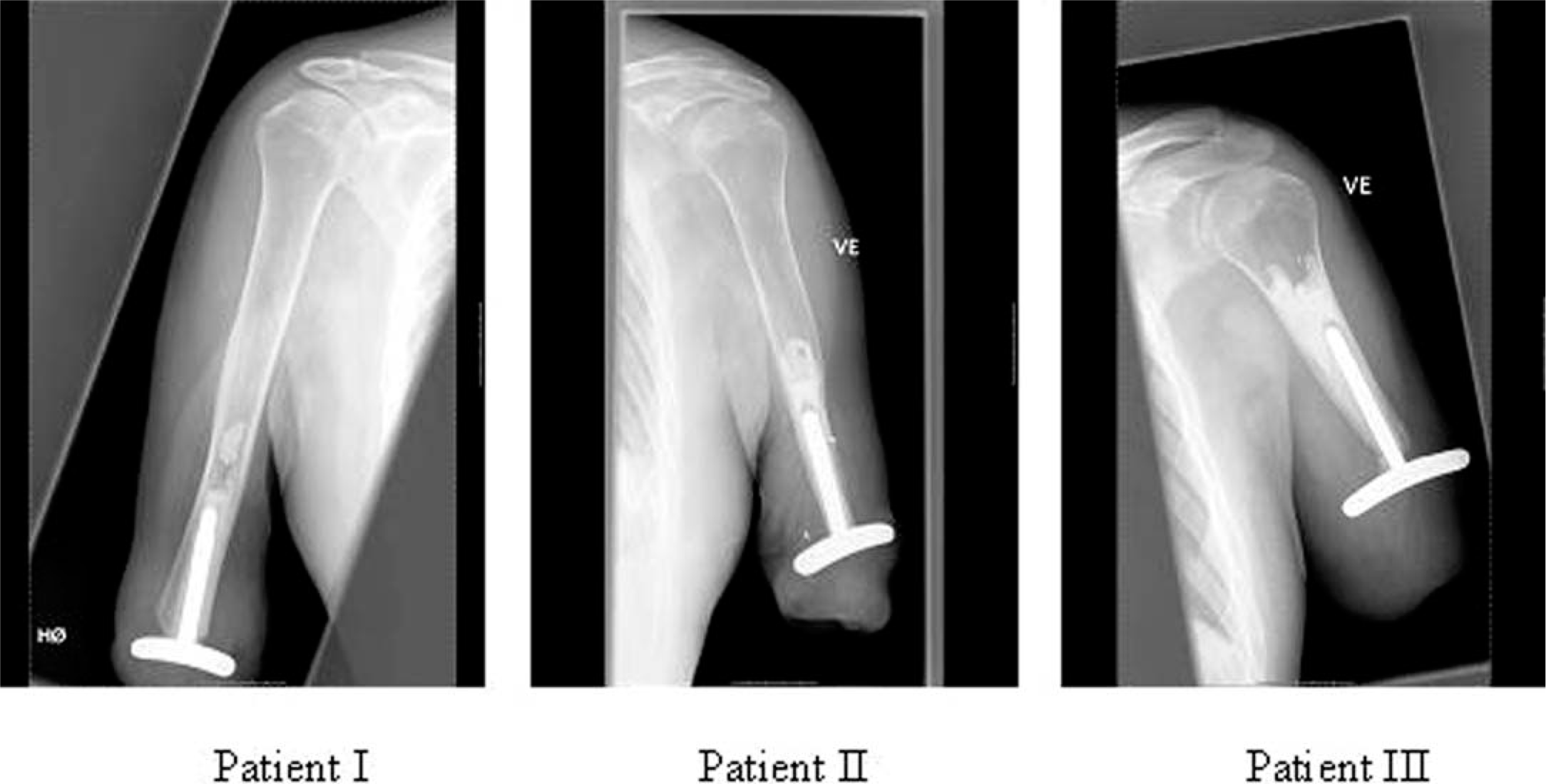
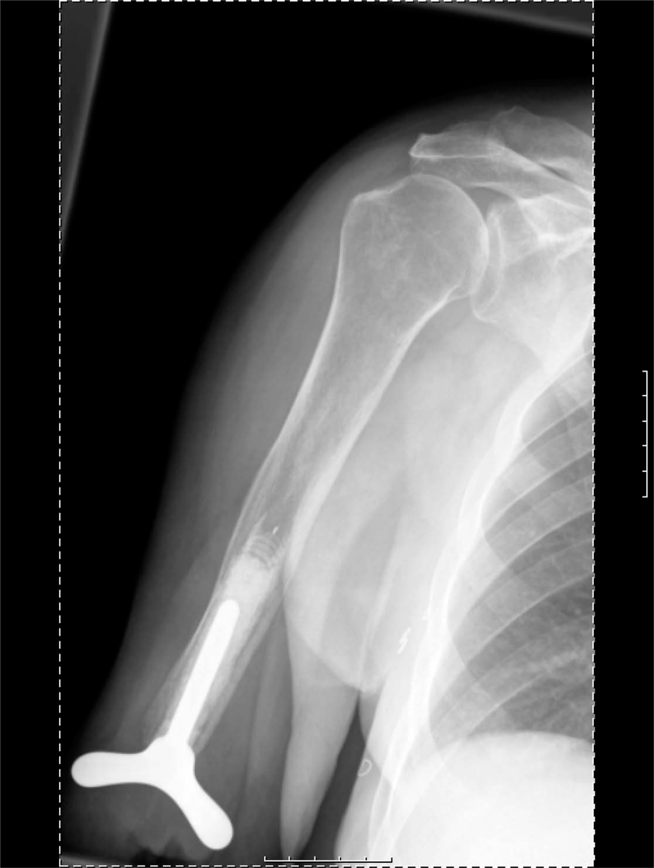
Each patient received a Humerus-T-Prosthesis with a similar stem (length 75 mm, proximal diameter = 10 mm, distal diameter = 9 mm). In Patient III a plateau was made at the basis of the stem to allow lengthening of the amputation stump. The osteotomy of the humerus was made 5 mm proximal to the amputation level in Patients I and III, and 50 mm in Patient II. Post-operative x-rays are shown in Figure 3.
Patients I, II and III received an HTP-prosthesis with an intercondylar diameter of 55, 60 and 65 mm, respectively. In Patient I the amputation stump was elongated by 5 mm, in Patient II the amputation stump was shortened by 40 mm, and in Patient III the amputation stump was elongated by 12.5 mm.
Due to the quality of the soft tissue, the implants could easily be placed under the deep fascia in all three patients. There were no per- or post-operative complications. Post-operatively the patients were discharged after two, three and two days, respectively. Adaptation to the new arm prosthesis started at 3 – 4 weeks. Different designs of the new arm prosthesis were evaluated. Common to all of them was that the prosthesis was suspended and stabilized by the humerus and implant only with a socket that terminated inferior to the deltoid. The rotation and traction stability of the arm prosthesis were secured through tight adaptation around the new condyles. The clinical and functional status pre- and post-operatively is listed in Table I.
Active motion and pain with arm prosthesis.
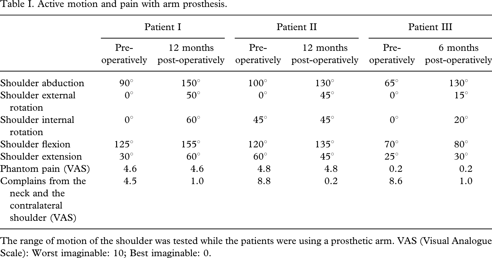
The range of motion of the shoulder was tested while the patients were using a prosthetic arm. VAS (Visual Analogue Scale): Worst imaginable: 10; Best imaginable: 0.
When adapting to the new arm prosthesis the patients recognized pain over the new condyles. Patient III even resulted with a superficial pressure wound over the lateral condyle. The wound healed with scarring over the lateral condyle. In Patients I and II, the pain disappeared after the design of an arm prosthesis with adjustable pressure control over the new condyles (Figure 4). At the final control, 36 months after the operation, Patients I and II had no discomfort or pain when using their new prostheses, they had a similar range of motion in both shoulders and they were full-time prosthetic users. Some 24 months after the operation, Patient III was subject to a new trauma to his left shoulder with a fracture of the scapula. Until now, this has limited the use of the new arm prosthesis.
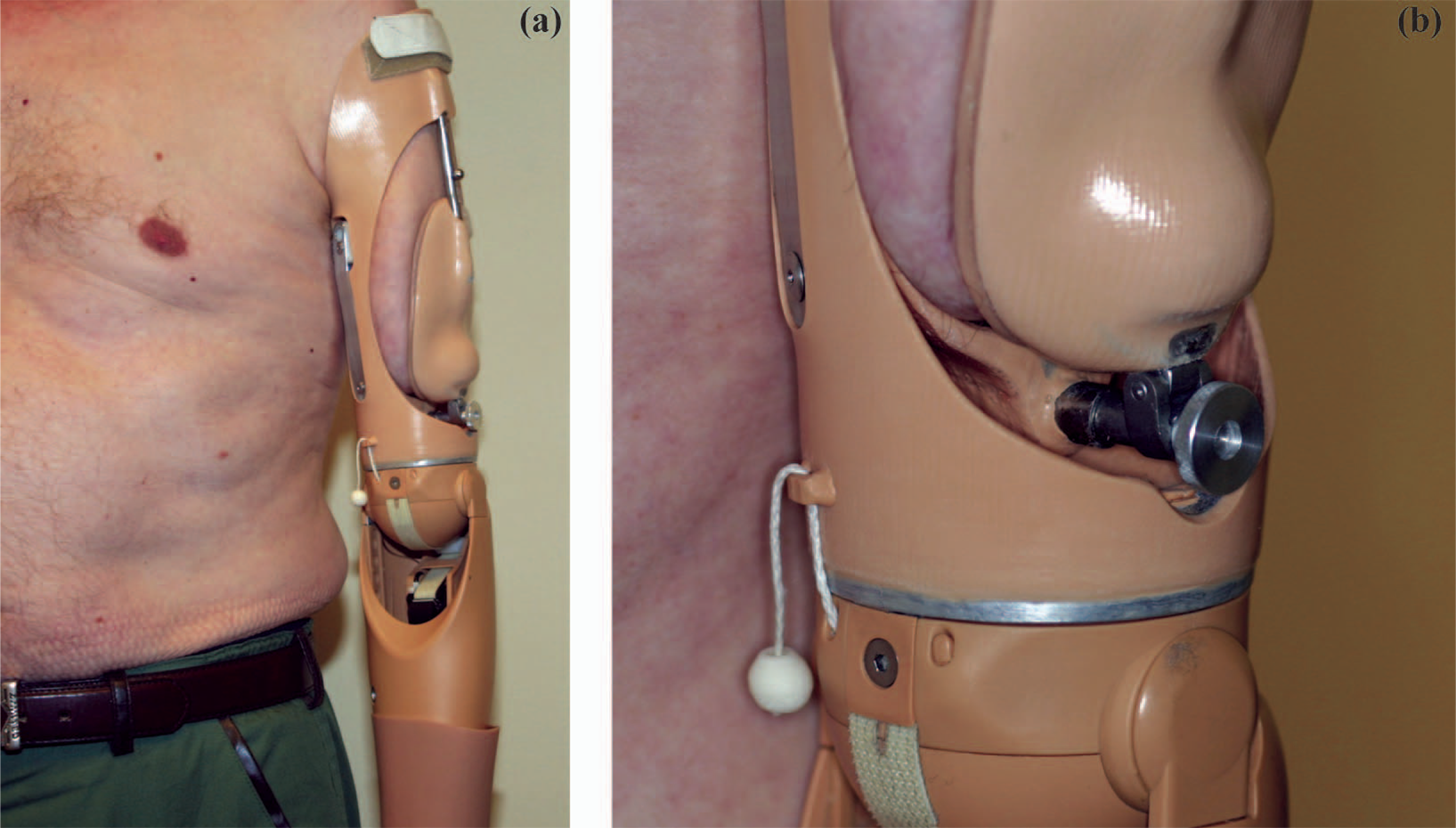
(a) shows the new arm prosthesis socket. (b) shows the wheel by which the patient can adjust the pressure over the artificial humeral condyles.
Discussion
The basic idea of this study was to create an amputation stump similar to that in elbow-disarticulated amputees, where the humerus condyles allow secure fit of the arm prosthesis socket.
Based on the present pilot study, it is difficult to make any conclusions regarding the optimum design of the implant. All three patients reported pain over the new condyles when adapting to the new arm prosthesis. In Patients I and II these problems later disappeared and it seems that the use of the new type of arm prosthesis socket with adjustable pressure over the condyles has solved this problem. However, it is important to notice that Patient number III had a superficial pressure wound over the lateral condyle. This patient had the shortest amputation stump and he got the pressure wound while using an arm prosthesis with myoelectric elbow and hand. Obviously the high torque and pressure applied by the new arm prosthesis resulted in skin breakdown. He is now using a cosmetic arm to allow a more gradual adaptation to the new arm prosthesis socket.
To optimize the concept the authors have changed the design of the implant (Figure 5) and four patients have been operated on with implantation of this second generation HTP. So far it has been the experience that the rehabilitation time for prosthetic fitting has been shorter in these patients, compared with the patients who received the first generation HTP. Different designs of the implant and different designs of the arm prosthesis socket should be tested using an in vitro model.
The second generation of the HTP prosthesis.
The in vitro mechanical testing demonstrated that both the interfaces between the prosthesis and the cement and between the cement and the bone could withstand high loads. A pull of more than 2700 N could be applied to the implant before a permanent displacement occurred. Furthermore, the interfaces tolerated a torsional load of 52 Nm without any measurable migration of the stem. If it is assumed that the width of the symmetrical condyles of the prosthesis is 10 cm, a torque of 50 Nm on the stem-cement interface implies that the construct can withstand a torsional force of 1000 N applied to the apex of the condyle. In all likelihood, the fixation of the arm prosthesis to the soft tissues of the trans-humeral amputation stump would fail at far lower loads compared with the loads tolerated by the internal HTP prosthesis.
At the distal half of the humerus diaphysis, the use of an uncemented prosthesis with a hydroxyapatite coated stem could be an alternative to a cemented implant. At the proximal part of the humerus diaphysis the increasing diameter of the intramedullary canal makes a cemented implant more preferable. In vitro mechanical testing on cadaver bone should be performed where traction and torsional loading are applied to cemented and uncemented implants at different amputation levels.
Most upper limb amputations are due to an accident (Hunter 1996). In most patients, a shortening of the amputation stump is not acceptable and reduced soft tissue quality may be a major limitation for application of the authors' technique. This problem could be solved by a two-stage procedure where a sub-fascial tissue expander is applied to create acceptable soft tissue conditions.
Trans-humeral amputees using a myoelectric prosthesis will probably benefit most from this new concept for prosthetic fitting. However, even in industrialized countries trans-humeral amputees are using both myoelectric and body-powered arm prosthesis (Millstein et al. 1986). Hence, studies should be performed on the use of the new arm prosthesis as a body-powered prosthesis. Furthermore, surgical modification of the amputation stump by use of an implant could be applied to other amputation levels. The Regional Ethical Committee has given the authors permission to implant a Femoral-Condyle-Prosthesis in trans-femoral amputees.
In conclusion, two out of three patients operated on with implantation of the Humerus-T-Prosthesis were successfully rehabilitated with the use of a new type of trans-humeral prosthesis without a harness and straps around the contralateral shoulder. Further studies should be performed, giving priority to the testing of different designs of the implant.
Footnotes
Acknowledgement
The authors would like to thank Cypromed, Hamar, Norway for funding. None of the authors have received other funding for this study from industrial, commercial or government interests.