
Abstract
Introduction
Intervertebral disc (IVD) degeneration is characterized by a loss of cellularity and matrix biosynthesis for resident cells, along with a loss of the demarcation between nucleus pulposus (NP) and anulus fibrosus (AF) regions.1,2 Cell therapy is an increasingly attractive strategy to regenerate disc structure and restore function based on delivery of both autologous and allogeneic cells.3-5 The potential for NP cells to achieve disc-appropriate matrix regeneration may change with aging, however, due to a poorly defined NP-specific phenotype that relates to matrix regeneration in poorly understood ways. Our previous studies discovered a set of novel cell surface proteins (CD24, CD54, CD155, CD166, and CD221), one transcriptional factor (Brachyury, T), and three neuronal-related proteins (brain abundant membrane attached signal protein 1, Basp1; Neurochondrin, Ncdn and Neuropilin, Nrp1) in healthy, immature rat and human NP tissues that may serve as a subset of NP phenotypic markers.6,7 The goal of this study is to evaluate the potential for human IVD cells to express these NP-specific molecular markers using a laminin-rich pseudo-3D culture system previously shown to be supportive of NP cell phenotype.8
Materials and Methods
Tissue and Cell Isolation
Human NP tissues were procured from patients undergoing surgery for scoliosis (“juvenile,” n = 4, 6-16 years) and degenerative disc disease (“adult,” n = 4, 39-69 years). NP cells were isolated by a non-enzymatic protocol from tissues as described previously.9
A Pseudo-3D System for Cell Culture
Cells were allowed to form 3D, multi-cell clusters spontaneously in a laminin-rich pseudo-3D culture system.8 Briefly, trypsinized cells were seeded upon Transwell (0.5 × 10⁶ cells per well) precoated with Matrigel (60 μL/well, n = 3 per patient) and cultured in F-12 basal medium (supplemented with 2.5% Matrigel for free laminin ligand access, 10% FBS, and 2.5 mg/mL L-ascorbic acid-2-phosphate) for up to 28 days.
Immunohistochemistry
At the end of the culture time, cell constructs were harvested for cryo-sectioning to determine NP-specific molecular marker expression and proteoglycan and collagen matrix production. Each tissue construct was analyzed by histology (H&E and Safranin O) and immunostaining. Frozen sections of cell constructs were fixed and permeabilized as needed, and then incubated with specific antihuman antibodies and isotype controls for selected NP markers (CDs, Brachyury T, Basp1, Ncdn, Nrp, and type II collagen) and appropriate secondary antibodies.
Results
Immunostaining demonstrated positive staining for CD24, CD54, CD90, CD221, Basp1, Ncdn, and Nrp1 in juvenile NP cell constructs when cultured in a laminin-rich pseudo-3D culture system for 28 days (selected markers shown in the figure). These results are consistent with our previous finding for expression of these markers in juvenile NP tissues. Among these proteins, Basp1, Ncdn, and Nrp1 displayed more intense staining in the juvenile group as compared with the adult group (figure). CD155, CD166, and Brachyury T were not detected in NP cell constructs of either age group (data not shown), although they were expressed in juvenile NP tissue previously. CD54 and CD90, but not CD24, were detected in adult NP cell constructs (figure) similar to our previous finding for their expression pattern in adult NP tissues. Interestingly, positive expression for CD221, Basp1, Ncdn, and Nrp1 was detected in adult NP cell constructs (figure), although these proteins are absent in adult NP tissue. In contrast, CD54 and CD166, known to be expressed in adult NP native tissue, were not detected in cell constructs formed from adult NP cells (data not shown). In addition, staining for proteoglycan and collagen II was more intense in juvenile NP cell constructs than in adult (data not shown).
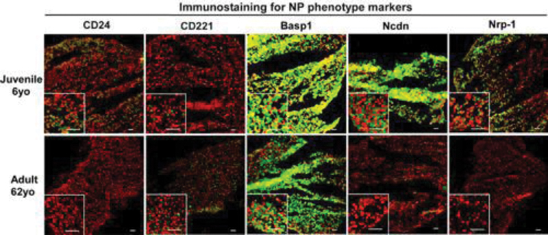
Immunostaining of CD proteins, neuronal-related proteins for human NP cells of different ages (6 vs. 62 years) after culture in a laminin-rich pseudo-3D system (28 days). Bar = 50 µm.
Conclusion
The results of this study demonstrate that NP cell culture in a laminin-rich “pseudo-3D” culture system can promote the expression of some juvenile human NP-specific markers (CD24, CD90, CD221, Basp1, Ncdn, and Nrp1). Importantly, a subset of these molecular markers was reexpressed in human NP cells in vitro, despite lost marker expression in the native adult tissue. This culture system may be useful for developing cell-based therapy for IVD regeneration.
None declared
Freemont AJ. The cellular pathobiology of the degenerate intervertebral disc and discogenic back pain. Rheumatology (Oxford) 2009;48(1):5–10
Urban JP, Roberts S. Degeneration of the intervertebral disc. Arthritis Res Ther 2003;5(3):120–130
Sakai D, Mochida J, Iwashina T, et al. Differentiation of mesenchymal stem cells transplanted to a rabbit degenerative disc model: potential and limitations for stem cell therapy in disc regeneration. Spine 2005;30(21):2379–2387
Smith LJ, Nerurkar NL, Choi KS, Harfe BD, Elliott DM. Degeneration and regeneration of the intervertebral disc: lessons from development. Dis Model Mech 2011;4(1):31–41
Risbud MV, Albert TJ, Guttapalli A, et al. Differentiation of mesenchymal stem cells towards a nucleus pulposus-like phenotype in vitro: implications for cell-based transplantation therapy. Spine 2004;29(23):2627–2632
Tang X, Jing L, Chen J. Changes in the molecular phenotype of nucleus pulposus cells with intervertebral disc aging. PLoS ONE 2012;7(12):e52020
Tang X, Jing L, Setton LA, et al. Novel molecular phenotype of notochordal-like cells in nucleus pulposus of human intervertebral disk. Global Spine J 2012;2(Suppl 1):S95
Chon BH, Lee EJ, Jing L, Setton LA, Chen J. Human umbilical cord mesenchymal stromal cells exhibit immature nucleus pulposus cell phenotype in a laminin-rich pseudo-three-dimensional culture system. Stem Cell Res Ther 2013;4(5):120
Tang X, Richardson WJ, Fitch RD, Brown CR, Isaacs RE, Chen J. A new non-enzymatic method for isolating human intervertebral disc cells preserves the phenotype of nucleus pulposus cells. Cytotechnology 2013