
Abstract
Introduction
Fibrin gels are considered to be a promising biomaterial for tissue engineering because of its role as a “physiological scaffold” in tissue regeneration. It has been reported that fibrin gels could lead to cell migration into a wound/tissue-engineered construct and induce cell proliferation/growth.1-5 Due to the limited self-repair capability of degenerated intervertebral disc, cell-based therapy such as mesenchymal stem cell (MSC) application is becoming an attractive strategy in disc regeneration.6 However, interactions between fibrin and MSCs during disc regeneration are unknown. The objective of this study is to explore the potential value of fibrin gel as a delivery system for MSC and nucleus pulposus cell (NPC) co-culture therapy. We tested different fibrin formulations (fibrinogen and thrombin concentrations) and characterized fibrin gel structure and extracellular matrix production under hypoxic conditions in vitro. The findings will help to find an effective delivery system for cell-based therapy in disc regeneration.
Materials and Methods
Cell Isolation
Commercially available HMSCs (Lonza, MD) were expanded to the fifth passage, and NPCs were isolated from bovine tail discs using a collagenase/protease cocktail digestion.
Histology and RT-PCR
After 7 day's culture, fibrin gels were collected for H&E, Safranin-O staining. In addition, matrix genes, collagen II, aggrecan, and MMP13 expression of cells within gel were detected with qRT-PCR and normalized to universal GAPDH. Post hoc multiple pair-wise comparison tests (Dunn) were performed to determine differences between treatment groups with significance of p < 0.05
Results
After 7 days, fibrin gels demonstrated much more compact and condense network structure where cells entrapped and spread as compared with day 1 (Figs. A and B). Safranin O staining showed enriched proteoglycan around cells (Fig. C). Under hypoxia condition, 9 mg/mL fibrin gel increased type II collagen expression at day 7 by 2.9-fold as compared with day 1 (p < 0.05, Fig. D), whereas no significant difference between normoxia and hypoxia was observed. Similar expression differences were observed for aggrecan. Interestingly, MMP13 expression increased significantly at day 7 in normoxia group, while it decreased dramatically in hypoxia group (p < 0.01, Fig. D). Overall, the 9 mg/mL (9:50 and 9:100) fibrin gel displayed more desirable effects on matrix gene (collagen II, aggrecan) expression than on 18 mg/mL (18:50 and 18:100) fibrin gel (Fig. E).
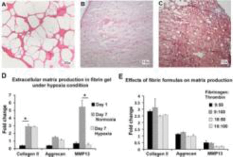
(A) Fibrin gel structure at day 1 (fibrinogen/thrombin, 9:50), 20×. (B) Fibrin gel structure at day 7 (9:50, H&E staining), 10× (C) SO staining at day 7 (9:50), 10×. (D) Effects of hypoxia on matrix production (9:50). (E) Effects of fibrin formulas on matrix production under hypoxia condition.
Conclusion
Fibrin gel promoted collagen II and aggrecan gene expression as well as proteoglycan production under both normoxia and hypoxia conditions. Among the formulation studies, 9 mg/mL fibrin exerted more desirable effects on cell spreading and matrix production. The findings suggesting fibrin gel may be a good delivery system for cell-based therapy in disc regeneration by promoting matrix production.
None declared
Ikari Y, Yee KO, Schwartz SM. Role of alpha5beta1 and alphavbeta3 integrins on smooth muscle cell spreading and migration in fibrin gels. Thromb Haemost 2PO.072;84(4):701–705
Nomura H, Naito M, Iguchi A, Thompson WD, Smith EB. Fibrin gel induces the migration of smooth muscle cells from rabbit aortic explants. Thromb Haemost 1999;82(4):1347–1352
Liu J, Tan Y, Zhang H, et al. Soft fibrin gels promote selection and growth of tumorigenic cells. Nat Mater 2012;11(8):734–741
Eyrich D, Brandl F, Appel B, et al. Long-term stable fibrin gels for cartilage engineering. Biomaterials 2007;28(1):55–65
Man AJ, Davis HE, Itoh A, Leach JK, Bannerman P. Neurite outgrowth in fibrin gels is regulated by substrate stiffness. Tissue Eng Part A 2011;17(23-24):2931–2942
Sakai D, Mochida J, Yamamoto Y, et al. Transplantation of mesenchymal stem cells embedded in Atelocollagen gel to the intervertebral disc: a potential therapeutic model for disc degeneration. Biomaterials 2003;24(20):3531–3541
Bensaïd W, Triffitt JT, Blanchat C, Oudina K, Sedel L, Petite H. A biodegradable fibrin scaffold for mesenchymal stem cell transplantation. Biomaterials 2003;24(14):2497–2502