
Abstract
Magnetic resonance imaging (MRI) has been applied to visualize monocyte infiltration with the use of intravenously injected ultrasmall superparamagnetic iron oxide (USPIO). However, USPIO uptake in vivo remains elusive, and the heterogeneous enhancement patterns observed by MRI point to multiple pathophysiological events. This study focused on specific imaging of monocyte infiltration into the brain by transfusion of superparamagnetic iron oxide (SPIO)-labeled monocytes in a rat model of neuroinflammation, experimentally induced photothrombosis (PT). At day 5 after lesion induction, animals were transfused with SPIO-labeled monocytes (5 × 10⁶ cells) or free USPIO (17 mg Fe/kg). MRI was performed 24, 72 and, 120 h later. To investigate temporal changes directly after intravenous USPIO administration, MRI was performed repeatedly up to 8 h. Relaxation measurements showed that rat monocytes were efficiently labeled in vitro using SPIO (R2=12±0.9 s−1). After transfusion of SPIO-labeled monocytes, a significant increase in contrast enhanced area (340%±106%) in the PT lesion was observed not before 72 h. Contrast enhancement after USPIO injection increased up to 407%±39% at a much earlier point of time (24 h) and diminished thereafter. Repetitive MRI directly after USPIO injection showed significant contrast enhancement in the lesion within 2 h. Our study shows that MRI enables in vivo tracking of SPIO-labeled monocytes longitudinally. Moreover, our data suggest that contrast enhancement after injection of free USPIO does not primarily represent signals from peripherally labeled monocytes that migrated toward the inflammatory lesion. The use of SPIO-labeled monocytes provides a better tool to specifically assess the time window of monocyte infiltration.
Introduction
Cellular infiltration in the central nervous system (CNS) is a key event during neuroinflammation and contributes to brain damage in neurological diseases such as multiple sclerosis (MS) and stroke (Lassmann, 1997; Price et al, 2003; Stoll et al, 1998). CNS inflammation is characterized by increased blood—brain barrier (BBB) permeability and expression of cell adhesion molecules on brain endothelial cells, which mediate cellular infiltration. It is generally believed that destruction of brain tissue and lesion development associated with inflammation is predominantly mediated by the infiltration of activated monocytes into the CNS (Dirnagl et al, 1999; Stoll et al, 1998). Previous work in animal models showed that selective depletion of the monocyte population during CNS inflammation reduced disease severity (Huitinga et al, 1995; Polfliet et al, 2002). Therefore, inhibition of monocyte infiltration is considered an attractive therapeutic strategy to limit neuroinflammation. A number of treatment studies have focused on diminishing migratory capacity of peripheral blood mononuclear cells by either immunomodulation (Corsini et al, 1997; Floris et al, 2002) or by direct blocking of cell adhesion molecules, as has been shown for the α-4 integrin blocking molecule natalizumab (Niino et al, 2006; Polman et al, 2006). However, continuous blocking of adhesion receptors may increase the risk of inflammation in peripheral organs. To design an effective treatment, it is important to specifically intervene within the time window of monocyte infiltration into the CNS. So far, the temporal pattern of monocyte recruitment during CNS inflammation is largely unknown. There is an urgent need for noninvasive and accurate tracking tools to elucidate monocyte recruitment in the CNS longitudinally.
The development of small and ultrasmall superparamagnetic iron oxides (SPIO and USPIO, respectively) extended the use of magnetic resonance imaging (MRI) to study cell dynamics in vivo (Arbab et al, 2003; Weissleder et al, 1997). Iron oxide particles shorten the transverse relaxation times T2 and T2∗, and clusters of cells that have taken up USPIO appear hypointense on T2(∗)-weighted images (Yeh et al, 1993). So far, intravenous administration of USPIO has been used to study monocyte infiltration in animal models with a neuroinflammatory component like stroke (Saleh et al, 2004b; Schroeter et al, 2004) and MS (Dousset et al, 1999a; Floris et al, 2004). Recently, this methodology has been applied in clinical trials to investigate macrophage activity in stroke (Saleh et al, 2004a) and MS patients (Dousset et al, 2006). It was shown in these studies that signal changes in the brain observed 24 h after USPIO administration differed from gadopentetic acid (Gd-DTPA)-enhanced areas, which marks BBB breakdown. This difference in contrast enhancements points to distinct pathophysiological events and USPIO-enhanced MRI is suggested to provide specific information on the cellular component of neuroinflammation.
Nevertheless, the exact route of USPIO uptake and distribution in vivo remains unclear, which hampers the interpretation of USPIO-related signals. USPIO enhancement in the CNS is generally believed to reflect infiltrated monocytes that have taken up USPIO in the blood circulation. However, MRI signal changes may originate from sources other than the migration of these peripherally labeled monocytes. USPIO may enter the CNS passively by leakage over a damaged BBB or by transcytosis across the brain endothelial cells (Xu et al, 1998). Moreover, it is suggested that cellular USPIO incorporation may occur outside the vasculature by activated microglia (Dousset et al, 1999a). Transfusion of monocytes that have been specifically labeled ex vivo may, therefore, be a better tool to address exclusively monocyte infiltration during an inflammatory response in the brain. This approach has recently been reported by Stroh et al (2006), in which they labeled spleen-derived mononuclear cells with very small SPIO. Labeled cells were intravenously injected in splenectomized mice after middle cerebral artery occlusion, and were found to be engrafted at the lesion border zone.
The main goal of our study is to monitor exclusively brain infiltration of SPIO-labeled monocytes and compare contrast enhancement to the injection of free USPIO. We used photothrombosis (PT) (Watson et al, 1985) as a rat model for neuroinflammation. In this animal model, focal illumination of the brain after intravenous injection of a photosensitive dye induces a well-defined cortical lesion that is characterized by the presence of infiltrating monocytes (Lee et al, 1996). Previous studies using this model have shown that MRI 24 h after USPIO administration allows detection of the inflammatory lesion in the brain (Kleinschnitz et al, 2003; Saleh et al, 2004b). However, the USPIO uptake mechanism in vivo remains elusive, and other factors such as BBB leakage may confound the interpretation of USPIO-enhanced MRI. To address this issue, we assessed the spatiotemporal profile of USPIO enhancement in the CNS directly after intravenous administration.
Materials and methods
Isolation of Monocytes
Rat monocytes were freshly isolated by perfusion as reported previously (Floris et al, 2002). Briefly, male adult Lewis Hannover rats were anesthetized intraperitoneally with an overdose of pentobarbital and fixed in the supine position. The thorax was opened, 300 μL heparin (5,000 IE/mL) was injected in the left ventricle (apex), and two cannulae, size 16 and 20 G, were inserted into the left and right ventricles, respectively. Rats were perfused with 500 mL medium (RPMI-1640), supplemented with 0.5% (w/v) Bovine Serum Albumin and 20 mmol/L Hepes (pH 7.4), and the effluent was collected. Peripheral blood mononuclear cells were isolated by centrifugation (400g, 40mins at room temperature) on a Ficoll density gradient (Lymfoprep; Fresenius Kabi Norge, Oslo, Norway) and incubated for 30 mins at 4°C with alexa488-coupled monoclonal antibodies directed against T and B cells (R.7.3 and OX33 respectively, 1 μg/mL mAb; 1 μL/106 target cells). Monocytes were purified from the peripheral blood mononuclear cells by negative selection using fluorescence activated cell sorting (Mo-Flo, Dakocytomation, Inc., Fort Collins, CO, USA). Monocyte viability was routinely checked by 7AAD (Molecular Probes, Eugene, OR, USA) exclusion and the purity of the isolate was 80% to 90% as determined by the number of ED8 (produced at the Department of Molecular Cell Biology and Immunology, VUMC)-positive cells using a FACScan flow cytometer (Calibur, Becton Dickinson, San Jose, CA, USA).
Labeling of Monocytes with Iron Oxides
Freshly isolated monocytes were incubated in the absence (vehicle-treated) or presence of USPIO (Sinerem, Guerbet, France) or SPIO (Endorem, Guerbet, France) as described previously for human monocytes (Oude Engberink et al, 2007). The optimal iron concentrations and incubation time from that study were used as a starting point to achieve efficient labeling of primary rat monocytes. Briefly, iron concentrations in culture medium (supplemented RPMI-1640) were adjusted to 1 mg and 4 mg Fe/mL containing 2 × 10⁶ cells. Cells were incubated for 1.5 h and viability was determined for all samples by Trypan blue exclusion.
To assess the effect of iron uptake on the migratory capacity of SPIO-labeled monocytes, their migration over a monolayer of rat brain endothelial cells was monitored as described previously (Floris et al, 2002; Hendriks et al, 2004; Van der Goes et al, 2001). Briefly, 7.5 × 10⁵ monocytes suspended in serum-free culture medium were allowed to migrate for 4 h over brain endothelial monolayers (established from rat brain endothelial cell line GP8/3; Floris et al, 2002; Van der Goes et al, 2001). Monocyte migration was analyzed for labeled cells (SPIO, 4 mg Fe/mL) and compared with vehicle-treated monocytes and freshly isolated monocytes. In addition, monocytes were activated with lipopolysaccharide (100 ng/mL) for 24 h. The level of migration was quantified as the percentage of migrated cells relative to the total number of monocytes present within a field of 200 μm2.
Animal Procedures
All animal procedures were approved by the local ethical committee and were performed in accordance with international guidelines on handling laboratory animals.
Intracerebral Injection of Labeled Cells
To validate the potential for in vivo MR detection of SPIO-labeled monocytes, intracerebral injections were performed. Lewis Hannover rats (n=6, 3 weeks of age) were anesthetized with 2% isoflurane in a N2O/O2 mixture (70/30) and allowed to breath spontaneously. Animals were placed in a stereotactic frame. A hole was drilled in the cranium at 0.5 mm anterior and 2.5 mm lateral from bregma. Unlabeled monocytes or SPIO-labeled monocytes (ex vivo labeled in 1 mg Fe/mL) were injected into the caudate putamen at a depth of 4 mm from the cortical surface over a period of 5 mins through a 26 G needle attached to a 50 μL syringe (Hamilton Co., Reno, NV, USA). A total of 2 × 10⁶ cells in a volume of 10 μL were injected. MRI was performed immediately after cell injection and animals were allowed to recover thereafter. Follow-up scans were performed 24, 72, and 120 h after cell injection.
Transfusion of SPIO-Labeled Monocytes/USPIO in the PT Model
To study monocyte infiltration in vivo, cortical lesions were induced by photochemically initiated thrombosis (PT) (Hoff et al, 2005; Watson et al, 1985). Lewis Hannover rats (n=15, 275 to 300 g) were anaesthetized as described above. For skull illumination, the scalp was incised and the periost was removed. The light source (2.4 mm diameter) was positioned perpendicular to the skull surface at 2.7 mm anterior and 2.7 mm lateral to bregma for lesion induction in the frontal cortex. Each animal was intravenously injected with erythrosin B (20 mg/kg), followed by injection of 0.9% sodium chloride to a total volume of 1 mL over a 2 mins time period. The skull was then illuminated for 2.5 mins (300 mW/cm2).
On day 5 after PT, animals were anaesthetized as described above. A tail vein was cannulated for intravenous administration of contrast agents. Rats received no injection (n=4, controls), free USPIO (n=4, 17 mg Fe/kg), or SPIO-labeled monocytes (n=4, 5 × 10⁶ cells ex vivo labeled in 4 mg Fe/mL). MRI was performed before and 24, 72, and 120 h after transfusion. At 72 h, two animals per group were killed for histological examination. In a subset of animals (n=3), MRI was performed repeatedly from immediately up to 8 h after intravenous USPIO administration.
Magnetic Resonance Imaging
All experiments were performed using a 4.7 T horizontal bore nuclear magnetic resonance spectrometer (Varian, Palo Alto, CA, USA), equipped with a high-performance gradient insert (12 cm inner diameter, maximum gradient strength 500 mT/m).
In Vitro MRI
To determine the efficiency of monocyte labeling with either USPIO or SPIO, we performed T2 measurements (9 × 0.5 mm slices, repetition time (TR)=3,200 ms, 10 echoes with echo time (TE) spacing=17.5 ms, field of view (FOV)=4 × 4 cm; matrix=128 × 128, receiver bandwidth=42.5 kHz, number of experiments (NEX)=2) using a birdcage coil (Varian, Palo Alto, CA, USA). Relaxation times were measured from agarose gel (0.4%) suspensions containing labeled monocytes (0.5 × 10⁶ cells per 250 μL) in 96-well plates. T2 maps were calculated from monoexponential fitting of MRI signal intensities as a function of TE.
In Vivo MRI
Animals were anesthetized as described above and were prepared for mechanical ventilation by endotracheal intubation. A tail vein was cannulated for injection of Gd-DTPA (Magnevist, Schering, Berlin, Germany). Animals were immobilized in a specially designed stereotactic holder and placed in an animal cradle that was inserted into the nuclear magnetic resonance spectrometer. During MR experiments, animals were mechanically ventilated with isoflurane (2%) in N2O/O2 (70/30). Expiratory CO2 was continuously monitored and body temperature was maintained at 37°C using a heated water pad. An infrared sensor (Nonin Medical Inc., Plymouth, MN, USA) was attached to the hind paw for monitoring heart rate and blood oxygen saturation. A homebuilt Helmholtz volume coil (∅ 85 mm) and an inductively coupled surface coil (∅ 35 mm) were used for radio frequency transmission and signal detection, respectively.
T2 maps were calculated as described above from T2 measurements (21 × 1 mm slices, TR=3,200 ms, TE-spacing=17.5 ms, receiver bandwidth=54.4 kHz, FOV=3.2 × 3.2 cm; matrix=128 × 128, NEX=4). For animals with a PT lesion, T2∗-weighted (T2∗W) images were acquired using a gradient echo MRI (TR=2,500 ms, TE=12.5, NEX=2). At the end of the MR session, T1-weighted (T1W) spin-echo images (TR=300ms; TE=11.5 ms, NEX=4) were collected before and 10 mins after 0.5 mmol/kg Gd-DTPA injection. Post- and pre-Gd-DTPA T1W images were subtracted for detection of Gd-DTPA enhancement. Total scanning time was 62 mins for each animal. In a subset of animals, T2W and T2∗W MRI was repeated for 16 cycles of 30 mins directly after intravenous USPIO administration.
Immunohistochemistry
Cell Samples
Immediately after incubation with either USPIO or SPIO, cell samples were washed, centrifuged (50g., 5 mins) onto glass slides, and air-dried. The presence of (U)SPIO was detected by Prussian blue staining of iron and cell spots were counterstained with nuclear fast red as previously described (Oude Engberink et al, 2007). Briefly, cell spots were fixed in acetone and incubated with a 1:1 mixture of 2% potassium hexacyanoferrat (II) and 2
Brain Sections
At 72 h after lesion induction, PT animals were killed for immunohistochemistry. For detection of free USPIO- and SPIO-labeled monocytes, brains were rapidly removed after the final MR scans, snap-frozen in the vapor phase of liquid nitrogen, and stored at −80°C. Serial 10 μm coronal cryosections (−20°C) were cut at the level of the cortical lesion and fixed in acetone for 10 mins. Sections were preincubated in phosphate-buffered saline with 10% fetal calf serum (Biowhittaker Europe, Verviers, Belgium), followed by incubation with monoclonal antibody ED1 (1.5 μg/mL; Serotec, Oxfordshire, UK) for 1 h at room temperature to detect infiltrated monocytes. As secondary antibody, a rabbit antimouse IgG-peroxidase conjugate (1 μg/mL) was used. Peroxidase activity was shown by incubation with 0.5 mg/mL 3,3′-diaminobenzidine-tetrahydrochloride in Tris-HCl buffer containing 0.03% H2O2. Sequential sections were stained for the presence of iron as described above.
Magnetic Resonance Image Analyses
In Vitro MRI
For analysis of the transverse relaxation rate R2 (1/T2), regions of interest (ROI) (circular, 7.5 mm in diameter) were placed in the center of a well, and special care was taken to exclude areas with air- susceptibility artifacts. R2 values were obtained by averaging the middle five ROI of the phantom volume. These procedures generated stable relaxation time values with minimum noise levels as described previously (Oude Engberink et al, 2007).
In Vivo MRI
Magnetic Resonance images obtained from PT animals were quantitatively analyzed for the contrast-enhanced area before and after transfusion. Brains were coregistered and analyzed using the Medical Image NetCDF package (MINC; McConnell Brain Imaging Centre Montreal Neurological Institute, McGill University, Canada). Slices with a clear T2 lesion determined 5 days after lesion induction were selected for further analysis. On corresponding T2∗W images, we calculated the number of hypointense voxels within the lesion defined by a signal intensity lower than the mean signal intensity −3 × standard deviation of the signal intensity in a cortical ROI (125 voxels) placed just outside the lesion.
To assess contrast enhancement in the lesion directly after USPIO injection, a different approach was used. The lesion area was manually outlined on a precontrast T2 image and the T2 in this ROI was calculated as a function of time after USPIO injection. To determine contrast enhancement in unaffected tissue, an ROI with the same dimensions was placed in the contralateral hemisphere.
Statistics
Data are expressed as mean±standard error of the mean (s.e.m.). Statistical analyses were performed using the statistical software package Sigmastat (version 3.11, 2004). Labeling efficiency and migratory capacity were analyzed using one-way analysis of variance followed by the Student—Newman—Keuls post hoc test. In vivo MR data were evaluated by two-way repeated measures analysis of variance, followed by the Student—Newman—Keuls test. P<0.05 was considered statistically significant.
Results
Labeling of Monocytes with SPIO Increases R2 without Affecting Migratory Capacity
To assess monocyte-labeling efficiency using USPIO or SPIO, in vitro MRI was performed on agar phantoms containing cell suspensions (Figure 1A). Uptake of iron by freshly isolated monocytes is correlated with a decrease in T2 relaxation time (s), that is, an increase in R2 relaxation rate (s−1). Significant increase in R2 was found for monocytes incubated with SPIO (Figure 1B). Incubation of monocytes with 4 mg Fe/mL SPIO resulted in a R2 of 12±0.9 s−1, which was significantly higher than values found for vehicle-treated cells or cells incubated with 1 mg Fe/mL SPIO (P<0.05). In contrast, no increase in R2 was observed for monocytes incubated in the presence of 1 mg or 4 mg Fe/mL USPIO.
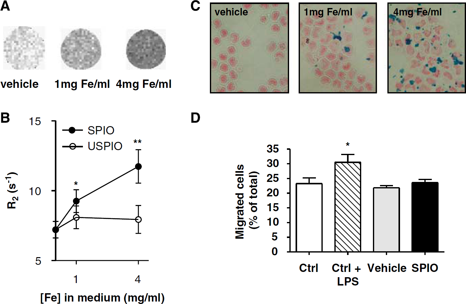
SPIO-labeled monocytes appear hypointense on T2-maps and their migratory capacity to cross a monolayer of brain endothelial cells is not affected. (
Prussian blue staining (Figure 1C) revealed the presence of intracellular iron clusters in cells incubated with SPIO, which were absent in cells incubated with USPIO. Semiquantitative analysis showed that incubation for 1.5 h with 4 mg Fe/mL SPIO resulted in iron-positive staining in 50% to 70% of the cells. Incubation of monocytes in concentrations over 4 mg Fe/mL SPIO resulted in the formation of large extracellular iron clusters (data not shown). Cell viability was unchanged (data not shown) and the migratory capacity of iron-labeled monocytes to cross a brain endothelial monolayer was not affected after incubation with 4 mg Fe/mL SPIO, since the numbers of migrated cells were at the same levels as for freshly isolated (ctrl) monocytes (Figure 1D).
Detection of SPIO-Labeled Monocytes after Intracerebral Injection
T2W images of rat brains after injection of SPIO-labeled monocytes into the brain parenchyma revealed a hypointense area at the injection site, which was absent in animals injected with unlabeled monocytes (Figure 2). With time, the hypointense area decreased but remained detectable after 5 days.
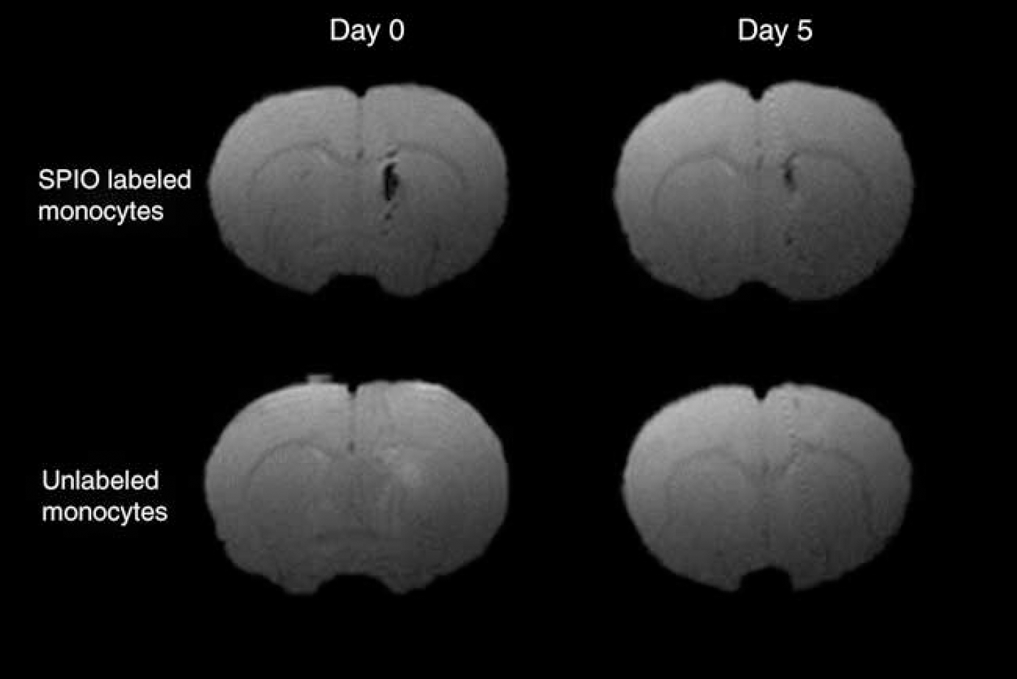
Intracerebral injection of SPIO-labeled monocytes resulted in strongly reduced signal intensity on T2W images. Immediately after injection (day 0), a large hypointense area was present at the injection site in the caudate putamen. After 5days, the hypointense area at the injection site had declined. After injection of unlabeled monocytes, a slightly hyperintense area was observed, most likely the result of the injected fluid.
SPIO-Labeled Monocytes are Detected in the Photothrombosis Lesion after Transfusion and Contrast Enhancement Differs from Intravenous USPIO Injection
To address monocyte infiltration upon neuroinflammation, SPIO-labeled monocytes were transfused in rats with a PT lesion. Contrast enhancement in the lesion after cell transfusion was compared with enhancement patterns in USPIO-injected rats and control rats. In the control group, lesions were induced but rats did not receive labeled cells or USPIO. Figure 3A shows coronal T2∗W images of brain sections through the center of the lesion. In rats transfused with SPIO-labeled monocytes, a well-defined area of signal loss was observed after 72 h but not at 24 h after transfusion. In contrast, the lesion in USPIO-injected rats revealed a large hypointense area 24 h after administration, which diminished after 72 h. Moreover, contrast enhancement was present in the vasculature at 24 h. In control animals, small signal voids in and around the lesion were detected at all time points.
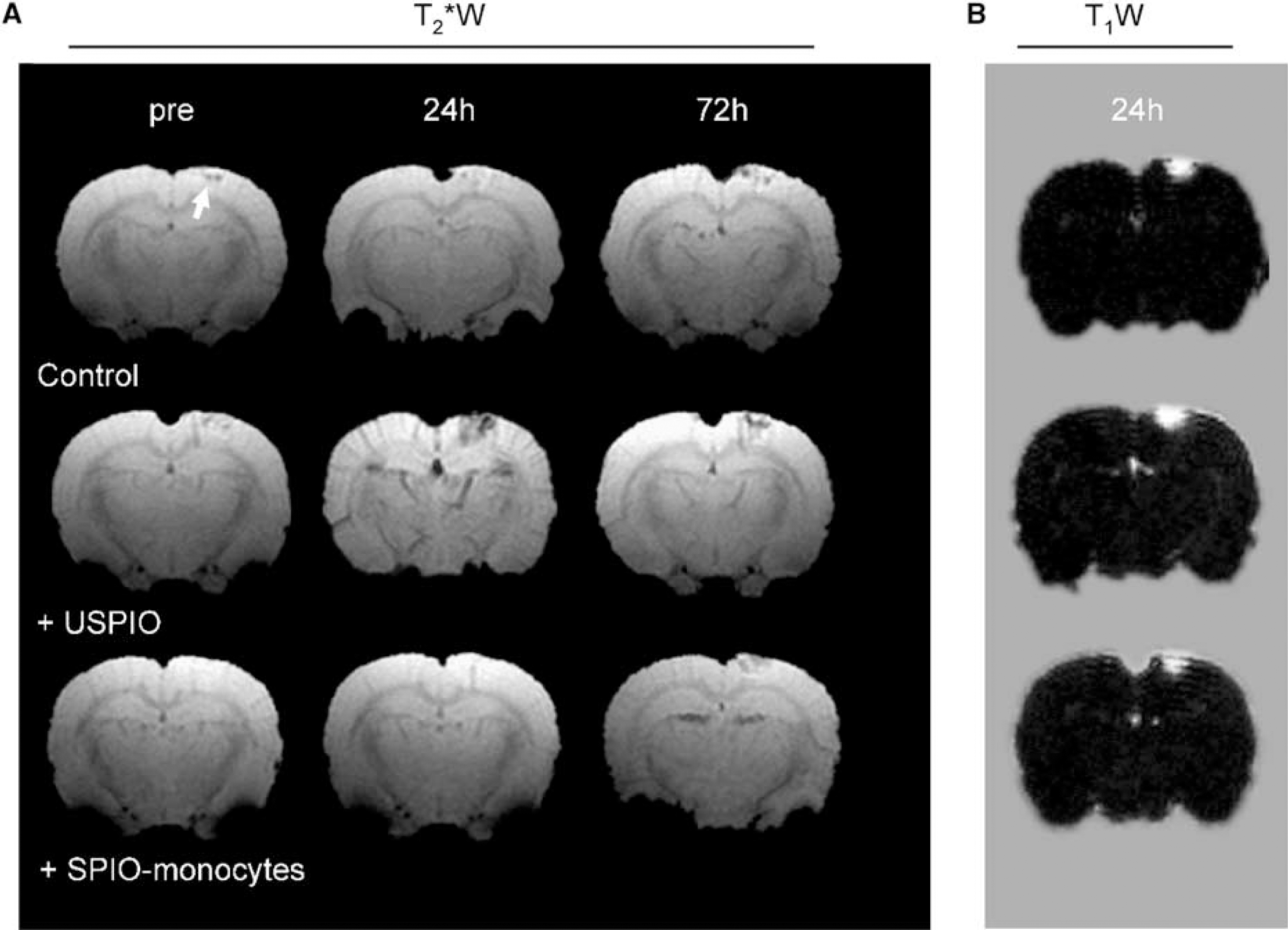
Transfusion of SPIO-labeled monocytes resulted in a different spatiotemporal enhancement pattern compared with injection of free USPIO. (
To assess BBB damage, animals received an injection of Gd-DTPA at the end of each scan session. T1W subtraction images at 24 h showed a clear positive signal that covered the total lesion size. The area of increased signal intensity was spatially similar to the area of signal loss 24 h after USPIO injection.
The area of iron oxide-induced contrast enhancement was quantitatively assessed by determining the number of hypointense voxels present within the lesion (Figure 4A) and was expressed as relative increase with respect to the preinjection image (Figure 4B). Figure 4A shows a T2∗W image 72 h after monocyte transfusion and the area within the lesion that was defined as hypointense. The contrast enhanced area slightly increased with time in control animals. In USPIO-injected rats, the number of contrast-enhanced voxels in the lesion increased up to 400% (407%±39%) at 24 h, which declined thereafter. In contrast, after transfusion of SPIO-labeled monocytes, there was a significant increase in the area of contrast enhancement after 72 h (340%±106%), which remained significantly elevated up to 120 h.
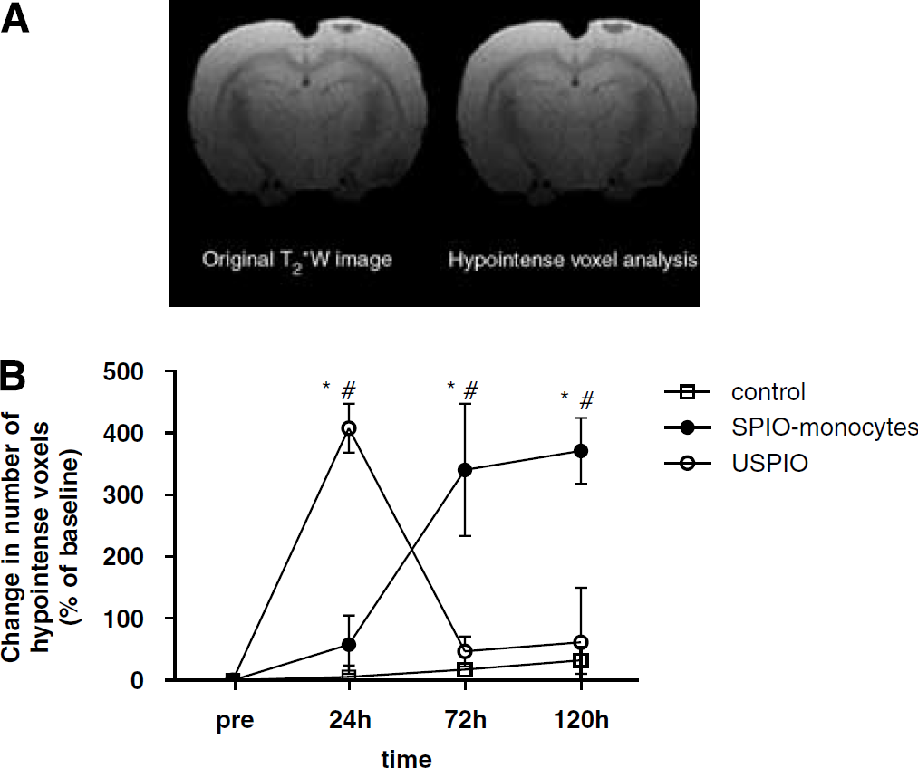
Temporal changes in contrast enhancement in the lesion. (
Immunohistochemical Analysis Shows Distinct Distribution of Iron Oxides in the Lesions after Transfusion of SPIO-Labeled Monocytes Compared with Free USPIO
Serial cryosections were analyzed to detect iron in the lesion and the presence of infiltrated monocytes (Figure 5). In all groups ED1-positive cells (monocytes) were observed throughout the lesion at 72 h. In subsequent sections, Prussian blue analysis revealed small iron-positive spots inside lesions of animals transfused with SPIO-labeled monocytes, reflecting infiltration of labeled monocytes. In contrast, in animals that received intravenous USPIO injections, Prussian blue stainings showed a large blue rim covering part of the lesion site.
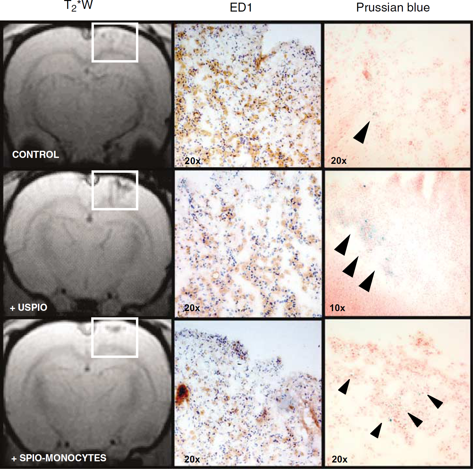
ED1+ cells and distinct patterns of iron accumulation are present throughout the lesion. Subsequent brain sections of animals killed at 72 h were stained either for infiltrated monocytes (ED1+ cells indicated by dark brown color) or iron (Prussian blue; presence of iron (blue) is indicated by arrowheads). The presence of ED1+ cells is shown for each group of animals. In USPIO-injected rats, a large iron-positive rim is present in the lesion. In contrast, in animals transfused with SPIO-labeled monocytes, the lesion showed a distinct pattern of iron-positive areas, reflecting the presence of infiltrated SPIO-labeled monocytes.
Time Course of Ultrasmall Superparamagnetic Iron Oxide Enhancement in Photothrombosis Lesion
To investigate the time course of contrast enhancement after USPIO injection in more detail, PT rats were scanned repeatedly from 0 up to 8 h after USPIO injection and again after 24 h. T2 maps (Figure 6A) revealed hypointense areas in the lesion site at an early stage, increasing in size up to 8 h. Loss of T 2 in the lesion (Figure 6B) was significantly different from that in the contra-lateral hemisphere 2 h after USPIO injection, with a maximum reduction at 6 to 8 h. The T 2 in the lesion was only partially restored after 24 h. The contralateral hemisphere showed a smaller decrease in T 2 , which remained constant over 0.5 to 8 h, indicating the presence of USPIO in circulation. Contralateral T2 was normal again after 24 h.
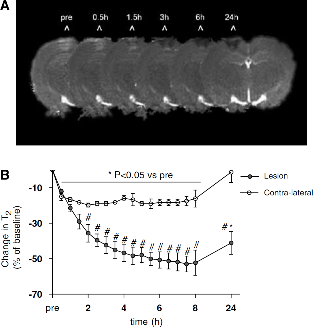
Contrast enhancement in the lesion differs from the contra-lateral site within 2 h after USPIO injection. (
Discussion
Our MRI study shows that monocyte trafficking toward areas of neuroinflammation can be monitored longitudinally and non-invasively using monocytes that are ex vivo labeled with iron oxides. Importantly, in a PT cortical lesion that is accompanied by massive monocyte infiltration several days after lesion induction, the enhancement pattern in the lesion differed from MRI signal changes after intravenous administration of free USPIO. Moreover, USPIO enhancement was already present in the lesion within 2 h after injection, whereas SPIO-labeled monocytes were visually detected after 72 h. This suggests that other mechanisms, such as BBB leakage or endothelial trapping, contribute to early USPIO enhancement in the brain.
Monocyte infiltration is a key event in ongoing inflammation and tissue destruction in many CNS pathologies (Nilupul Perera et al, 2006; Price et al, 2003; Stoll et al, 1998). To improve the efficacy of therapeutic compounds that limit cellular infiltration, it is important to elucidate the pattern of monocyte recruitment in the CNS. Ex vivo labeling of monocytes with iron oxides enables MR visualization and may provide an accurate and non-invasive tracking method.
We found that freshly isolated rat monocytes can be labeled ex vivo more efficiently using SPIO than USPIO, which is in line with our previous observations on human monocytes (Oude Engberink et al, 2007). Uptake of SPIO and the labeling procedure did not affect cell viability and the capacity of monocytes to cross a monolayer of brain endothelial cells. The low uptake of USPIO by monocytes compared with SPIO may be explained by the relatively small size of these iron oxides (30 nm), which excludes the endosomal pathway by receptor-mediated uptake (Raynal et al, 2004). In this study, we used USPIO rather than SPIO to investigate the effect of free iron oxide particles, as the blood pool half-life of USPIO (5 to 6 h in rats) is much longer compared with SPIO (less than 6 min) (Weissleder et al, 1989). Earlier studies have used intravenous administration of free USPIO and performed MRI 24 h later to study macrophage activity in vivo (Dousset et al, 1999a; Rausch et al, 2001; Weissleder et al, 1989). However, the pathway of free particle uptake and delivery into the brain parenchyma is not fully understood.
Therefore, we compared intravenous administration of free USPIO to transfusion of SPIO-labeled monocytes in rats with a PT lesion. Photochemically induced thrombosis elicits a strong inflammatory response resulting in a large area of monocyte infiltration, which occurs 5 to 8 days after lesion induction (Schroeter et al., 1997; Stoll et al, 1998). With MRI we detected the presence of SPIO-labeled monocytes in the lesion 72 h after transfusion. Histochemical analyses showed the presence of iron-positive cells in the lesion. However, MR images of control animals also showed small hypointense areas in the lesion that were present at all MR time points. These results are in line with previous studies (Saleh et al, 2004b) and may point to residues of erythrocytes accumulating in phagocytes that are clearing the lesion from debris. To overcome these confounds introduced by the PT model, we quantitatively analyzed the MR images and showed that 72 h after transfusion of SPIO-labeled monocytes signal changes in the lesion were significantly increased compared with control animals. Alternatively, in future studies the introduction of a fluorescent label should be considered as this may facilitate discrimination between nonspecific signal voids in the lesion from infiltrated SPIO-labeled monocytes. An earlier study has reported on MR tracking of labeled mononuclear cells into the ischemic brain of splenectomized mice (Stroh et al, 2006). In agreement with our results, the authors reported MRI signal changes in the brain within two to four days after cell transfusion. This relatively late contrast enhancement in the lesion is different from findings from earlier studies, in which free USPIO injection was applied to detect infiltration of monocytes (Dousset et al, 1999b; Saleh et al, 2004b). In these studies contrast enhancement was maximal 24 h after USPIO injection. To further address this discrepancy in the time course of contrast enhancement, we directly compared enhancement patterns after injection of free or intracellular iron oxide particles.
In contrast to transfusion of SPIO-labeled monocytes, the intravenous USPIO injection resulted in strong contrast enhancement at 24 h, which subsequently declined at 72 h. Previous studies in this animal model reported that such USPIO enhancements in a PT lesion are in spatial agreement with ED1+ areas (Saleh et al, 2004b; Schroeter et al, 2004), suggesting incorporation of USPIO into infiltrated monocytes. The time course of contrast enhancement has been described to correspond to the time window of monocyte influx (Kleinschnitz et al, 2003). In contrast, our study showed that the spatiotemporal profile of USPIO enhancement (24 h) differed from MRI signal changes after transfusion of SPIO-labeled monocytes (72 h).
So far, the use of free USPIO has been suggested to specifically image macrophage infiltration. Interestingly, in an earlier study, it was shown that after increasing the circulation time of free SPIO by elimination of blood-borne macrophages, similar contrast enhancement is observed as after free USPIO injection (Oweida et al, 2005). This supports the possibility that leakage of iron oxides over an impaired BBB contributes to contrast enhancement in the brain. This concept is also supported by our findings. We showed that contrast enhancement in the lesion increased significantly 2 h after USPIO injection and was maximally enhanced after 6 to 8 h. Moreover, we showed that Gd-DTPA enhancement, a conventional marker for BBB breakdown, was in spatial agreement with USPIO enhancement. Within the short time frame, it is unlikely that monocytes incorporate USPIO in circulation as the labeling capacity of USPIO is very low, as discussed earlier. Intravascular trapping of iron oxide particles, because of the presence of occluded vessels, may be an alternative explanation for the observed acute enhancement in the lesion. However, intravascular trapping of USPIO has only been observed in this model on intravenous administration of USPIO during lesion induction (Kleinschnitz et al, 2005). Previously, non-specific USPIO uptake was reported after permanent middle cerebral artery occlusion (Rausch et al, 2001; Wiart et al, 2007). In these studies, USPIO was intravenously injected 5 h after occlusion and the early signal enhancement observed in the lesion similarly supports the assumption that USPIO can penetrate a damaged brain area independent of monocyte/macrophage infiltration. In our study, USPIO were injected 5 days after lesion induction, reducing the possibility that USPIO are trapped in occluded vessels and most likely enter the brain parenchyma via transcytosis or passive leakage.
In conclusion, we have shown that cellular MRI can be used to specifically monitor homing of monocytes, labeled ex vivo with SPIO, toward areas of neuroinflammation in the rat brain. This strategy may provide an important tool for the evaluation of drugs that limit cellular infiltration into the CNS, thereby reducing neurological deficits. In addition, our study suggests that contrast enhancement after intravenous injection of free USPIO may not solely represent the migration of peripherally labeled cells, but may also be the result of leakage over a damaged BBB. This report may help to elucidate the heterogeneous enhancement patterns after USPIO administration for the assessment of inflammatory response in stroke and MS patients.
Footnotes
Acknowledgements
The authors thank Guerbet France for supplying the MR contrast agents.