
Abstract
Despite aggressive therapy, existing treatments offer poor prognosis for glioblastoma multiforme patients, in part due to poor penetration of most drugs across the blood–brain barrier (BBB). We propose a minimal-invasive combined treatment approach consisting of local BBB disruption in the tumor in parallel to systemic drug administration. Local BBB disruption is obtained by convection-enhanced delivery of a novel BBB disruption agent, enabling efficient/targeted delivery of the systemically administered drug by the tumors own vasculature. Various human serum albumin (HSA) analogs were synthesized and screened for BBB disruption efficacy in custom
INTRODUCTION
Although not highly prevalent, brain tumors are among the most lethal types of cancer. In the United States alone, more than 700,0 people are currently living with a primary brain or central nervous system (CNS) tumor diagnosis and it is reported that only 5% of diagnosed patients survive beyond 5 years (Parkin
One of the major obstacles in treating brain tumors is the blood–brain barrier (BBB) that restricts the penetration of most drugs into the brain/tumor tissue. Consequently, attempts have been made to administer systemically high doses of chemotherapeutics to reach therapeutic concentrations in the brain, resulting in systemic toxicity and serious adverse effects. 4 Although the BBB is compromised to some extent in malignant gliomas, making it more permeable for drugs, this disruption is not enough to enable therapeutic doses of systemically administered drugs to reach the tumor tissues. Moreover, the BBB in the infiltrating zone surrounding the tumor mass remains mostly intact restricting the penetration of drugs into these regions. For treatment to be effective, therapeutic drug doses should be delivered to the entire tumor and surrounding infiltrating zone, since survival of even a few cells may lead to cancer reoccurrence as typically takes place with high-grade gliomas. 5
Cationized albumin was found to induce BBB disruption by absorptive-mediated transendocytosis
6
when systemically administered in rats. Recently, we have confirmed these findings in an
Convection-enhanced drug delivery (CED) is a novel approach for direct delivery of therapeutic agents into brain tumors, obtained by delivering continuous infusion of substances via intracranial catheters, leading to convective distribution within the tissue. This approach yields efficient drug distributions at high concentrations in brain tumors, orders of magnitude higher than those obtained by systemic administration. 10 Despite the controversial results of initial clinical trials stemming from the complexity of this type of treatment, CED of therapeutic agents remains a promising strategy for treating malignant gliomas and is extensively studied. Multiple earlier stage trials have addressed only a fraction of the myriad of technical and technological issues accompanying this novel approach revealing its complexity. Development of CED has been further limited by the fact that both new technologies and novel therapeutic agents are being developed simultaneously. 11 Despite the understanding that the efficacy of CED is determined by its ability to provide significant volumes of drug distribution, utilization of real-time imaging of the drug distribution has only recently been applied to clinical studies. 12 The enormous potential of CED has been shown in these trials by depicting therapeutic concentrations distribution volumes in the range of 20 to 40 mL per implanted catheter.13–16 After determination of the specific challenges of this approach, new trials are being planned/initiated with computerized planning of catheter location, real-time imaging, novel therapeutic agents, and novel catheters that are expected to provide more reliable drug distributions. 11
Methotrexate (MTX), a folic acid analog widely used as a chemotherapeutic drug, was chosen in this study as an example of a potent antineoplastic agent which despite its effectiveness against glioma, has failed to provide clinical benefit due to its low brain penetrability. 17
The goal of this study was to develop a combined approach, consisting of inducing significant local BBB disruption in the tumor and surrounding infiltration zone in parallel to systemic delivery of a chemotherapeutic agent. Since high-grade gliomas are highly vascularized tumors,18,19 the presented approach uses the tumor's own vasculature for delivering the systemically administered drug to the target in the most natural way. In parallel, the BBB is disrupted efficiently and rapidly in the local vicinity of the tumor and infiltrating zone by local administration of the BBB-opening agent via CED.
Candidate BBB-opening agents were screened
MATERIALS AND METHODS
MATERIALS
Dulbecco's Modified Earl's medium (DMEM) was purchased from Gibco (Life Technologies, Carlsbad, CA, USA). Gentamicin, glutamine, new born calf serum, and penicillin/streptomycin were obtained from Biological Industries (Kibbutz Beit Haemek, Israel). Earl's Medium 199, Hoechst reagent, and hydrocortisone were purchased from Sigma (St Louis, MO, USA). For the immunocytochemistry, we used the following antibodies: mouse anti-occludin and zonula occludens-1 (ZO-1) (Zymed, Life Technologies). Cy and Alexa Fluor-conjugated secondary antibodies were acquired from Jackson Immunoresearch (West Grove, PA, USA) and Molecular Probes (Life Technologies), respectively, and used for immunocytochemistry. Alexa Fluor 488-conjugated phalloidin for the detection of F-actin was purchased from Molecular Probes. Unless otherwise mentioned, all other materials used were purchased from Sigma-Aldrich Israel Ltd (Rehovot, Israel).
Preparation of Cationized and Neutralized (Ethylamine) Human Serum Albumin
Human serum albumin (67 mg, 1 μmole) dissolved in 2.0 mL—containing 1 mol/L of 1.3 diaminopropane-2HCl, hexamethyldiamine-2HCl, Dicystamine-2HCl, argininamide-2HCl, or ethylamine-HCl. The pH was adjusted to pH 6.0±0.1. Solid EDC (100 mg, 526 μmoles) was then added, and the reaction was carried out with stirring for 4 hours at 25°C. The derivatives thus obtained were dialyzed against H2O for 2 days with several changes of H2O and lyophilized. In all analogs prepared by this procedure 45 to 85 carboxylate moieties of HSA (out of 99) were derivatized. This was quantitated by reacting an aliquot of each analog (~2 mg) with 1 mol/L glycinamide, excess EDC, in 8 mol/L urea. After dialysis, the additional glycine moieties were quantitated by amino-acid analyses after acid hydrolyses. The protein concentration was calculated according to alanine (62 residues) and valine (41 residues).
Media
Plating medium was composed of newborn calf serum (10%), L-glutamine (2 mmol/L), penicillin (100 units/mL), streptomycin (0.1 mg/mL), and gentamicin (0.1 mg/mL), all dissolved in Earl's Medium 199 (Sigma, St Louis, MO, USA). The assay medium consisted of L-glutamine (2 mmol/L), penicillin (100 units/mL), streptomycin (0.1 mg/mL), gentamicin (0.1 mg/mL), and hydrocortisone (550 nmol/L) in DMEM diluted 1:1 in Ham's F12 medium (Biological Industries).
Mono and Coculture
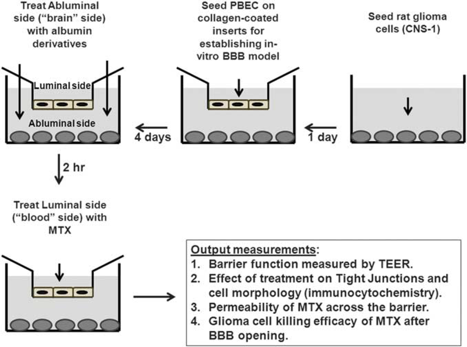
Schematic description of the blood–brain barrier reflecting
Permeability of Methotrexate In Vitro
To evaluate the efficacy of MTX to permeate the BBB, we used the
Immunocytochemistry
Porcine brain endothelial cells were grown on Transwell inserts for several days until confluence was reached (TEER >300 Ωcm2). After 2 hours treatment with EA–HSA (abluminal side, 11.2 μmol/L) cells were fixed with ice-cold 4% paraformaldehyde for 10 minutes at 25°C and exposed to a blocking solution (20% horse serum/0.1% Triton/phosphate-buffered saline) for 2 hours. The PBEC were then incubated with mouse antioccludin and rabbit anti ZO-1 antibodies at a 1:200 dilution, overnight at 4°C, washed with phosphate-buffered saline and stained with Cy3-labeled anti-rabbit or Alexa-Flour 488 anti-mouse secondary antibodies (1:200, 1 hour, room temperature). Nuclei were counterstained with Hoechst reagent for 20 seconds. After mounting (Aqua Poly/Mount), the inserts were observed and photographed using a BX43 Olympus fluorescent microscope with a DP73 Olympus camera (Olympus America Inc., Center Valley, PA, USA). Actin filaments were stained with Alexa Fluor 488-conjugated phalloidin (3 μL/insert, incubated together with the secondary antibody). Experiments were repeated at least three times.
Blood–Brain Barrier Disruption Evaluation in Normal Rat Brain—Experimental Outline
Several forms of cationized albumins and a neutralized analog (EA–HSA) of albumin were administered by CED into naïve rat brains under full anesthesia. The concentrations tested ranged from 10 μg/rat to 160 μg/rat and solvent used for the infusate varied. A list of the different derivatives, number of rats, and types of solvents are summarized in Supplementary Table 1 (Supplementary Information). The magnetic resonance imaging (MRI) contrast agent Gd-DOTA was administered intraperitoneally before CED (1 mmol/kg body weight) and BBB disruption was assessed by MRI shortly after administrating the BBB-opening agent by CED. A second MRI scan was performed on day 7 to assess possible tissue damage.
Efficacy experiment design: groups, treatments, and time line
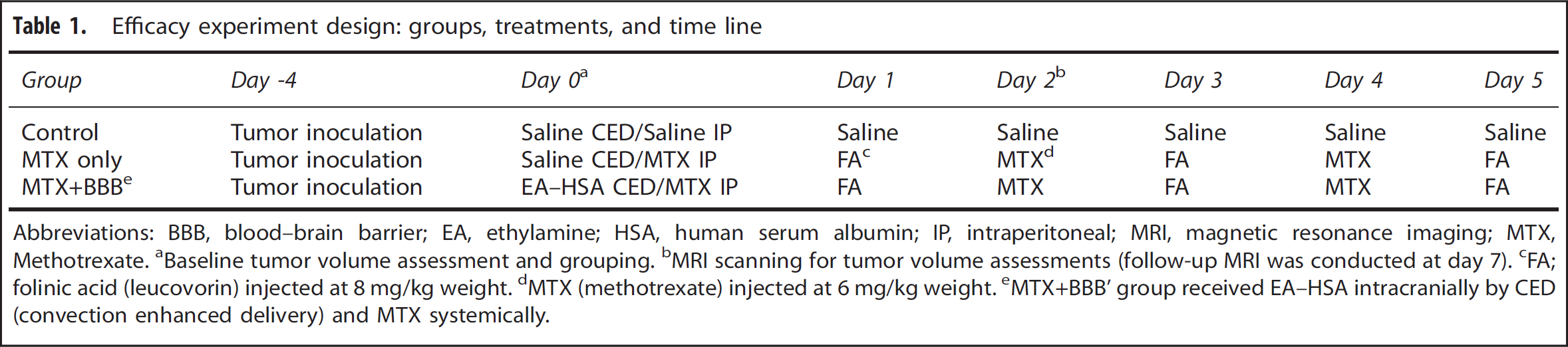
Abbreviations: BBB, blood–brain barrier; EA, ethylamine; HSA, human serum albumin; IP, intraperitoneal; MRI, magnetic resonance imaging; MTX, Methotrexate.
Baseline tumor volume assessment and grouping.
MRI scanning for tumor volume assessments (follow-up MRI was conducted at day 7).
FA; folinic acid (leucovorin) injected at 8 mg/kg weight.
MTX (methotrexate) injected at 6 mg/kg weight.
MTX+BBB’ group received EA–HSA intracranially by CED (convection enhanced delivery) and MTX systemically.
Convection Infusates for the Efficacy Study
To increase the distribution efficacy of the infusates, the viscosity of the solutions delivered by CED was increased by adding 10% sucrose. 24 The following solutions were prepared: Solution #1—10% sucrose in saline, Solution #2—EA–HSA [HSA-(CONH-C2H5)85] at 0.5 mg/mL and 10% sucrose in saline.
Efficacy Study in a Rat Glioma Model—Experimental Outline Glioma cells (rat CNS-1, 2 × 10⁵ cells) were intracranially inoculated in three groups of rats (
Animals and Ethics
The experiments were conducted according to the recommendations of the declarations of Helsinki and Tokyo and to the ARRIVE Guidelines for the use of experimental animals approved by the Animal Care Committees of Sheba Medical Center. Lewis male rats weighing 250 to 300 g (8 to 10 weeks, Harlan Biotech, Rehovot, Israel) were fed with Purina Chow and water available
Intracranial Tumor Inoculation
A midline scalp incision was made under general anesthesia to identify the bregma. A burr hole (1 mm) was then made on the right side, 3 mm anterior, and 2 mm lateral to the bregma. A 33-gauge needle attached to a 1000 μL syringe (Gastight; Hamilton, Reno, NV, USA) was placed 5.5 mm deep into the striatum. A pellet of 2 × 10⁵ CNS-1 rat glioma cells precipitated in 10 μL phosphate-buffered saline buffer was infused into the striatum using a BASI syringe pump at a rate of 2 μL/min over a period of 5 minutes. The burr hole was then sealed with bone wax to avoid the tumors from growing out of the skull.
Convection-Enhanced Drug Delivery Procedure A midline scalp incision was made under general anesthesia to identify the bregma. A burr hole (1 mm) was then made on the right side, 3 mm anterior, and 2 mm lateral to the bregma. For the tumor-bearing rats, the previously made burr hole was reopened. A 33-gauge needle attached to a 1,000-μL syringe was placed stereotactically 5.5 mm deep into the striatum. The infusion was carried out with a BASI syringe pump at a rate of 2 μL/min for a period of 20 minutes. The burr hole was then resealed with bone wax.
Magnetic Resonance Imaging Data Acquisition Rats were scanned under general anesthesia using a clinical GE 1.5T MRI system (Optima MR450w, General Electric, Milwaukee, WI, USA) with a clinical phased array knee coil and the following sequences: contrast-enhanced T1-weighted MRI for depiction of BBB disruption and assessing tumor volumes, T2-weighted MRI for assessment of early and late toxicity and gradient-echo MRI for depiction of possible hemorrhages. All sequences were acquired with a field of view of 10 × 7 cm, 256 × 224 pixels and a slice thickness of 1 mm. T1-weighted MR images were acquired with a fast spin-echo sequences, bandwidth of 15.6 kHz, echo time of 16 ms, and repetition time of 494 ms. T2-weighted MR images were acquired with a fast spin-echo sequence, bandwidth of 20 kHz, echo time of 85 ms, and repetition time of 4,639 ms. Gradient-echo MR images were acquired with a flip angle of 15°, a bandwidth of 15.63 kHz, echo time of 15 ms, and repetition time of 300 ms.
Calculation of Blood–Brain Barrier Disruption Volumes and Tumor Growth Rates
The volume (in mm3) of BBB disruption or enhancing tumor volume was calculated from the contrast-enhanced T1-weighted MR images. Regions of interest were defined over the entire enhancing region for each slice (excluding the ventricles). The number of pixels in the regions of interest was then counted and multiplied by the volume of a single pixel. Tumor growth rates were calculated by dividing the tumor volumes at days 2 and 7 after treatment with the baseline tumor volumes measured at day 0.
Survival
The three groups of rats were monitored daily for survival and killed when they lost > 20% of body weight and were unable to eat or drink.
Statistics
Statistics were calculated using Prism version 4.0 (GraphPad Software, CA, USA). Student's
RESULTS
Engineering a ‘Brain-Cancer Related’
To fit with this study, we further modified this system to a model defined by us as the ‘brain-cancer-related-three component
Screening Albumin Analogs for Inducing Blood–Brain Barrier Disruption In Vitro and In Vivo
A family of cationized albumins was prepared and optimized
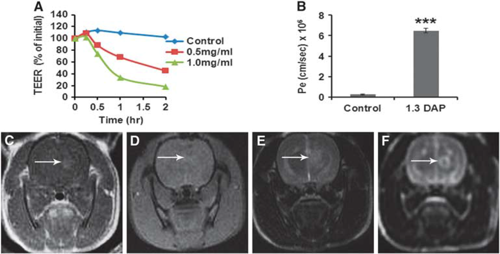
All cationized albumins were subsequently studied
An exception was an analog of HSA in which the carboxylates were derivatized with EA, turning them into noncharged residues. This neutralized N-hexyl amidated albumin analog [HSA-CONH(C2 H5)85], which we termed EA–HSA, was found suitable in all respects. It reduced TEER values of PBEC-M in a dose and time-dependent manner and yielded a Pe value for the penetration of MTX of 11.74±1.3 × 10⁻⁶cm/s which is a 48-fold increase over control (Figures 3A and 3B). In addition, EA–HSA showed a similar pattern of dose-dependent decrease in TEER with slower kinetics attributed to the buffering capacity of the astrocytes when applied at the abluminal side in the contact coculture
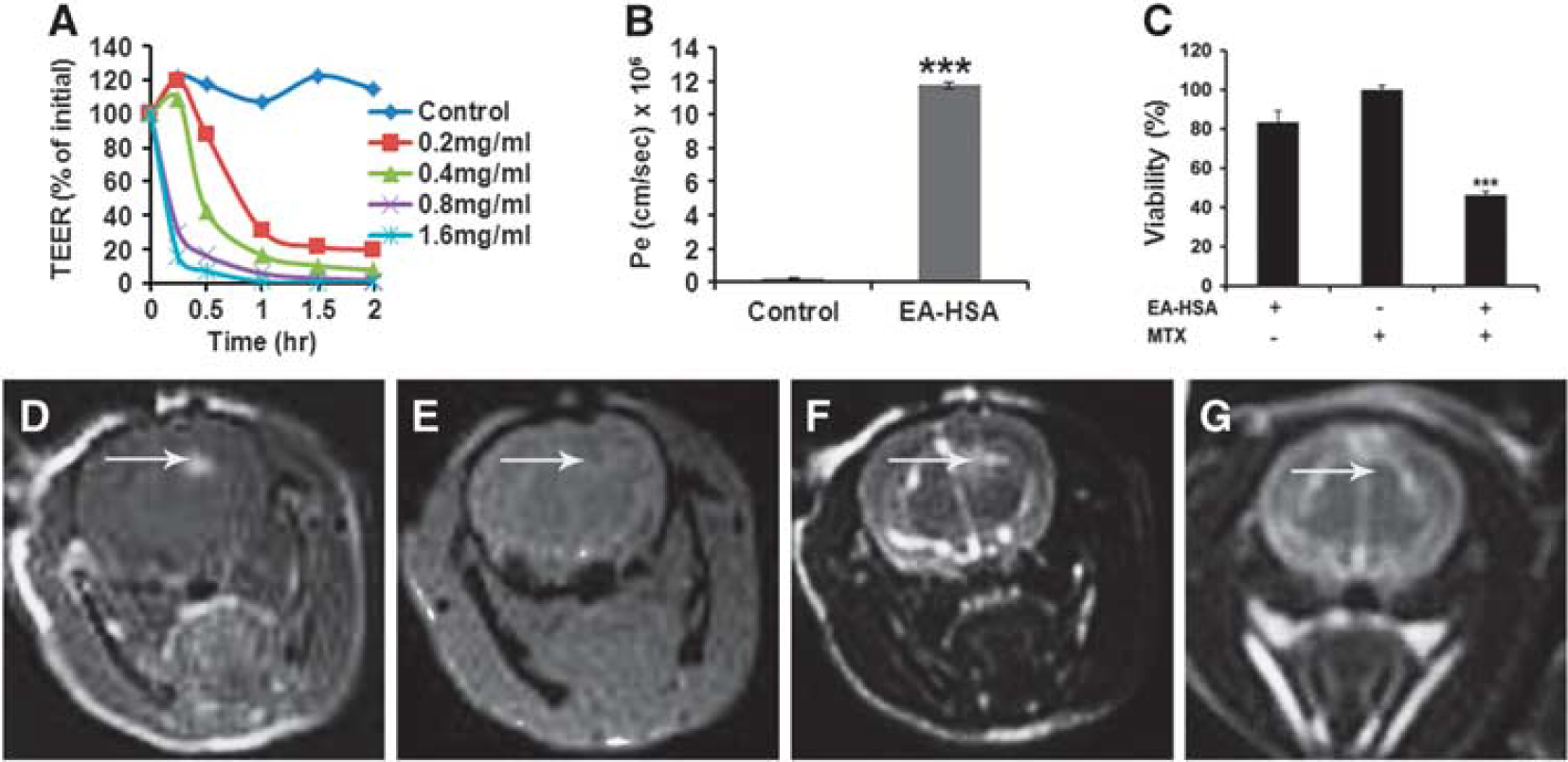
The antineoplastic efficacy of this analog against glioma cells located at the brain side was validated in the ‘brain cancer-related’ experimental system (Figures 1 and 3C). Therefore, EA–HSA at a concentration as low as 5.6 μmol/L, reduced TEER by 90% to 98% within a short period (Figure 3A) thus enabling MTX entry at a sufficient rate (Figure 3B) to yield 50% abluminal located glioma cell killing, within a period of 48 hours (Figure 3C).
Ethylamine–human serum albumin passed the
Alterations in the Porcine Brain Endothelial Cells Monolayer after Treatment With Ethylamine–Human Serum Albumin Figures 4 and 5 show typical results of our immunocytochemical studies, which aimed to identify alterations in tight junction (TJ)-related membrane protein(s). These studies were performed at a stage when TEER has been reduced by EA–HSA to a level permitting the paracellular passage of impermeable substances.
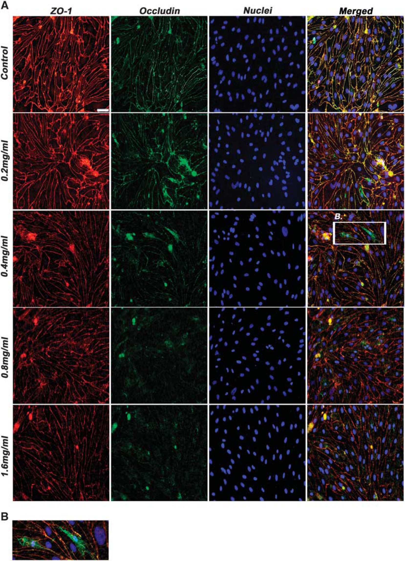
Expression of tight junction proteins after exposure to EA–HSA. (
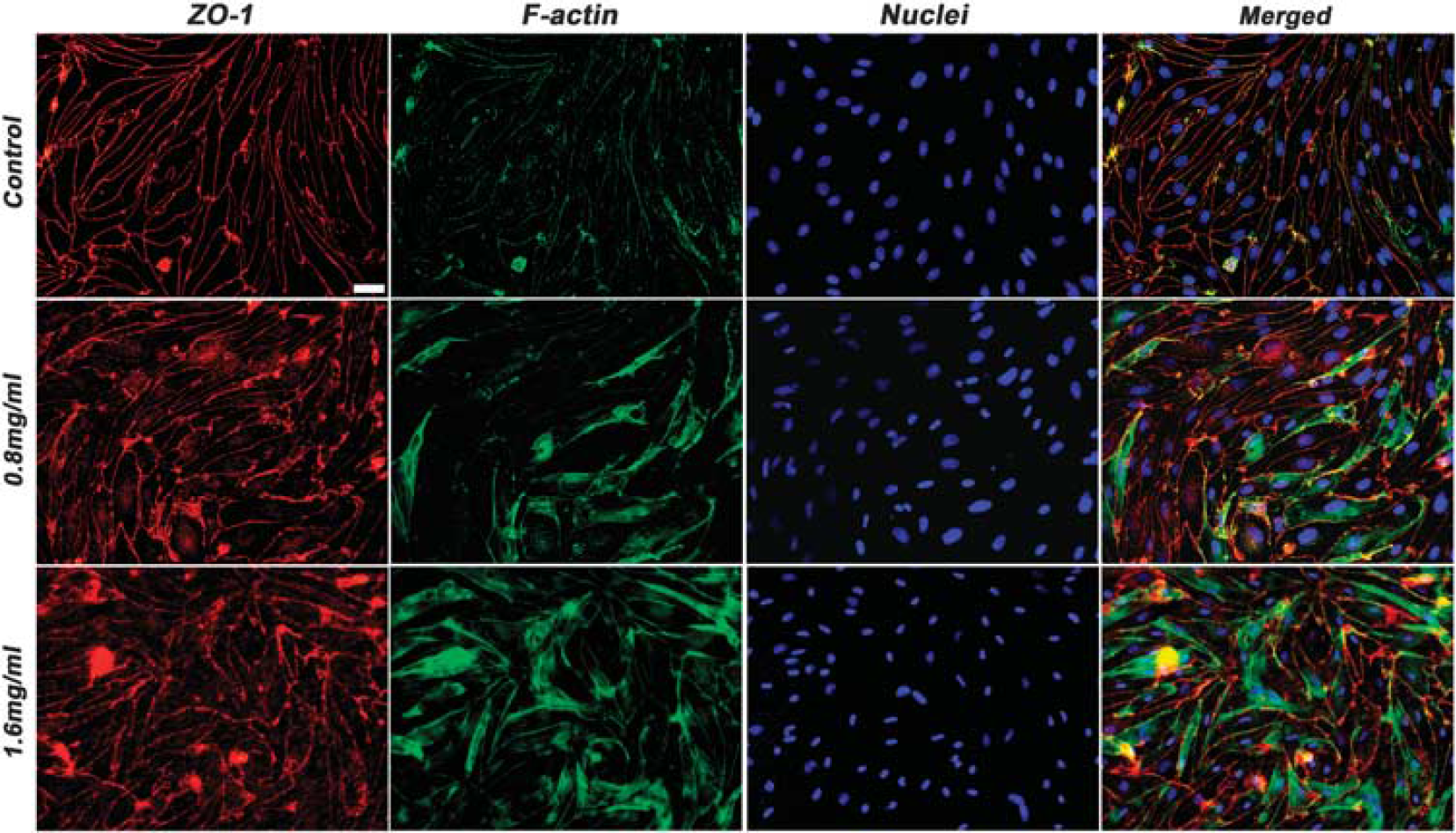
Stress fibers formation after exposure to EA–HSA. Porcine brain endothelial cells monolayer (PBEC-M) inserts were treated with 11.2 pmol/L EA–HSA at the abluminal side for 2 hours. Immunostaining of zonula occludens-1 (ZO-1) and actin filaments was then performed. Nuclei were counterstained with Hoechst reagent. Representative pictures are displayed from four different experiments. Bar 20 pm. EA, ethylamine; HSA, human serum albumin.
Occludin and ZO-1 act as the membrane cell to cell connecting proteins and the assembly of the TJ proteins to the cytoskeleton, respectively.25,26 Occludin is a major TJ protein responsible for the blockade of paracellular passage of molecules. Figure 4 shows that indeed the expression of this protein was significantly altered after incubation with EA–HSA while the expression pattern of ZO-1 was only slightly altered. Occludin has migrated from its location at the cell borders into the cytoplasm and degraded there (Figures 4A and 4B). zonula occludens-1, however, preserved its membrane location and showed minor alterations only at high concentrations of this BBB disrupting agent (Figure 4A).
Actin reorganization has an important role in the cells structural support and may also have an active role in the formation and maintenance of TJ expression and patterns of distribution. 27 To examine whether actin fibers have a role in the process of EA– HSA-induced BBB opening, we treated PBEC-M at the abluminal side with increasing concentrations of EA–HSA. Cells were immunostained for ZO-1 and for fibrous actin. As shown in Figure 5, disorganization of actin filaments did take place to a certain extent. Thus, the observed reduced PBEC-M tightness and the passage of impermeable agents such as MTX (Figures 3A and 3B) appear to be manifested by disruption of occludin that is followed or accompanied by the reorganization of actin.
Overcoming Systemic Toxicity of Peripherally Administered Methotrexate in the Naive Rat Model
Initially, we have attempted to administer MTX
Combined Convection-Enhanced Drug Delivery of Ethylamine–Human Serum Albumin and Methotrexate Therapy Suppressed Tumor Growth and Prolonged Survival in Rats Bearing Intracranial Glioma Tumors Figure 6 summarizes the outcome of the combined treatment in the glioma rat model. In this model, sham-treated rats showed increased average tumor volumes of 2.4±0.3 folds within the first 2 days and 12.8±2.8 folds within 1 week after CED. Systemically administered MTX alone did not facilitate significant effect in suppressing tumor growth rate in the first 2 days after treatment (mean values 2.2±0.4 and 2.5±0.3 for the MTX-treated and control groups, respectively, Figure 6A). Methotrexate alone however facilitated a significant effect between days 2 and 7 (mean values 2.2 ±0.4 and 12.8±1.5 in the MTX and the control groups, respectively, Figure 6B).The combined treatment of systemic MTX together with CED of EA–HSA fully suppressed tumor growth, leaving it nearly at the volume measured at day 0 (0.9±0.1 and 1.0 ± 0.1 for day 2 and day 7, respectively). The effect of MTX
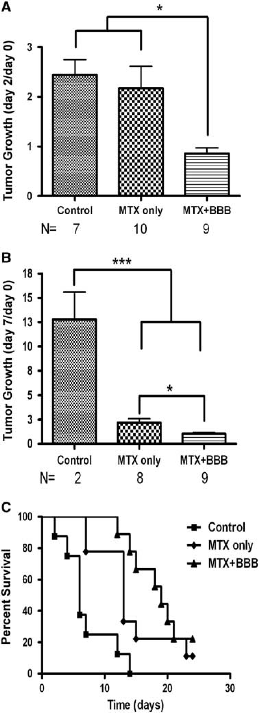
Rates of tumor growth and survival after the combined EA–HSA-MTX therapy in the glioma rat model. Tumor growth rates (A, B) for the three treatment groups: Control (sham treated,
In agreement with the above findings, survival of the rats that received the combination therapy was significantly extended. Median survival times reached 5, 12, and 19 days for the control, MTX-treated, and the combination-treated groups, respectively (Figure 6C).
The CED administration of a neurtralized HSA analog without parallel systemic treatment of MTX showed no beneficial effects over control (Supplementary Results).
Leukocytes Transmigration Across Blood–Brain Barrier after Ethylamine–Human Serum Albumin Treatment To study whether BBB disruption induced by EA–HSA allows for increased leukocyte migration over the BBB, thus potentially increasing the antineoplastic effects of the combined treatment, we studied such migration with/without EA–HSA treatment using our
DISCUSSION
In spite of aggressive therapy, existing treatments offer poor prognosis for glioblastoma multiforme (GBM) patients. The reasons for limited therapeutic effects include tumor infiltration into the surrounding brain parenchyma as well as poor penetration of most therapeutic agents across the BBB. In addition, GBM cells are highly resistant to therapeutic apoptotic stimuli. However, they exhibit a paradoxical propensity for extensive cellular necrosis.28,29 Due to the extreme adaptability of GBM cells, residual tumor, including the infiltrating zone surrounding the tumor, should be treated with high efficacy as well, to prevent sublethal hits of tumor cells leading to the growth of more malignant clonal cell populations. 30
Blood–brain barrier disruption obtained by intracarotid and intraarterial injections of high concentrations of Mannitol has been studied extensively by the BBB disruption (BBBD) consortium initiated by Prof Neuwelt from Ohio State University. 31 This type of therapy indeed showed promising results mainly in patients with CNS lymphoma but less in patients with GBM. We believe that the results of the BBBD consortium indeed show the potential of BBBD for the treatment of brain tumors. Still, a major limitation of BBBD by intracarotid/intraarterial administration of the BBB disruption agent may be that BBBD obtained by this method covers large regions of the vascular tree in the treated hemisphere with no specific targeting to the tumor region. 32 We believe that the combined approached presented in our manuscript may overcome this limitation since targeting of the tumor and surrounding infiltrating zone is obtained by implanting the source of the BBB disruption agent within the tumor mass. Using this approach, the BBB disruption agent is efficiently delivered to the tumor mass and surrounding infiltrating zone with limited effects to more distant brain regions.
To overcome some of these challenges, we propose a minimal invasive combined treatment approach for GBM consisting of local disruption of the BBB in the tumor and infiltrating zone in parallel to systemic drug administration. Since gliomas are highly vascular tumor, 18 we hypothesized that by applying the disrupting agent to the tumor mass, efficient BBB disruption will be induced in the tumor mass as well as in the infiltrating zone, thus enabling efficient delivery of the systemically administered therapeutic agent by the tumors own vasculature to the target regions. To obtain maximal BBB disruption at the vicinity of the tumor and surrounding infiltrating zone, we administered the EA–HSA by CED. Convection-enhanced drug delivery is advantageous both in allowing efficient delivery of high EA–HSA concentrations over large volumes of the target tissue and in fully avoiding systemic toxicity.
We hypothesize that the overall volume of BBB disruption should be larger than the volume of infusate distribution due to leakage of the BBB-opening agent into the tumor vascular. This way, the EA–HSA may be further carried by the tumor vasculature into the infiltrating zone, enabling efficient penetration of the systemically administered therapeutic agent into further regions of infiltrating tumor. To test this hypothesis, further studies are required.
Taking into account these considerations, the study was designed to determine an efficient and safe BBB-opening agent and establish the added value of the combined approach for the treatment of brain tumors. The study was carried out in four stages: (1) Chemical synthesis of a variety of candidate BBB opening albumin derivatives, (2)
The chemotherapeutic agent a priory selected was MTX, a folic acid analog used as a chemotherapeutic drug. As folate receptors are over expressed on the cell membranes of many types of cancer cells, MTX is one of the most widely used drugs for the treatment of many forms of cancer, including tumors of the brain, breast, ovaries, and several leukemias. However, MTX is limited by its low solubility, dose-related toxicity, lack of selectivity, rapid diffusion throughout the body, short half-life in the bloodstream, drug resistance by target cells and low BBB penetration. 17 The combined approach showed here may provide means for overcoming these common limitations thus taking advantage of potent drugs such as MTX for obtaining significant treatment effects in GBM patients.
To proceed to the animal efficacy experiment
The results of the efficacy study in the rat glioma model showed that despite the rapidly growing tumors in the control group, the combined therapy completely suppressed tumor growth and significantly prolonged survival by nearly a factor of 3 compared with control. Interestingly, systemically administered MTX seemed to show no therapeutic effect in the first 2 days after treatment (no significant change in tumor growth versus control) while the combined approach showed significant antitumor effects (complete arrest in tumor growth). Later on, 7 days after treatment, systemically administered MTX did show therapeutic benefits, although still significantly lower than those obtained by the combined approach. This delayed effect of MTX may be explained by increased BBB disruption as the tumor matures or by an accumulated effect on the tumor vasculature induced by repeated treatments with MTX. It also suggests that in the group receiving the combination therapy, where the tumor failed to grow, MTX entry was highly dependent on the BBB-opening efficacy of EA-HSA. To improve treatment efficacy, it may be possible to repeat the CED treatment (using the same implanted catheter) more than once.
From a cellular mechanistic point of view, the mode of action by which EA–HSA induces BBB opening was investigated in a set of immunocytochemical studies. We have shown that out of the several transmembrane and cytosolic TJ proteins responsible for connecting neighboring endothelial cells to each other, occludin expression was particularly altered upon incubation of PBEC-M with EA–HSA. Zonula occludens-1 is a scaffolding protein responsible for the linkage between the intracellular actin cytoskeleton and the outer membrane TJ's proteins (claudin-5, occludin). This interaction is postulated to provide additional rigidity to the structures and allow for rapid alterations in barrier integrity in response to a variety of stimuli.
34
One can assume that the relatively unchanged ZO-1 may be explained by the cells attempt to maintain BBB functionality under the stress induced by the EA-HSA, also manifested by the formation of stress fibers. It should be mentioned however that increased leakiness of the BBB is not always correlated with visible changes in major structural component of TJ.
35
Although occludin trafficking away from TJ's complexes was found to be a sensitive, early, and reliable sign for TJ opening and BBB disruption in osmotically-affected rat BBB,
36
Youakim
An important aspect of disrupting the BBB is the possible induction or enhancement of an immune response, which may contribute significantly to the antineoplastic effects of the treatment. It has been previously shown that significant BBB disruption, observed after acute traumas such as head trauma or stroke, was accompanied by immune cells that were recruited to the inflamed/damaged area inducing significant immune response (NK, neutrophils, monocytes, and other types of leukocytes. For example: stroke;
39
and traumatic brain injury
40
). Our observations that leukocytes migration across the
In summary, the results of this study present a new compound, EA–HSA, for inducing efficient, transient and safe local BBB disruption that may be explained by alterations in occludin expression.
In addition, the study showed the feasibility of using this analog in a combined minimal-invasive treatment approach for inducing significant treatment effects reflected by tumor growth arrest and prolonged survival in a rat glioma model.
DISCLOSURE/CONFLICT OF INTEREST
The authors declare no conflict of interest.
Footnotes
References
Supplementary Material
Please find the following supplemental material available below.
For Open Access articles published under a Creative Commons License, all supplemental material carries the same license as the article it is associated with.
For non-Open Access articles published, all supplemental material carries a non-exclusive license, and permission requests for re-use of supplemental material or any part of supplemental material shall be sent directly to the copyright owner as specified in the copyright notice associated with the article.