
Abstract
Hyperlipidemia is a major cardiovascular risk factor associated with progressive cerebrovascular dysfunction and diminished collateral perfusion in stroke. Rho-associated kinase (ROCK) may be an important mediator of hyperlipidemic vascular dysfunction. We tested the efficacy of acute or chronic ROCK inhibition on the size of dynamic perfusion defect using laser speckle flowmetry in hyperlipidemic apolipoprotein E knockout mice fed on a high-fat diet for 8 weeks. Mice were studied at an age before the development of flow-limiting atherosclerotic stenoses in aorta and major cervical arteries. Focal ischemia was induced by distal middle cerebral artery occlusion (dMCAO) during optical imaging. The ROCK inhibitor fasudil (10 mg/kg) was administered either as a single dose 1 hour before ischemia onset, or daily for 4 weeks. Fasudil decreased both baseline arterial blood pressure and cerebrovascular resistance (CVR) by ∼15%, and significantly improved tissue perfusion during dMCAO. Interestingly, peri-infarct depolarizations were also reduced. Chronic treatment did not further enhance these benefits compared with acute treatment with a single dose. These data show that ROCK inhibition improves CVR and ischemic tissue perfusion in hyperlipidemic mice.
Keywords
INTRODUCTION
Hyperlipidemia is among the most prevalent vascular risk factors associated with stroke. Vascular endothelial and smooth muscle dysfunction has been shown in hyperlipidemic animal models as well as in patients.1, 2 In cerebral vasculature, hyperlipidemia impairs endothelium-dependent relaxations, 3 diminishes resting perfusion, and blunts major vasodilator reflexes, such as neurovascular coupling, hypercapnic hyperemia, and autoregulation. 4 We have recently shown that hyperlipidemic vascular dysfunction is associated with markedly larger perfusion defects on focal cerebral arterial occlusion. 4
A large body of evidence suggests that upregulation of Rho-associated kinase (ROCK) activity is one mechanism by which hyperlipidemia leads to vascular dysfunction.5, 6, 7, 8 Numerous studies have also shown that ROCK inhibition improves stroke outcome in otherwise normal animals, 9 and that the mechanism involves endothelium-dependent improvement in ischemic tissue perfusion presumably by augmenting the collateral blood supply. 10 Because hyperlipidemia is associated with upregulation of ROCK activity, we tested the efficacy of ROCK inhibitors in improving cortical perfusion on focal arterial occlusion in hyperlipidemic apolipoprotein E knockout (ApoE KO) mice.
MATERIALS AND METHODS
All experimental procedures were performed in accordance with the Guide for Care and Use of Laboratory Animals (NIH Publication No. 85-23, 1996), and were approved by the institutional review board (MGH Subcommittee on Research Animal Care, SRAC). Male ApoE KO mice (Jackson Laboratories, Bar Harbor, ME, USA) were housed under diurnal lighting conditions and allowed food and tap water ad libitum. Mice were fed western-type high-fat diet (42% of total calories from fat and 0.15% cholesterol; Harlan-Teklad, Indianapolis, IN, USA) for 8 weeks starting at 4 weeks of age. At 12 weeks of age, mice were randomized to receive vehicle (saline, 0.1 mL/day for 4 weeks; n=6), or fasudil (10 mg/kg; Tocris Bioscience, Bristol, UK) either as a single dose 1 hour before ischemia (n=6), or daily for 4 weeks (n=8). In a smaller group, we also tested hydroxyfasudil, the active metabolite of fasudil, administered 1 hour before ischemia onset (10 mg/kg, n=5). All drugs were administered via intraperitoneal injection. In chronic groups, treatment started after 4 weeks of high-fat diet, and last dose was administered 1 hour before ischemia onset.
Mice were anesthetized with isoflurane (2% for induction, 1% for maintenance, in 70% N2O and 30% O2) and intubated via a tracheostomy. The depth of anesthesia was adjusted to suppress cardiovascular responses to the tail pinch. Femoral artery was catheterized for the measurement of mean arterial blood pressure (MABP; ETH 400 transducer amplifier; CB Sciences, Inc., Milford, MA, USA). Rectal temperature was kept at 37°C using a thermostatically controlled heating mat (FHC, Brunswick, ME, USA). Mice were then paralyzed (pancuronium bromide, 0.4 mg/kg per hour, intraperitoneally), mechanically ventilated (SAR-830; CWE, Ardmore, PA, USA), and placed on a stereotaxic frame (David Kopf, Tujunga, CA, USA). Arterial blood gases and pH were measured every 20 minutes (Corning 178 blood gas/pH analyzer, Ciba Corning Diagnostics, Medford, MA, USA) and ventilation adjusted to maintain them within physiologic range. The data were continuously recorded using a data acquisition and analysis system (PowerLab, AD Instruments, Medford, MA, USA) and stored in a computer.
Focal cerebral ischemia was induced by distal middle cerebral artery occlusion (dMCAO) as previously described. 11 After general surgical preparation, mice were placed in a stereotaxic frame, and skull surface was prepared for optical imaging. Temporalis muscle was separated from the temporal bone and removed, and a burr hole (2 mm diameter) was drilled under saline cooling in the temporal bone overlying the middle cerebral artery just above the zygomatic arch. The dura was kept intact. Cortical spreading depression accidentally triggered during the craniotomy, as detected by the appearance of the characteristic long-lasting oligemia, was an a priori exclusion criterion, but did not occur in this cohort. Mice were allowed to stabilize for 30 minutes after surgical preparation before MCAO. A steel microvascular clip with sharpened tips was gently placed to occlude the middle cerebral artery branch over the lateral cortical convexity just distal to the base of the zygomatic arch and the inferior cerebral vein.
Laser speckle flowmetry was used to study the spatiotemporal characteristics of cerebral blood flow (CBF) changes starting 5 minutes before dMCAO and continuing until 60 minutes after occlusion. The technique for laser speckle flowmetry has been described in detail elsewhere. 11 Cortical perfusion defect was quantified two dimensionally by calculating the area (mm2) of the hypoperfused cortex with residual CBF 20%, 30%, or 40% of the preischemic baseline using a thresholding paradigm. The impact of peri-infarct depolarizations (PIDs) on ischemic perfusion defect was calculated as % change in the area of cortex with residual CBF30% from immediately before a PID to just after its recovery, as described in detail previously.12, 13, 14 In addition, using the preischemic speckle contrast images we determined the τc−1 values as a measure of resting CBF, as described previously,4, 15 and calculated the resting cerebrovascular resistance (CVR) using the formula CVR (%)=MABP/CBF, where both MABP and CBF were expressed relative to the vehicle group. In a separate cohort of ApoE KO mice treated with vehicle or fasudil for 4 weeks (n=3 each), we determined the plasma lipid profile (Table 1).
Plasma lipid profile and systemic physiologic parameters in 12-week-old ApoE KO mice on high-fat diet for 8 weeks
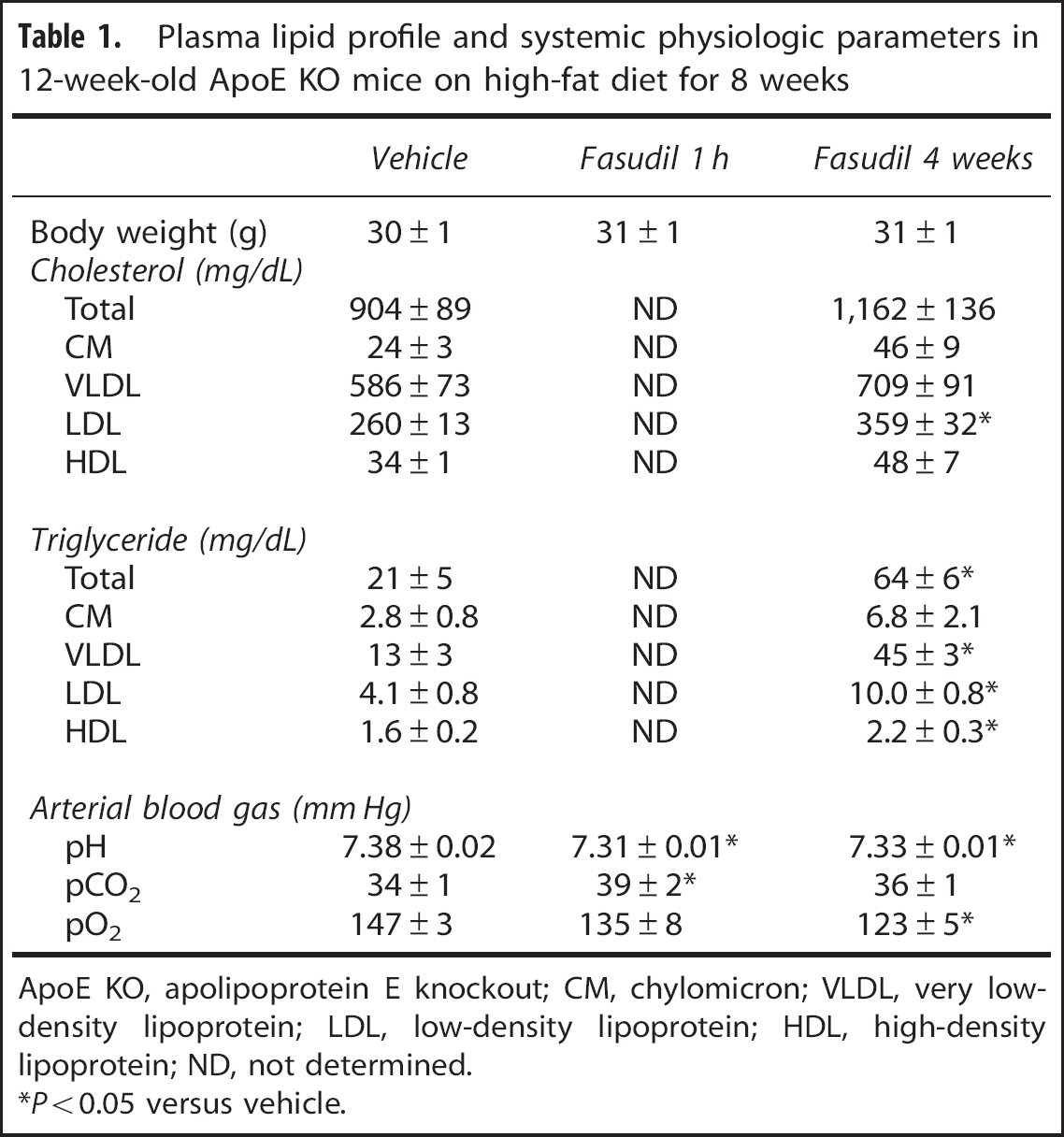
ApoE KO, apolipoprotein E knockout; CM, chylomicron; VLDL, very low-density lipoprotein; LDL, low-density lipoprotein; HDL, high-density lipoprotein; ND, not determined.
∗P<0.05 versus vehicle.
Data were expressed as mean±standard error, or box-whisker plots in which median, 25% to 75% and full ranges, and the mean (+) were shown. Statistical comparisons were performed using two-way ANOVA followed by Tukey's test to correct for multiple comparisons. P<0.05 was considered as statistically significant.
RESULTS
Distal occlusion of the middle cerebral artery created a focal cortical hypoperfusion over the dorsolateral hemisphere (Figure 1A). Fasudil reduced the area of perfusion defect by approximately half (Figure 1B). Treatment with a single dose 1 hour before ischemia was as effective as chronic daily treatment for 4 weeks. Hydroxyfasudil, the active metabolite of fasudil, also decreased the perfusion defect when administered as a single dose (10 mg/kg) 1 hour before ischemia onset, with an efficacy comparable to fasudil (1.9±0.4, 4.5±0.7, and 8.4±0.8 mm2 for 20%, 30%, and 40% residual CBF thresholds, respectively; P<0.05 versus vehicle; n=5).
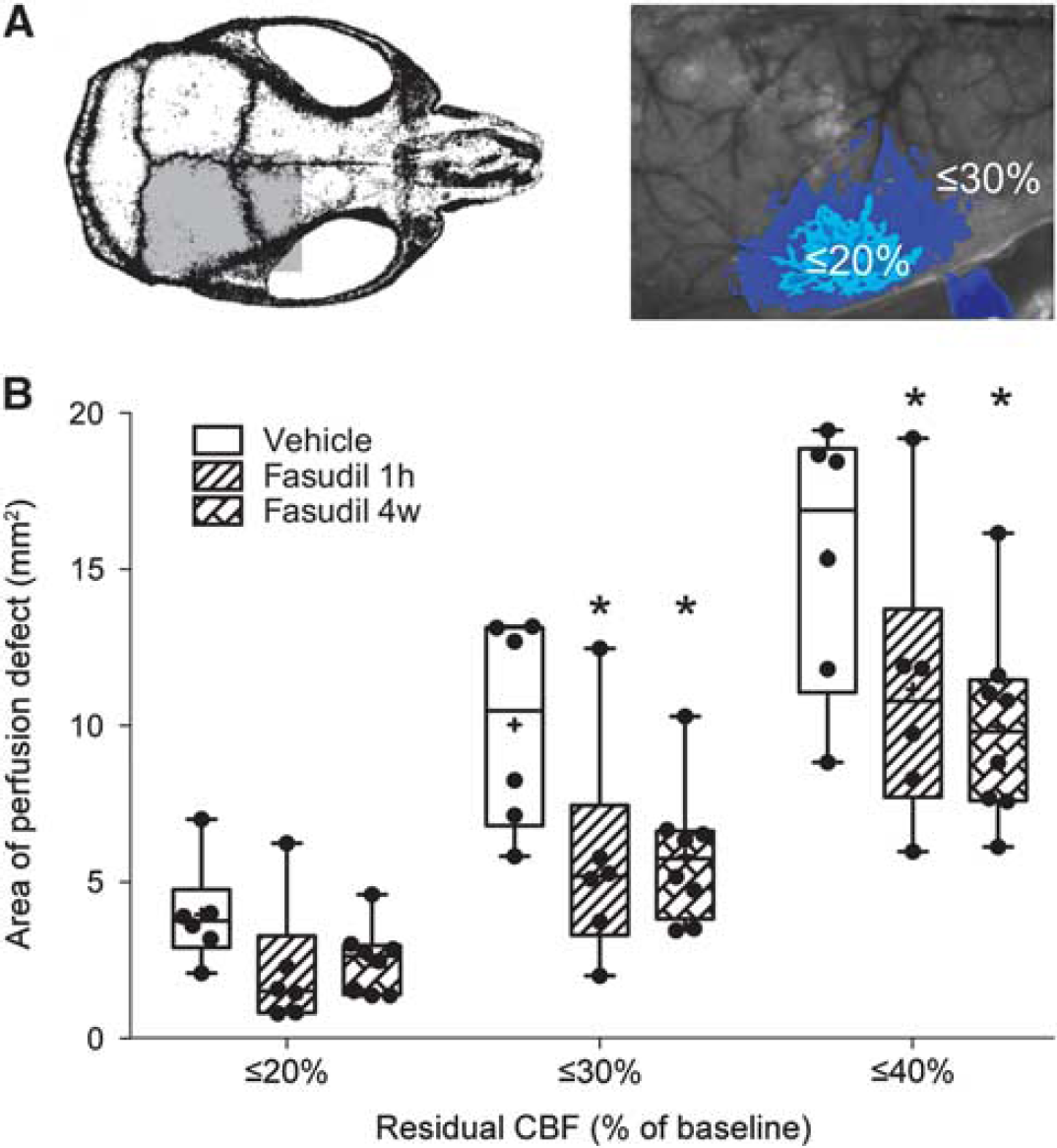
Effects of fasudil on ischemic tissue perfusion after distal middle cerebral artery occlusion (dMCAO) in hyperlipidemic apolipoprotein E (ApoE) knockout mice. (
One mechanism that could account for improved ischemic perfusion was cerebral vasodilation. We assessed this by measuring resting (i.e., preischemic) CBF in dorsal cortex using laser speckle contrast images, and found no difference among groups (Figure 2). However, we also noted that improved ischemic tissue perfusion occurred despite 10% to 15% lower resting MABPs in fasudil-treated mice compared with the vehicle group (Figure 2). When calculated from resting MABP and CBF values, resting CVR values were 10% to 15% lower in fasudil-treated groups (Figure 2). Once again, 4 week daily treatment did not enhance this effect compared with a single dose administered just 1 hour before ischemia onset.
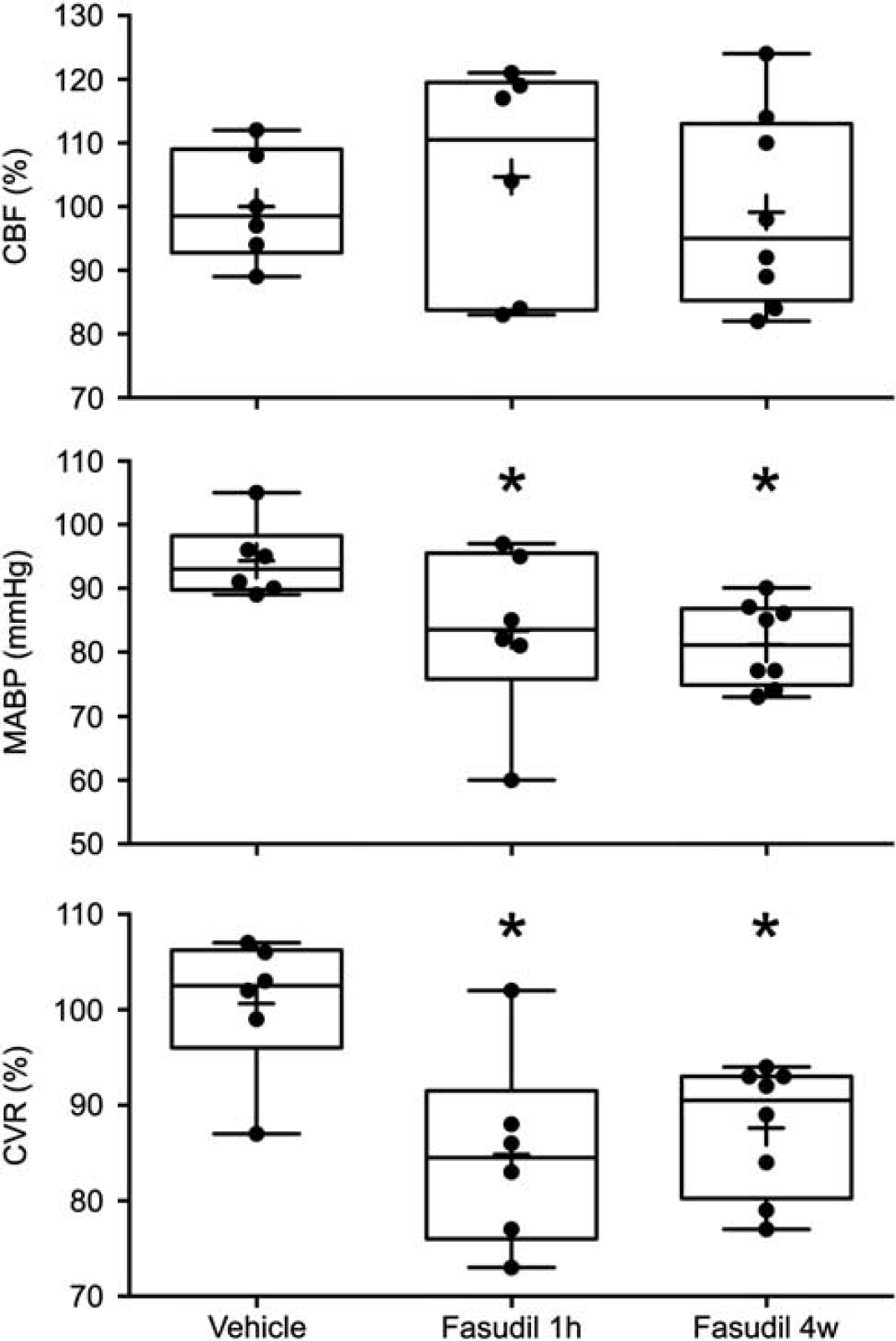
Effects of single dose or chronic fasudil treatment on resting cortical blood flow (CBF), mean arterial blood pressure (MABP), and cerebrovascular resistance (CVR) in hyperlipidemic apolipoprotein E (ApoE) knockout mice. Data are from preischemic baseline obtained at the beginning of experiments before distal middle cerebral artery occlusion (dMCAO). CBF was measured by calculating the τc−1 values using laser speckle contrast images, which were then normalized to the vehicle group average and expressed as %. CVR was calculated using the MABP and CBF, and expressed as % of vehicle group average. Please see Materials and Methods for details. Whiskers, minimum and maximum values; boxes, 25% to 75% range; horizontal line, median; ‘+’, mean. Individual data points are also shown. ∗P<0.05 versus vehicle.
As an additional mechanism to explain improved cortical perfusion in focal ischemia, we examined the effect of ROCK inhibition on the occurrence and cerebral hemodynamic impact of PIDs. Peri-infarct depolarizations typically induced a transient hyperemia in mildly ischemic cortex, and hypoperfusion in more severely ischemic regions (Figure 3A). Because high-fat diet is known to diminish PID occurrence, 4 we usually detected only 1 PID during the 60-minute imaging period in vehicle controls (Figure 3B). We, therefore, pooled all ROCK inhibitor treatment groups to gain statistical power for nonparametric testing, and found that ROCK inhibition was associated with fewer PID occurrence. As reported previously, 12 each PID expanded the area of severely hypoperfused cortex (30% residual CBF) by ∼10% in the vehicle group (Figure 3C). Inhibition of ROCK did not ameliorate this adverse vasoconstrictive effect of PIDs on cortical perfusion.
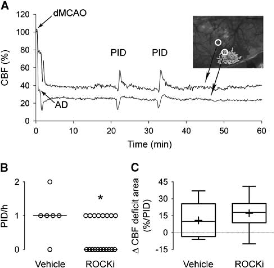
Effects of Rho-associated kinase (ROCK) inhibition on peri-infarct depolarization (PID) occurrence and its impact on the size of ischemic cerebral blood flow (CBF) deficit. (
Body weight did not differ among the treatment groups (Table 1). Plasma lipid profile showed markedly elevated plasma cholesterol but not triglyceride levels, as reported previously in ApoE KO mice. 4 Fasudil appeared to increase the cholesterol and triglyceride fractions, including low-density lipoprotein, after 4 weeks of treatment. Fasudil treatment also had mild but statistically significant effects on arterial blood pH, pCO2, and pO2; however, all values remained within previously reported normal physiologic range for anesthetized and mechanically ventilated mice. 16 Moreover, we did not find a significant correlation between systemic physiologic parameters and the area of perfusion defect (R=−0.06 to 0.38; P>0.05), suggesting that drug effects on systemic physiology were unlikely to explain the cerebral hemodynamic effects of fasudil at baseline or during dMCAO.
DISCUSSION
In this study, we show that ROCK inhibition improves perfusion in focal ischemic cortex in hyperlipidemic mice. The area of cortex with 20% and 30% residual CBF was smaller by 37% to 51% and 42% to 55%, respectively, in ROCK inhibitor-treated ApoE KO mice compared with vehicle. In an identical experimental paradigm, we have previously shown that hydroxyfasudil and Y27632 decreased the area of cortex with 20% and 30% residual CBF by 41% to 55% and 25% to 39%, respectively, in wild-type mice on normal diet. Therefore, ROCK inhibition appears to be at least as efficacious in hyperlipidemic mice as in wild type. Whether ROCK inhibition is more efficacious in the setting of hyperlipidemia5, 6, 7, 8 can only be directly addressed by performing side-by-side comparisons of efficacy in the same study.
The most likely mechanism by which ROCK inhibition decreased the size of perfusion defects is enhanced collateral flow. Because acute treatment 1 hour before ischemia was as efficacious as chronic treatment for 4 weeks, it is unlikely that vascular hypertrophy or neovascularization, or mechanisms dependent on altered gene expression, have a role. Instead, reduced CVR at resting state suggests a functional improvement in collateral flow. Mechanisms by which ROCK inhibition can decrease the CVR include diminished vascular tone, blood viscosity,17, 18 leukocyte adhesion19, 20 and platelet activation,21, 22 as well as improved erythrocyte deformability. 23 It should be noted, however, that relative CVR calculations make certain steady-state assumptions that can be directly influenced by the drug treatment, such as normal cardiac stroke volume and intracranial pressure, and should be interpreted cautiously.
There are several explanations for the lack of enhanced efficacy on chronic treatment. For example, the selected dose level may be supramaximal for ROCK inhibition, or there may be a ceiling for collateral flow augmentation in acute ischemia. It is also possible that the relatively short half-life of fasudil does not permit continuous plasma and tissue exposure on single daily dosing, and that more frequent dosing paradigms may be necessary to recruit mechanisms related to gene expression and structural alterations for a larger effect.
Interestingly, we also found that ROCK inhibition was associated with fewer PIDs. We and many other groups have shown that PIDs worsen ischemic tissue perfusion and expand the perfusion defect in a stepwise manner.12, 24 Therefore, fewer PIDs may indeed be an additional mechanism by which ROCK inhibition improves ischemic tissue perfusion. However, decreased PID occurrence may also be a consequence of smaller perfusion defects; this needs further testing. Finally, fasudil did not decrease plasma lipid levels ruling out this as a potential mechanism of improved perfusion, although the data should be interpreted with caution because of the small sample sizes.
In summary, ROCK inhibition is at least as efficacious in hyperlipidemic mice as in wild type in improving collateral perfusion in focal cerebral ischemia. Reduced CVR and suppressed PID occurrence are two mechanisms that can explain the improved perfusion in ischemic tissue.
Footnotes
The authors declare no conflict of interest.