
Abstract
The positron emission tomography (PET) radiotracer [11C]Pittsburgh Compound B (PIB) is a marker of amyloid plaque deposition in brain, and binding potential is usually quantified using the cerebellum as a reference where the specific binding is negligible. The use of the cerebellum as a reference, however, has been questioned by the reported cerebellar [11C]PIB retention in familial Alzheimer's disease (AD) subjects. In this work, we developed a supervised clustering procedure for the automatic extraction of a reference region in [11C]PIB studies. Supervised clustering models each gray matter voxel as the linear combination of three predefined kinetic classes, normal and lesion gray matter, and blood pool, and extract reference voxels in which the contribution of the normal gray matter class is high. In the validation with idiopathic AD subjects, supervised clustering extracted reference voxels mostly in the cerebellum that indicated little specific [11C]PIB binding, and total distribution volumes of the extracted region were lower than those of the cerebellum. Next, the methodology was applied to the familial AD cohort where the cerebellar amyloid load had been demonstrated previously, resulting in higher binding potential compared with that obtained with the cerebellar reference. The supervised clustering method is a useful tool for the accurate quantification of [11C]PIB studies.
INTRODUCTION
Positron emission tomography (PET) with [11C]Pittsburgh Compound B (PIB) has been utilized for the
Automatic clustering was first proposed to extract the reference region for brain PET studies in which the anatomic definition of a region without specific binding was difficult, for example, in the case of the neuroinflammatory marker [11C]-(R)-PK11195 given its unpredictable distribution pattern. 12 In this context, the automatic method segmented brain voxels into classes on the basis of their time–activity courses and selected as reference the class of voxels that exhibited the kinetic behavior closer to that of gray matter in healthy controls. The approach was subsequently refined to increase the reliability of reference extraction for [11C]-(R)-PK11195 study using a supervised approach, that modeled each pixel's time–activity curve (TAC) as the sum of six predefined kinetic classes, normal gray matter, white matter, pathologic peripheral benzodiazepine receptor binding, vascular binding, muscle, and the skull. 13 Multicenter validation of the approach has further confirmed that supervised reference region extraction is indeed the optimal reference approach for [11C]-(R)-PK11195 quantification. 14
In the present study, we optimized the supervised procedure for the extraction of a reference region in [11C]PIB PET studies, and validated this method in the [11C]PIB studies of idiopathic AD subjects where a plasma input function was available. In particular, we wished to demonstrate that in these AD subjects, the algorithm was able to select reference voxels that lacked a specific-binding component that was identified through the plasma input. The method was then applied to a cohort of familial AD patients where the cerebellar uptake had been previously demonstrated.
MATERIALS AND METHODS
Algorithm Implementation of Supervised Clustering for [11C]PIB
The supervised clustering approach for [11C]PIB PET study (Super-PIB) was constructed as an extension of the one developed for [11C]PK11195. 13 This algorithm consists of three elements: (1) an input normalization procedure to scale each frame of the dynamic sequence, (2) a set of predefined kinetic classes, (3) a regression procedure to calculate the contribution of each kinetic class to the signal of each voxel. These weights, and in particular those of the gray matter class, are then used to calculate the reference input TAC.
Normalization of positron emission tomography dynamic sequence
In the PET dynamic images, the radioactivity of each frame was normalized for every voxels in the brain by subtracting the mean frame activity and dividing it by the frame standard deviation (SD) to create a unit input.
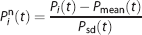
where
Predefined kinetic classes
Three kinetic classes of normal gray matter (class 1), high specific-binding gray matter (class 2), and blood pool (class 3), were predefined from the normalized PET dynamic sequence. The kinetic class of normal gray matter was extracted from the average of normalized time course (
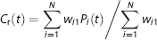
Estimation of each class contribution
The supervised clustering algorithm modeled the normalized kinetic of voxel

where
The weights

Extraction of reference region
Voxels in which the ratio of class 1 (
where
Note that the threshold value of
Positron Emission Tomography Study
Eight healthy control subjects, 10 patients with idiopathic AD, and eight patients with familial AD (presenilin 1 mutation carriers AD) participated in this study. Patient data have been reported in the previously published investigation. 7 Elevated [11C]PIB binding was reported in some elderly normal volunteers,16-20 but this was not the case in healthy control subjects enrolled in this study. Ethical approval was granted by the Hammersmith Hospitals Trust Ethics Committee, and permission to administer radioisotopes was granted by the Administration of Radioactive Substances Advisory Committee of the Department of Health, UK. Informed written consent was obtained from all patients and healthy volunteers. All scans were performed in accordance with the guideline of Hammersmith Hospitals Trust Ethics Committee.
PET studies with [11C]PIB were performed on an ECAT EXACT HR + (CTI/Siemens) PET camera with 15.5-cm axial field of view, 63 transaxial planes. A transmission scan was acquired before an emission scan using a single rotating photon point source of 150 MBq of 137Cs for subsequent attenuation correction and scatter correction. ~ 370 MBq of [11C]PIB was administered by a bolus injection 30 seconds after the start of emission scan. The specific radioactivity was 20,235 (± 6240) MBq/μmol at the time of injection. Emission data were then acquired over 90 minutes in list mode and reorganized as 32 time frames (30-s background frame, 1 × 15-s frame, 1 × 5-s frame, 1 × 10-s frame, 2 × 30-s frames, 9 × 60-s frames, 3 × 180-s frames, and 14 × 300-s frames). The data were reconstructed by a filtered back-projection using ramp filter set at 0.5 times the Nyquist frequency. The transaxial and axial spatial resolution of the reconstructed images were 5.6 and 5.4 mm full width at half maximum, respectively, at 10-cm distance from the center.
Arterial blood sampling was carried out for healthy controls and idiopathic AD subjects, not for the familial AD subjects, because it was not possible to obtain consent for this particular cohort. Arterial whole blood activity was monitored continuously for the first 15 minutes of the scan with a bismuth germinate coincidence detector at a flow rate of 5 mL/minute. At the same time, discrete arterial blood samples were withdrawn at 5, 10, 15, 20, 30, 40, 50, 60, 75, and 90 minutes after the injection into heparinized syringes, and the radioactivity concentration of the whole blood and plasma was measured. Eight plasma samples at 5, 10, 15, 30, 40, 60, 70, and 90 minutes were analyzed for metabolites using an on-line system with a reverse-phase high-performance liquid chromatography linked to radioactivity and absorbance detectors connected to a PC-based integrator.
21
The level of [11C]PIB and other 11C components at each sample time was calculated as a percentage of total radioactive component in each analyte, and the function of unmetabolized parent fraction in plasma was obtained from the fit of sigmoidal function for the parent fraction to the eight measurements as shown previously by Edison
Volumetric T1-weighted MR images were obtained with a 1.5 Tesla GE Signa scanner.
Data Analysis
Magnetic resonance images were segmented and coregistered to the summation PET images by SPM2 (Functional Imaging Laboratory, Wellcome Department of Imaging Neuroscience, University College London, London, UK). Gray matter and white matter maps were obtained from the segmented MR images and thresholded (only map value>90% of maximum value were retained) to minimize the effect of partial volume. The eight healthy volunteers and three idiopathic AD subjects were used to predefine the three kinetic classes, and the test analysis for the Super-PIB application analysis was performed on the other seven idiopathic AD subjects and eight familial AD subjects. In these subjects, the weight of each class component was calculated for all voxels within the gray matter by equation (2), and TAC of the reference region was extracted by equation (4).
Regions of interest were manually drawn on the coregistered MR images for the cerebellum, prefrontal cortex, thalamus and pons, and ROIs for the cerebellum and prefrontal cortex were masked with the gray matter maps. The respective TACs were then extracted from the dynamic PET images.
Validation of Super-PIB Extracted Reference Region
The TACs of the Super-PIB extracted region were compared with that of the cerebellum in idiopathic AD subjects to investigate whether Super-PIB can exclude pathologic gray matter region with specific binding from the reference region. The total distribution volumes (
Next, the Super-PIB clustering was evaluated by investigating the distribution of Super-PIB extracted reference voxels in the cerebellum. All voxels in the cerebellum were divided into four segments according to the ratio of each kinetic class component estimated by equation (3); those with voxels regarded as a reference region (REF), those with voxels regarded as a non-reference region owing to the high specific-binding component (
Comparison between Plasma Input and Reference Input Graphical Analysis
First, binding potentials (
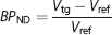
where
Next,

where
Binding Potential in Familial Alzheimer's Disease Patients
In the familial AD group,
Statistical Analysis
All analyses were carried out using Matlab (The Mathworks, Natick, MA, USA). Method comparison was carried out using either linear regression or Bland-Altman plots.
25
Statistical comparisons were performed using linear parametric models (Student's
RESULTS
Reference Extraction by Supervised Clustering
The normalized TAC of each class defined for use in the supervised clustering is shown in Figure 1. The weight ratio of normal gray matter class (
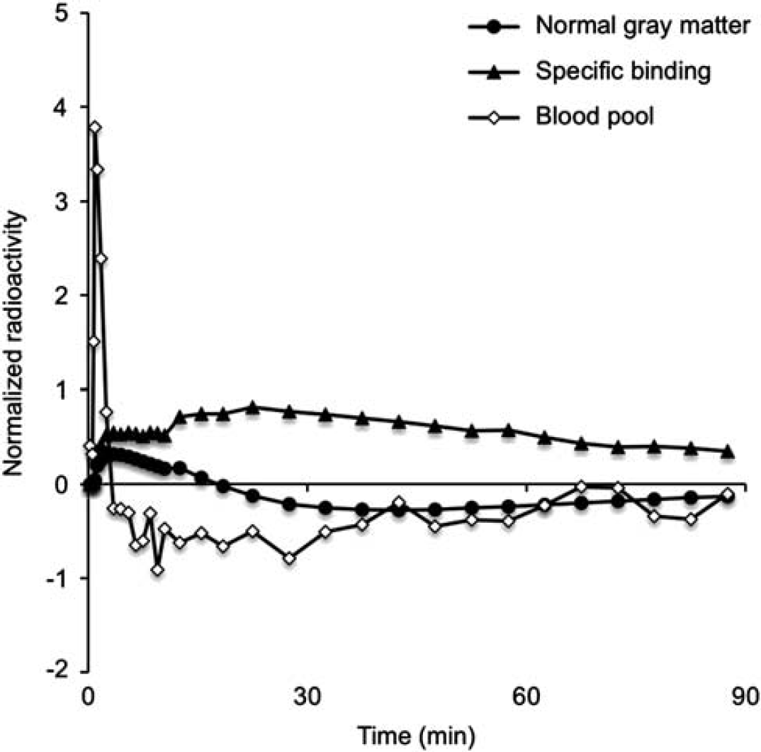
Normalized time–activity curve of the predefined three classes; normal gray matter, specific-binding gray matter, and blood pool.
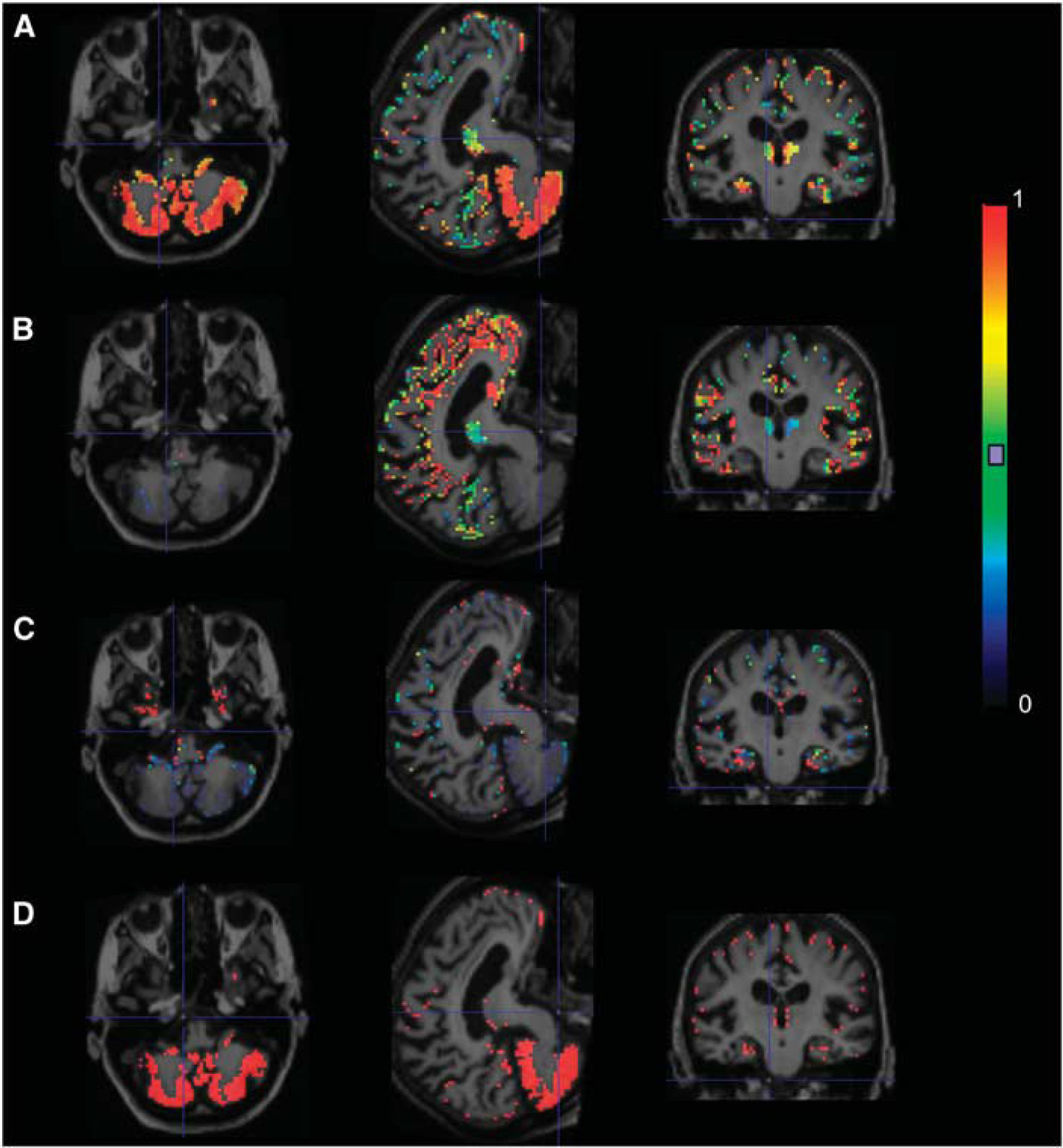
Maps of normal gray matter ratio (
The TACs of Super-PIB reference region were close to those of the cerebellum, though the radioactivity of Super-PIB reference region was slightly lower in later frames. The average
Evaluation of Super-PIB Segments in the Cerebellum
Application of Super-PIB clustering in idiopathic AD patients resulted in the cerebellum being divided into segments, i.e., REF, NREF-Binding, NREF-Blood, and NREF-Others. The case where the ratio of non-reference voxels in the cerebellum was largest among seven subjects is shown in Figure 3 and related TACs are shown in Figure 3D that clearly demonstrates the tracer retention at late times for the TAC of the NREF-Binding segment. In this instance, the
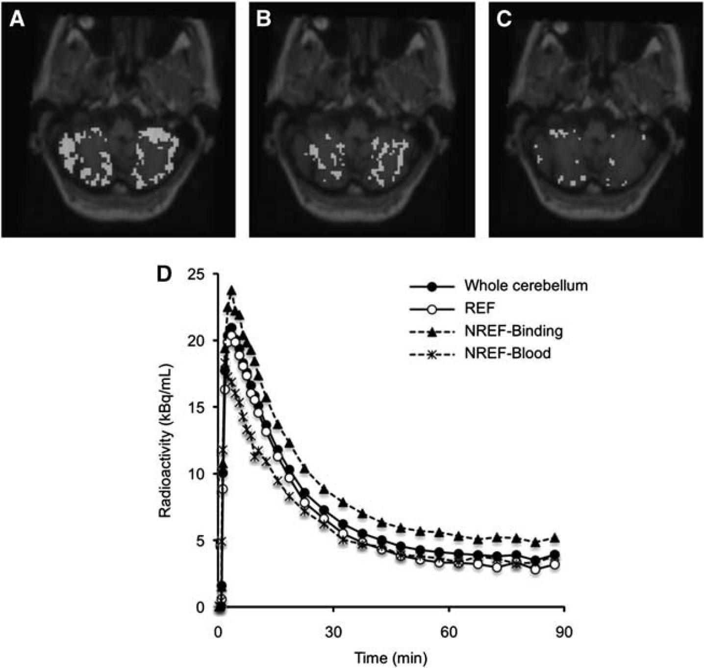
Example of cerebellar segmentation for the idiopathic Alzheimer's disease subject that demonstrated the higher non-reference percentage of voxels in the cerebellum; reference (A; REF), non-reference with high specific-binding fraction (B; NREF-Binding), and non-reference with high blood-pool fraction (C; NREF-Blood) superimposed on magnetic resonance imaging (upper), and averaged time–activity curve of the whole cerebellum and each cerebellar segment (
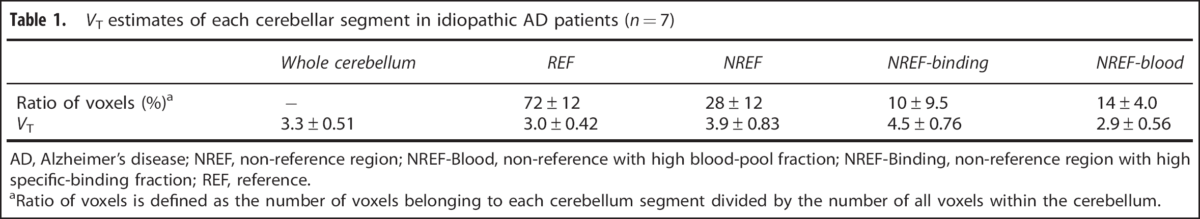
AD, Alzheimer's disease; NREF, non-reference region; NREF-Blood, non-reference with high blood-pool fraction; NREF-Binding, non-reference region with high specific-binding fraction; REF, reference.
aRatio of voxels is defined as the number of voxels belonging to each cerebellum segment divided by the number of all voxels within the cerebellum.
The histograms of the
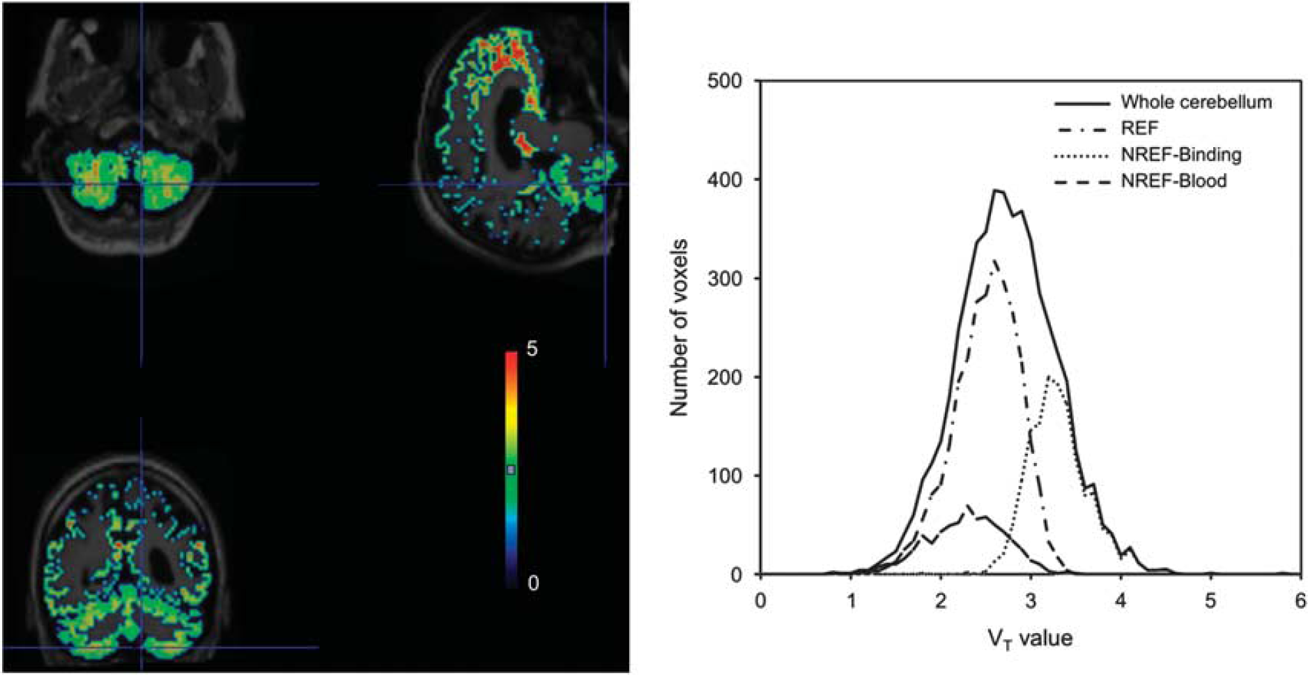
Parametric VT map of the same idiopathic Alzheimer's disease subject obtained using a plasma input function (left) and histogram of VT estimates for voxels within each cerebellar segment identified by the supervised algorithm. REF, reference; NREF-Binding, non-reference region with high specific-binding fraction; NREF-Blood, non-reference with high blood-pool fraction.
Comparison between Plasma Input and Reference Input Graphical Analysis
In the GA-plasma,
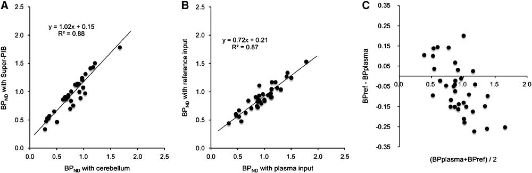
Relationship of binding potential (
Binding Potential in Familial Alzheimer's Disease Patients
In familial AD subjects,
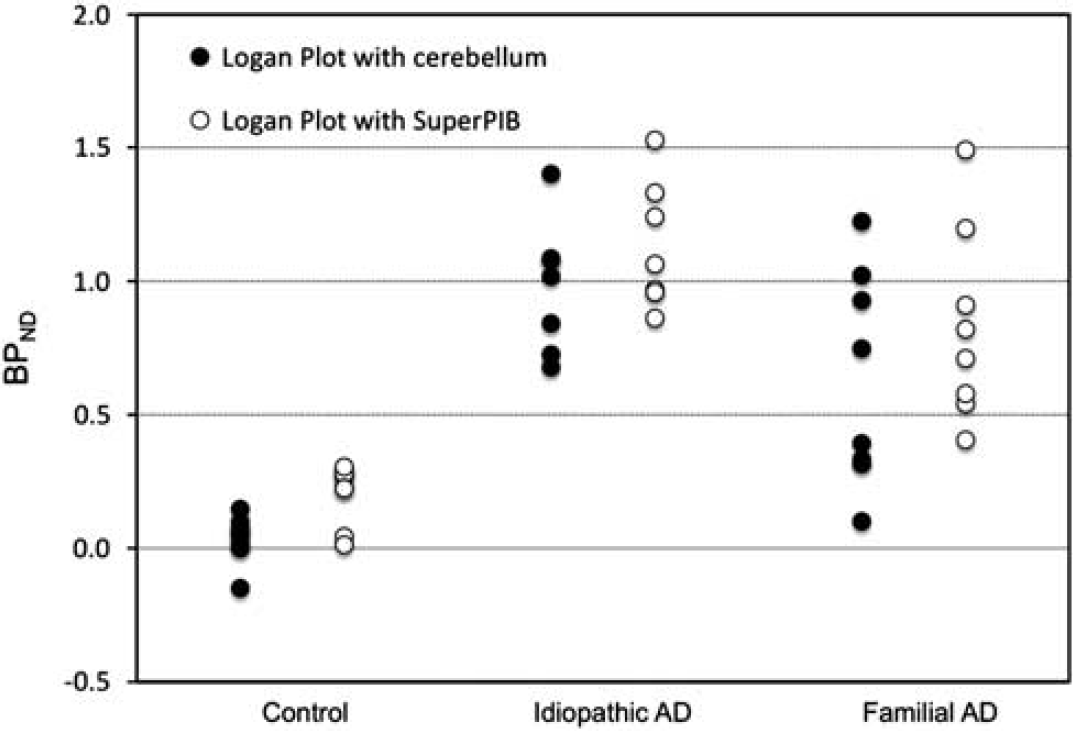
Binding potential (
Note that the use of the novel reference methodology results in an increase of the
DISCUSSION
The aim of this work was to develop and validate an automatic extraction algorithm for the accurate identification of a reference region in dynamic [11C]PIB studies.
The methodology is based on the principle that the kinetics of the gray matter voxel can be explained as a linear mixture of healthy gray matter plus gray matter with specific binding and a blood volume component. These classes can be readily identified given the negligible presence of specific binding due to amyloid in normal volunteers and, in contrast, the widespread and intense specific binding in the cortical regions of AD patients. Therefore, it is easy to obtain the typical kinetic pattern of a normal gray matter and a region with amyloid deposition. We acknowledge that the empirical definition of gray matter in control subjects as the tissue reference for our methodology may introduce a slight bias. A percentage of normal subjects (B30%) does seem to show low but significant amount of specific binding in gray matter regions (but not globally). However, the fact that our reference tissue was averaged on all gray matter and on all normal subjects (who, in our data set, did not seem to show any remarkable binding) renders the expected bias negligible. Differently from previous work with [11C]-(R)-PK11195, 13 we have excluded the use of normal white matter as a kinetic class given the unspecific accumulation of [11C]PIB in myelin.
Results in an idiopathic AD cohort confirmed the ability of the technology to recover a set of reference voxels, mostly cerebellar, that was away from areas of tracer accumulation as well as from areas close to or including large venous pools. Note that the inclusion of voxels with large contamination from the blood volume may introduce biases as well. In addition, in a familial AD cohort, the Super-PIB methodology demonstrated higher
The novel reference methodology, by selecting highly homogeneous gray matter voxels, avoids the spillover of specific-binding signal from white matter where [11C]PIB is bound to myelin. This results in the
The accuracy of the quantification achieved was confirmed by the good match between plasma and reference quantification. The underestimation of
As the previous application of supervised clustering has demonstrated, 14 the technique can be robustly applied across centers and scanners as long as injecting protocols, scanner resolution properties, and reconstruction parameters are similar. However, in case kinetic classes will have to be re-defined, as explained in the previous paragraph, in the case of [11C]PIB this is quite straightforward. Clinical use of the methodology may require the redefinition of the kinetic classes if scanner resolution and/or reconstruction parameters differ. In particular, given the experience with a similar methodology for [11C]-(R)-PK11195 studies, 14 we identified significant changes in the class profiles when moving to 5 to 2 mm resolution and from filtered back-projection to iterative reconstruction approaches.
In summary, the supervised clustering method automatically detects a reliable reference region in [11C]PIB PET dynamic studies, it is a useful and practical tool for the accurate quantification of the specific binding of the radiotracer and is a methodology of general applicability.
DISCLOSURE/CONFLICT OF INTEREST
The authors declare no conflict of interest.
Footnotes
ACKNOWLEDGEMENTS
FET was supported by the ‘PET Methodology Programme Grant’ from the Medical Research Council UK (G1100809/1).