
Abstract
An 8-month-old owned European cat showing abdominal pain, fever, distended painful bladder and urinary blockage was presented. Intravenous fluids were immediately administered and, after sedation, a urinary catheter was applied. Blood and urine analysis revealed cystitis and a moderate-to-severe degree of renal failure. About 20 thread-like nematodes, identified as Capillaria plica larvae and fragments of adult stages, were found in the urine sediment. After treatment with an oral formulation of fenbendazole at 25 mg/kg q 12 h for 10 days, urinary signs and bladder worms disappeared. Cases of Capillaria species bladder worms in cats are rarely reported and most infected cats show no clinical signs, presumably because of a low parasite burden. In the present study, feline capillariosis was associated to urethral obstruction, severe difficulties in urination, cystitis and renal failures.
The infection of the bladder of domestic cats by Capillaria (Pearsonema) has been reported in countries worldwide but appears to be uncommon.1–3 Capillaria plica and Capillaria feliscati are the species responsible for feline urinary capillariosis. 4 The adult worms are small and thread-like and either superficially attached to, or buried within, the bladder mucosa.5,6 The length of adult female worms ranges from 29–60 mm, while adult males are 13–30 mm long. 4 The life-cycle of both these parasites is indirect and involves, as intermediate host, an earthworm that ingests the eggs released with the urine. The definitive host becomes infected after ingestion of earthworms containing infective first-stage larvae. Larvae reside in the intestine for a short period until they migrate to the bladder where they moult into adults. The prepatent period, after which Capillaria species eggs can be isolated from the urine, is about 2 months after the ingestion of the infected earthworm. 2 On urinalysis, the finding of Capillaria species eggs is often incidental, but it has rarely been associated with signs of feline lower urinary tract disease (FLUTD).1,7 Infections can be self-limiting with a reduction of egg excretion until being undetectable in a few months. 5
In February 2010, an 8-month-old European male cat living outdoor in Pisa (Italy) and showing signs of low urinary tract disease, was examined. According to the owner, the animal had been intermittently straining as if to urinate for about 2 days but had no previous history of such signs nor of any other pathology. Clinical examination showed abdominal pain, fever (40.5°C) and distended painful bladder. Intravenous fluids were immediately administered to the cat. After sedation with intramuscular medetomidine hydrochloride (Domitor; Janssen Animal Health) at 50 μg/kg, the urine sample was collected by cystocentesis via a 23 G needle and a urinary catheter was applied in order to allow the cat to urinate. Thereafter, the antidote atipamezole at 200 μg/kg was intramuscularly administered. The cat was treated with amoxicillin (7 mg/kg) plus clavulanic acid (1.75 mg/kg) (Synulox; Pfizer), given subcutaneously q 12 h for 10 days to prevent any secondary bladder infections occurring as a result of catheterisation. After 3 days the catheter was removed.
Routine blood analyses (complete blood cell count and serumbiochemicalpanel)were performed using a photometric instrument (CGA EOS 880, Florence, Italy).
Urine chemical analyses were performed using a commercial reagent strip (Multistix10 SG; Siemens, UK).
An aliquot of the collected urine sample was centrifuged at 1200 rpm for 10 min and the sediment was microscopically examined.
Isolated parasites were examined immediately and after fixation in Looss liquid. For optical microscope observation, at 250x and 400x magnification, isolated parasites were mounted and measured with an ocular micrometer.
Results from blood analysis revealed increased blood urea nitrogen (BUN) 150 mg/dl (reference interval 20–50 mg/dl) and creatinine 4 mg/dl (reference interval 0.6–2 mg/dl) indicating a moderate-to-severe degree of renal failure. The other parameters were within reference interval, including glycaemia (95 mg/dl). The urine was brown red in colour with specific gravity of 1025, a pH of 6.0 and presence of blood (200 erythrocytes/μl), leukocytes (125 leukocytes/μl) and proteins (300 mg/dl). Microscopic examination of the urinary sediment confirmed haematuria and revealed a low leukocyte count (20 leukocytes/high power field) and the presence of a large number of thread-like nematodes (about 20 worms). Crystal precipitates were absent.
The nematodes were mostly represented by filariform immature stages (Fig 1), but few fragments of adult worms were also isolated. Eggs were not present in the urinary sediment nor inside the body of the two isolated fragments of adult females. The length of immature stages ranged from about 724–1547 μm, while they were about 20–25 μm in width. Adult stages were identified on the basis of the morphology of the male caudal end and the vulva of the female present in very few worm fragments (two males and two females fragments). The male caudal end showed two small digitiform papillae surrounded by a triangular caudal ala (Fig 2b), while the vulva of the female presented a funnel like appendage (Fig 2a). These features are typical of C. plica4,8 so isolated parasites were identified as larvae and fragments of adult stages of C. plica.
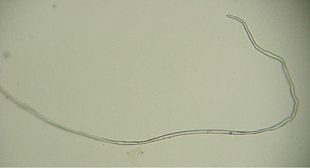
Larval immature stage of C. plica isolated from an 8-month-old symptomatic cat. 250x magnification. Scale bar: 50 mm.

(a) Vulva of a female of C. plica presenting a funnel like appendage. (b) C. plica male caudal end showing one of the two small digitiform papillae surrounded by a triangular caudal ala. 400x magnification. Scale bar: 25 mm.
Following this diagnosis the cat was treated with an oral formulation of fenbendazole (Panacur; Intervet) at the dosage of 25 mg/kg q 12 h for 10 days. 9
After the treatment the clinical signs disappeared, subsequent blood and urine analysis resulted within the normal range and neither Capillaria species worms nor eggs were found.
Cats with urinary capillariosis usually do not show clinical signs and the infection is frequently self-limiting. For these reasons this parasitic disease is considered to be of minor clinical significance. However, heavily infected animals may show signs of urinary tract disease with pollakiuria, dysuria and haematuria.1,7 Occasionally, C. plica has been found localised within the ureter and the renal pelvis. 5 The occurrence of renal failure in association with urinary capillariosis has been reported in two dogs.10,11
The examined cat was symptomatic showing severe difficulties of urination, while urine and blood analysis were indicative of cystitis and acute renal failure. The animal resulted heavily infected with a large number of immature worms; the eventual presence of several of these parasites in the urethra could have directly led to the urethral obstruction, thus explaining the severe difficulties with urination and the bladder distension observed in the cat. Alternatively, the large number of worms and the consequent bladder and possibly urethral inflammation with the submucosal oedema, could have been equally responsible for the urethral obstruction. The urinary blockage was probably the main cause of the acute renal failure, as frequently observed in the cat with FLUTD/feline urological syndrome (FUS). 12 Thus, the present report shows the possibility that in the cat a renal failure might be related to the urethral obstruction caused by a heavy C. plica infection. These findings are also indicative of the possibility that Capillaria species infections may represent an important concomitant or predisposing factor responsible for feline urinary tract disease in those infected cats in which other causes are diagnosed or suspected.1,13
The young age of the animal examined in this study, the presence of a large number of parasitic immature stages and the lack of Capillaria species eggs at the examination of the urine sediment are indicative of a recent infection, with the appearance of signs within the prepatent period, before the eggs could be found in the urine. In addition, data obtained in this study show that in symptomatic animals Capillaria species eggs may be not always present in the urinary sediment, thus complicating the diagnosis of the disease and specific parasitological skills are required. The life-cycle of this nematode species involves an earthworm as intermediate host and living outdoors, as in the cat examined in the study, may be a risk factor for Capillaria species infection. However, although the diet of the domestic cat includes invertebrates, these latter animals are only of secondary importance accounting for only a small percentage of the total food intake of feral animals.14,15 For this reason some authors 13 hypothesise the presence of paratenic hosts in Capillaria species life-cycle able to amplify the transmission of infection.
Successes in treating urinary capillariosis has been reported using benzimidazoles, ivermectin and levamisole.5,10,16 One to several doses of 50 mg/kg of fenbendazole per os (PO) have been administered to resolve Capillaria species infection in the dog. 17 The treatment with 25 mg/kg of fenbendazole q 12 h PO for 10 days 9 was effective in this study, as urinary signs and bladder worms disappeared in the examined cat.
Cases of Capillaria species bladder worms in cats are rarely reported, as most infected animals show no clinical signs, presumably because of a low parasite burden. Therefore, the prevalence of urinary capillariosis is difficult to estimate. In Italy, the prevalence of cat bladder capillariosis is unknown and to the best of our knowledge this is the first case reported in the cat in this country.
Footnotes
Acknowledgements
Financial support was provided by the University of Pisa (Fondi di Ateneo 2010).