
Abstract
Renal diseases are common in older cats. Decreased renal blood flow may be the first sign of dysfunction and can be evaluated by Doppler ultrasound. But previous studies suggest that the resistive index (RI) has a low sensitivity for detecting renal disease. Doppler waveforms of renal and intrarenal arteries demonstrate decreased blood flow before there are any changes in the RI. The purpose of this study was to evaluate the normal Doppler flowmetrics parameters of renal arteries (RAs), interlobar arteries (IAs) and abdominal aorta (AO) in adult healthy, Persian cats. Twenty-five Persian cats (13 females and 12 males with mean age of 30 months and an age range 12–60 months) with normal clinical examinations and biochemical tests and normal systemic blood pressure were given B-mode ultrasonographies in order to exclude all nephropathies, including polycystic kidney disease. All measurements were performed on both kidneys. Both kidneys (n=50) were examined by color mapping of the renal vasculature. Pulsed Doppler was used to examine both RAs, the IAs at cranial, middle and caudal sites, and the AO. The RI was calculated for all of the vessels. Early systolic acceleration (ESA) of RA and IA was obtained with Doppler spectral analysis. Furthermore, the ratio indices between RA/AO, and IA/RA velocities were calculated. The mean values of peak systolic velocity (PSV) and the diameter for AO were 53.17±13.46 cm/s and 0.38±0.08 cm, respectively. The mean RA diameter for all 50 kidneys was 0.15±0.02 cm. Considering the velocimetric values in both RAs, the mean PSV and RI that were obtained were 41.17±9.40 cm/s and 0.54±0.07. The RA had a mean ESA of 1.12±1.14 m/s2 and the calculated upper limit of the reference value was 3.40 m/s2. The mean renal-aortic ratio was 0.828±0.296. The IA showed PSV and RI values of 32.16±9.33 cm/s and 0.52±0.06, respectively. The mean ESA of all IAs was 0.73±0.61 m/s2. The calculated upper limit of the reference value was 2.0 m/s2. The mean renal-interlobar artery ratio was 1.45±0.57. The RI values obtained in this study were similar to values reported in the literature. Some conditions that lead to a decrease in compliance and to an increase in vascular resistance can affect the Doppler spectral waveforms without changes in RI. To our knowledge, there are no studies that were directed toward to the normal ESA values of the renal vasculature in Persian cats. This study introduced a new ratio between the PSV of the RA and the IA. This index was developed based on the well-known effects of Doppler on the detection of stenosis, regardless of the cause. Further studies are necessary to verify the hemodynamic behavior of this index under pathological conditions in cats as well as the effect of aging, nephropathies and systemic pressure on Doppler velocimetric parameters.
Renal failure has a high morbidity in cats, especially in older animals. 1 Color Doppler ultrasound is an important tool for evaluating the urinary system in cats. This complementary method of imaging diagnosis is capable of assessing renal diseases due to its ability to evaluate both anatomy and physiology from the visualization of blood flow. Doppler ultrasound is used in human medicine to study renal vascular architecture. 2 Color Doppler mapping can demonstrate renal perfusion and, using this technique, specific vessels can be selected for spectral analysis and to determine blood flow information. The docile temperament of Persian cats makes Doppler evaluation possible without sedation. Additionally, Persian cats present a high prevalence of polycystic kidney disease with positive ultrasound examination that is not always associated with renal dysfunction in the early stages. Decreased renal blood flow may be the first sign of dysfunction. Parenchymal diseases leading to a decrease in compliance and increased vascular resistance can affect Doppler spectral waveforms without changes in the resistive index (RI). The key to gaining a better understanding of Doppler findings and indications is to determine the reference values. Renal vascular RI values are described in the literature, but there are no reports in feline medicine that show early systolic acceleration (ESA), a Doppler parameter that is useful for demonstrating changes in Doppler waveforms. The purpose of this study was to demonstrate the normal Doppler parameters of renal arteries (RAs), interlobar arteries (IAs) and the abdominal aorta (AO) in adult, healthy, Persian cats.
Materials and methods
Fifty kidneys from 25 healthy Persian cats (13 females and 12 males ranging in age from 12 to 60 months) were selected after clinical examination, biochemical tests (the tests included complete blood count, serum biochemistry to quantify renal and hepatic functions, complete urinalysis, urinary protein/creatinine ratio) 3,4 ; systemic blood pressure measurement, 5 and ultrasonographies 6−8 using a triplex Color Doppler ultrasonography unit with a 6–10 MHz linear transducer. B-mode, Color Doppler and pulsed Doppler ultrasound were performed on both kidneys to examine the RAs, cranial, middle and caudal IAs, and AO (before the RA outgrowth). Additional attention was given to the insonation angle. 2,9,10 The AO and both RA diameters were determined with B-mode ultrasonography. These vessels were identified in longitudinal planes according to literature descriptions. 11−13 Color Doppler mapping was used to visualize renal vasculature. Subsequent pulsed Doppler interrogation from each one of the arteries was obtained with a sample width of 2 mm. The following indices were measured manually using the built-in calipers of the sonographic machine: peak systolic velocity (PSV), end diastolic velocity (EDV), RI and ESA. ESA and RI of RAs and IAs in both kidneys were obtained from spectral analysis (Fig 1) and the machine displayed the calculated values automatically. All data were obtained by averaging the values from three similar waveforms. All of the cats were examined without sedation or anesthesia. The data verified a normal distribution in the study population. Parametric tests were used to evaluate all of the data (mean, maximum, minimum, standard deviation). In order to establish normal values, 95% confidence intervals were calculated. A Student's t-test was conducted to compare the velocity values of each artery in both kidneys in order to evaluate significant differences. All differences were considered to be significant at a probability level of 95% (P<0.05) between the kidneys. The Pearson coefficient was determined to verify any correlations between velocities. The AR/AO and AI/AR velocity ratios were also calculated.

Pulsed Doppler mapping of RA showing ESA, PSV and EDV in a normal cat.
This study was approved by the Research Ethics Committee at INRAD (Institute of Radiology), at the Universidade de São Paulo. The pet owners signed an ‘informed consent’ form allowing for the collected data to be used in this research.
Results
As Table 1 demonstrates, the mean values for the AO were PSV 53.17±13.46 cm/s, EDV 20.73±7.17 cm/s and a diameter of 0.38±0.08 cm.
Diameter, peak systolic and EDVs of AO values mean and standard deviation in Persian cats.

AO=abdominal aorta; SD=standard deviation.
The results obtained from the RAs are summarized in Table 2. The mean RA diameter of 50 kidneys was 0.15±0.02 cm. The Student's t-test did not reveal any significant differences between the right and left sides. Considering the velocity values in both RAs, the mean PSV and RI were 41.17±9.40 cm/s and 0.54±0.07, respectively. The mean ESA of the RAs was 1.12±1.14 m/s2. The upper limit of the ESA value obtained in our study was 3.40 m/s2. Correlations between renal and aortic velocities were verified by Pearson's correlation coefficient. The mean renal-aortic ratio was 0.828±0.296, ranging from 0.387 to 1.518.
Mean and standard deviation of PSVs, EDV and RI of RAs in Persian cats.
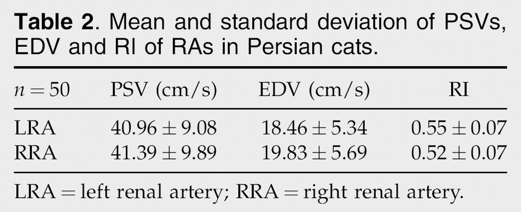
LRA=left renal artery; RRA=right renal artery.
The Student's t-test did not reveal statistical differences in the Doppler parameters of the IAs between both sides. The Doppler velocimetry findings of the IAs are described in Table 3. Statistical analysis did not show any significant differences between the IAs. Taken together, the mean PSV, RI and ESA values of the arteries were 32.16±9.33 cm/s 0.52±0.06 and 0.73±0.61 m/s2, respectively. The upper limit of ESA reference value was 2.0 m/s2.
Mean and standard deviation of PSVs, EDVs, RI and ESA of IAs in Persian cats.

Cr IAs=cranial interlobar arteries; Md IAs=middle interlobar arteries; Cd IAs=caudal interlobar arteries.
No differences in PSV between both the RA (right and left) and IA (cranial, middle and caudal) were detected with statistical analysis. Pearson's correlation coefficient showed a positive correlation between the RA and IA velocities and between right and left IAs. The mean PSV renal/interlobar ratio was 1.45±0.57 and the ratios ranged from 0.50 to 3.47.
Discussion
Renal diseases occur very often in small animals, especially in older cats. 1
Doppler ultrasonography is a contemporary method for diagnosis in veterinary medicine. In spite of the high prevalence of nephropathies in cats, there is a lack of information about the Doppler velocimetry parameters of renal vasculature in the veterinary literature.
The present study was performed in 25 healthy, unsedated Persian cats. However, the decision to use exclusively Persian cats is a limitation of this study, because there is no data to support that the resulted parameters could act as a reference range for all breeds. This breed was used mainly because of its docile temperament. Additional studies are needed to provide validation in different breeds of cats. There were almost no failures for detecting the main RAs in the animals. Only one of the interlobar vessels was not detected in one cat. The technical failure rate for these vessels in humans is 17.2%. 14,15 However, arcuate vessels were not easily mapped due to the impact of respiratory movements and the small diameter of the vessels, which resulted in a technical failure rate of 50%.
Time varied across examinations. It took about 60 min to obtain all of the data. After collecting half of the samples, the examination took 45 min. A routine examination would take about 30 min, as there is no need to take all of the measurements. The condition of the patient (fasting, hair clipping, respiratory pattern, and temperament) was considered when the failure rate was calculated.
AO assessment (diameter and systolic peak velocity)
Doppler ultrasound of the aorta has proved to be a very useful tool in human medicine. The ultrasound provides structural and hemodynamic information for diagnosis as well as for follow-up examinations of renal and vascular diseases. 14,15 In the veterinary literature, there are few studies that show morphometric, morphologic and hemodynamic parameters of the aorta in cats.
In dogs, the AO becomes narrower caudally. In a study with 131 dogs (divided into six groups according to ‘crown-rump length’), the mean diameter of the aorta before RAs outgrowth was 0.80±0.15 cm. 17 In a human study, the mean diameter of the aorta was 1.28±0.34 cm, 14 and in the present study, the mean diameter of the AO where the RAs branch off was 0.38±0.08 cm. These results (Table 1) confirmed the hypotheses about the correlation between vessel diameter and biometric factors such as the body length of the studied species. 17
As reported in humans 15 and in dogs, 16 aortic Doppler mapping showed a phasic waveform at the cranial to the origin of the RAs.
In dogs, the mean PSV measured was 104±22.04 cm/s 16 while in humans it was 103.20±51.50 cm/s. 14 The mean PSV in our study was 53.17±26.9 cm/s and the range was 26.25–80.08 cm/s. These findings suggest that blood flow velocity depends on the diameter and length of the studied vessels. The feline aorta is narrower and shorter compared to the aorta in humans and dogs.
RA assessment (diameter and Doppler parameters)
In dogs and cats, the RAs branch off from the lateral aspect of the AO, and the diameter of the RAs ranges from 0.30 to 0.40 cm. 18,19 Studies in humans 20 showed that the normal RA diameter is 0.39±0.86 cm. In this study, the diameters that were observed ranged from 0.12 to 0.17 cm for the RA and there was no statistical difference between the right and left sides.
The Student's t-test results for paired observations of PSV values revealed no statistical difference between the RAs. The mean PSV value was 41.17±9.40 cm/s with a range of 22.45–64.50 cm/s.
In dogs and cats, RAs are long and winding. It is not unusual to see a RA branching before entering the hilum. 19 The longer and more winding the primary vessels are, the slower the blood flow velocity is in small derivative vessels. 21 This study obtained higher PSV values in the aorta than in the RA.
In humans, the ESA of renal and intrarenal arteries provides information about blood flow alterations in the distal vascular bed. Parenchymal diseases that lead to a decrease in compliance and to an increased vascular resistance, affect Doppler spectral waveforms. 22 In our study, the mean ESA in RAs was 1.12±1.14 m/s2. The upper limit of the normal value obtained was 3.40 m/s2. There are currently no references in the veterinary literature.
Prior studies established that the RI of the RA in dogs is 0.64±0.04 for the right artery and 0.63±0.028 for the left artery, with no significant statistical differences between the sides. 21 The literature suggests that there is a mean RI value of 0.59±0.05 for the RA with an upper limit of 0.69 in cats. 23 In this study, a corresponding RI was obtained with a mean of 0.54±0.07. The upper limit of the reference value, which has been reported before, was 0.69. 23,24 All of the animals were healthy adults and had normal arterial blood pressure. It was known that RI values tend to be higher in children and the elderly. 25 Considering that this could also occur in cats, further studies with cats of different ages and under different clinical conditions are needed.
Renal/aortic ratio
In human medicine, the renal/aortic PSV ratio renal artery ratio (RAR) is considered a direct technique for using Doppler waveforms to detect RA stenosis. Studies in human patients 26 have reported a normal RAR lower than 3.5, others 14 have found 3.0 to be the upper limit of the normal range.
This study in cats found a RAR lower than in humans (around 50%), which can be explained by the proportional differences between the diameters and velocities of both vessels involved. Complementary studies are necessary to verify the hemodynamic behavior of these vessels in cats under different clinical conditions.
IAS assessment
Flow velocity changes in interlobar and arcuate arteries have the potential to signal differences in the gradient of the juxtaglomerular apparatus. 27 Although cats are unable to hold their breath during examinations, IAs were characterized in almost all of the animals, except for one artery in one of the kidneys, which was not depicted due to overlying bowel gas, respiratory and body movements.
ESA
Some diseases can change the arterial wall compliance and increase the vascular resistance of the distal renal vascular bed without changes in the RI. 22 ESA of the intrarenal arteries in humans provides poststenotic ‘tardus-parvus’ Doppler waveforms. 28−31 The appearance of a ‘tardus-parvus’ Doppler waveform may be understood by the analysis of the hemodynamic factors affecting the spectral characteristics. In particular, the elastic properties of the artery walls and changes in peripheral resistance within renal vascular circuits can be better understood. 26 The ESA does not exceed 3.0 m/s2 in the segmental arteries of human patients. 31 Our results revealed an upper limit of 2.0 m/s2. Other studies are necessary to compare this Doppler parameter under various clinical circumstances as there are currently no references in the literature for comparison.
PSV and RI of IAs
There are currently no references in the literature to normal values of PSV of IAs in cats.
Similar to other studies, 32 cats with normal kidney anatomy and function as determined by physical examination, biochemical profile, B-mode ultrasonography, and systemic blood pressure were selected for this study. However, our sample was larger than other studies as we analyzed 50 kidneys.
In another study, that used 14 normal domestic cats, 23 the mean RI value was 0.58±0.06, which is similar to previous results of unsedated patients before surgery. 32,33 As our data had no pharmacological influences, these results are consistent with the data from the studies cited above (0.52±0.07), and show no statistical differences between the kidneys or the IAs (cranial, middle and caudal).
Renal/interlobar ratio
There was a positive correlation between the renal and interlobar velocities as well as between the IAs (Pearson's correlation coefficient). In other words, the velocity increases or decreases throughout the arteries together. This was expected because all of these arteries branch off from the RA, which passes through the hilum and divides into interlobar branches that are situated between the renal papillae.
The literature suggests that RI has a low sensitivity for detecting renal disease. 34 In an attempt to improve the detection of changes in spectral waveforms, we established a new ratio between the RA and the IA PSV. This index was developed based on the well-known effects of Doppler on the detection of stenosis, regardless of the cause. 15
The renal segmental ratio (RA PSV divided by the segmental artery PSV) is an index developed to improve the detection of RA stenosis in human medicine (reference value 7.5). 14,15 Our upper limit of the reference value in cats was 3.5. Feline kidneys are unipyramidal and their IAs have the same anatomical relationship as the segmental arteries in humans. To our knowledge, this parameter has not been reported so far.
Conclusions
The main causes of RA stenosis in humans include atherosclerosis and dysplasia. Other studies have also reported less common causes such as traumas, thrombosis, embolism, neurofibromatosis and mechanical compression by masses. 14,15
The early detection of vascular lesions in cats is crucial for the diagnosis of stenosis; there are not many cases reported in the veterinary literature and most studies have been performed in dogs. 35−41 The prevalence of this kind of lesion in animals is not well known. Because cats, even those presenting hypertrophic cardiomyopathy, hyperthyroidism and systemic hypertension, do not undergo routine Doppler examination, we believe that the presence of such lesions is underestimated.
In conclusion, our results can be considered an appropriate model of the normal Doppler features of renal and intrarenal vasculature in Persian cats under physiological conditions. All of the parameters and correlations that were measured provide useful information for predicting stenosis and hemodynamic variables in Persian cats under pathological conditions. This information will also be valuable as a future reference for additional studies and requires validation in different breeds of cats.