
Abstract
Congenital limb deformities are rarely reported in cats. This paper describes the radiographic findings of congenital forelimb malformations in two cats. The radiographic changes were suggestive of an autopodium ectromelia associated with humero-ulnar synostosis in one case and zeugopodium ectromelia in the other case. Congenital feline limb deformities are poorly documented and, to the authors' knowledge, this is the first time that humero-ulnar synostosis has been reported in cats.
Abnormalities present at birth are structural or functional congenital defects arising from errors during development. 1 They can affect an isolated portion of a body system, the complete system or parts of several systems. 2 Malformations of the extremities, or parts of them, are varied in their manifestations, ranging from the absence of a single structure to partial or complete absence of the limbs. 3 Congenital limb deformities are rarely reported in cats. 4–9
The aim of this report is to describe the radiolographical findings of limb defects observed in two cats. The first case (1) was an 11-year-old female cat with left forelimb lameness. Externally, the limb was markedly reduced in size and characterised by the presence of only two digits. No movement was present at each of the articulations ( Fig 1 ).

Case 1: frontal (A) and lateral (B) views. Hypoplastic left forelimb characterised by deformity and the presence of only two digits.
The second case (2) was a 4-month-old male kitten that died of feline panleukopenia infection and was submitted to the department for necropsy. Radiographical evaluation was performed prior to the post-mortem examination. The anamnestic data revealed that the animal was born with a deformed right forelimb. Externally the right limb appeared ‘amputated’ at the level of the elbow. No previous skin damage was evident in the deformed limb ( Fig 2 ).
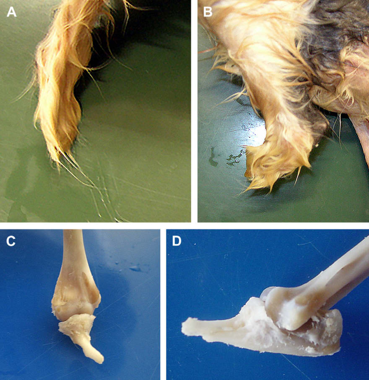
Case 2: frontal (A) and lateral (B) views of the right forelimb showing absence of the distal segment. Frontal (C) and lateral (D) views, gross appearance of the right elbow.
In both cases, radiographs of the antebrachium were taken, using two standard orthogonal projections (mediolateral and craniocaudal views). In case 1, radiography of both distal forelimbs was performed. The right foreleg was radiographically normal. The radiographs of the left foreleg are shown in Figs 3 and 4.

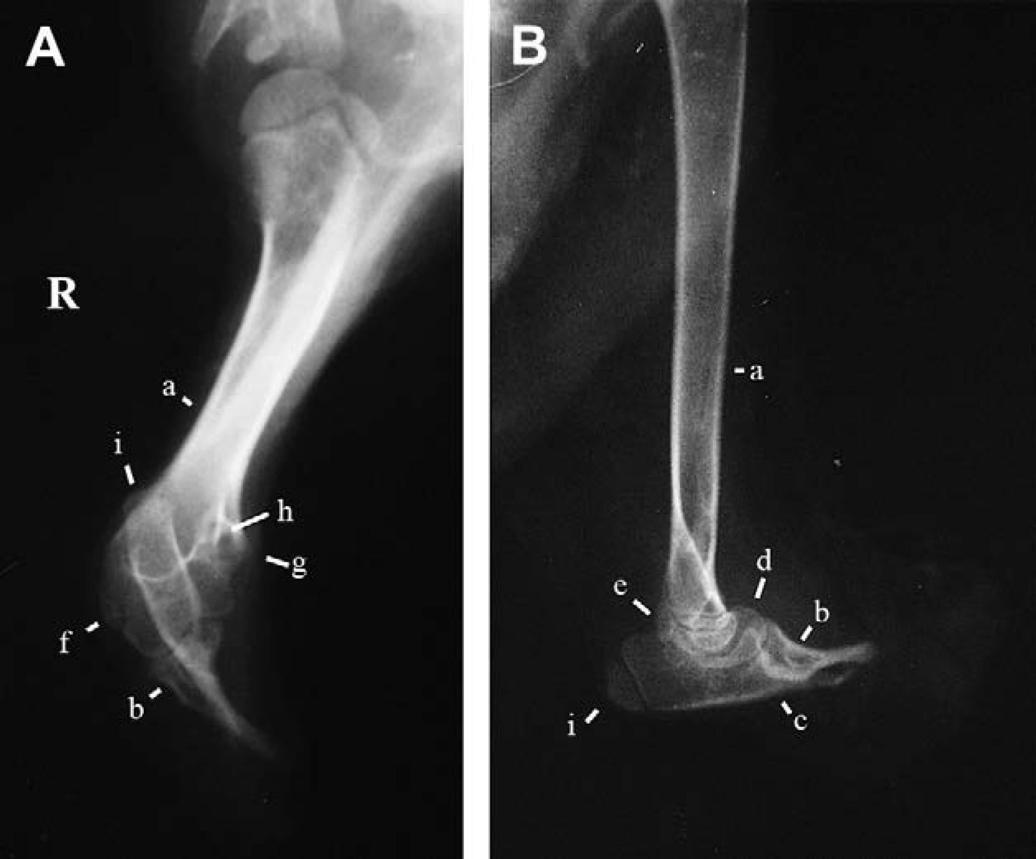
Case 1: mediolateral radiographs of the forelimbs. Ulna (a); radius (b); carpal bone (c); metacarpal bone (d); proximal phalanx (e); middle phalanx (f); distal phalanx (g); humero-ulnar synostosis (i).

Case 1: craniocaudal radiographs of the forelimbs. Ulna (a); radius (b); carpal bone (c); metacarpal bone (d); proximal phalanx (e); middle phalanx (f); distal phalanx (g); unknown, carpal bone or metacarpal bone (h).
In the elbow region there was a bony bridge between the distal humerus and the tuber olecranon of the ulna (humero-ulnar synostosis). There was malformation of the trochlea, capitellum, medial and lateral epicondyles of the distal humerus. The supracondylar foramen was absent and there was marked hypoplasia of the ulna and radius. Ectrodactyly was present between the radius and ulna. There was deformity of the proximal radius, which is displaced craniolaterally; the distal radius is deviated medially.
There was also deformity of the proximal ulna, absence of an anconeal process, medial and lateral coronoid process and trochlear depression; all the structures were rotated and medially deviated.
For case 2 radiographs were taken of the right antebrachium ( Fig 5 ). There was a relatively normal proximal ulna and elbow joint. The antebrachium was very short with complete loss of the distal ulna and radius and absence of the distal bone segments of the right forelimb. There was evidence of a tuber olecranon and the head of radius articulating with the distal humerus.
Case 2: craniocaudal (A) and mediolateral (B) radiographs of the right forelimb. Humerus (a); radius (b); ulna (c); condyle of humerus (d); caudal border of medial epicondyle of humerus (e); lateral epicondyle (f); medial epicondyle (g); supracondylar foramen (h); tuber olecranon (i).
Embryologically, the primary step in limb formation is the aggregation of the somatic mesoderm cells of the hypomere beneath the surface ectoderm, forming the limb bud. During development of limb buds, the superficial ectoderm becomes thicker along the distal ridge, the so-called ‘apical ectodermal ridge’ (AER). This persists until the condensation of the digital mesenchyme. 1 The critical period for a limb's development in the cat embryo is between the 16th and 28th day after fertilisation, when tissues are more susceptible to external influences. 1 Although little is known about the basic mechanisms of congenital limb deformities, several aetiological factors have been identified. 10
Among congenital anomalies, ectromelia is a limb malformation characterised by a severe hypoplasia or aplasia of one or more long bones of one or more limbs. 1 Ectromelic limbs are classified on the basis of the affected limb segment. For example, ectromelia of the stylopodium refers to a limb that terminates anywhere along the femur or humerus, and ectromelia of the zeugopodium where it terminates along the radius and ulna or tibia and fibula, and ectromelia of the autopodium where it terminates at the bones of the manus.
The radiographic appearances were suggestive of a diagnosis of autopodium ectromelia (ectromelia of carpal and metacarpal bones) associated with humero-ulnar synostosis in the first case and zeugopodium ectromelia in the second case. In both cases, the observed conditions were congenital.
In veterinary medicine, a case of ectromelia in an adult cat was cited in 1915 by De Lima, brachymelia and adactyly have been reported in cats9,11,12 while only a single case of bilateral synostosis, involving the radius and ulna, with secondary elbow malformation, has been described in this species. 13
In our cases there was no information concerning traumatic events, inbreeding and/or evidence of environmental or genetic influences and thus the aetiology of these abnormalities remains unknown.
Congenital feline limb deformities are rare and these two additional cases represent an important addition to the scant literature on this subject.