
Abstract
A 1.5-year-old female Persian cat was presented for inappetence and azotemia. Ultrasonography and urography revealed multiple abnormalities involving the genitourinary tract, including agenesis of the right kidney and ureter. Gross examination of the abnormal uterus revealed segmental aplasia of right caudal uterine horn causing cranial distension with fluid, a normal left uterine horn, and both normal ovaries. Microscopically, endometrial glands of the right uterine horn were markedly decreased in number. The right uterine horn was hemorrhagic suggesting estrus. This is the first report of this combination of urinary and uterus abnormalities in the veterinary literature.
Duplication of the female reproductive tract results from partial or complete non-fusion of the paired Müllerian ducts during embryological development. 1 These disorders result in segmental aplasia in the uterus, cervix, and vagina. Generally, this congenital abnormality of reproductive tract is not detected until first estrus in animals. 2–4 Segmental uterine atresia interrupts expulsion of endometrial secretions, which accumulate and enlarge the cranial portion of the obstructed segment. 5 Genital abnormalities are characterized by the congenital absence of the segment in the human reproductive tract. Recently, concomitant renal abnormalities have been linked to this condition in humans. 6 In this paper, we report concomitant abnormalities in the genitourinary tract in a Persian cat.
A 1.5-year-old female Persian cat was admitted to a local veterinary hospital due to anorexia, nervousness and estrus-related aggressiveness. Clinicopathologic testing revealed hemoconcentration (hematocrit 49.7%), azotemia (blood urea nitrogen 31.9 mg/dl, creatinine 1.6 mg/dl) and marked hyperammonemia (NH3 171 μg/dl) which failed to respond to fluid therapy. Urinalysis revealed proteinuria (+) and red blood cells (3+). Ultrasonography of the caudal abdomen failed to identify the right kidney and revealed a fluid-distended uterus. Excretory urography failed to opacify the right kidney and ureter (Fig 1). Exploratory laparotomy revealed an imperforate distended right uterine horn and a normal sized left uterine horn and confirmed the absence of the right kidney and ureter. After surgery the animal was treated with ranitidine (Ranitidine, SCD Pham, 0.1 ml/kg), enrofloxacin (Baytril, Bayer Animal Health, 0.2 ml/kg), tramadol HCl (Tramadon HCl, Shinpoong, 0.1 ml/kg), amoxycillin–clavamox (Amocra syrup, Kunil, 0.4 cc/kg BID), and fluid therapy for 2 weeks and laboratory examinations and urinalysis gradually showed normal values. The reproductive tract with ovaries was resected and submitted to Kyungpook National University for pathologic examination.
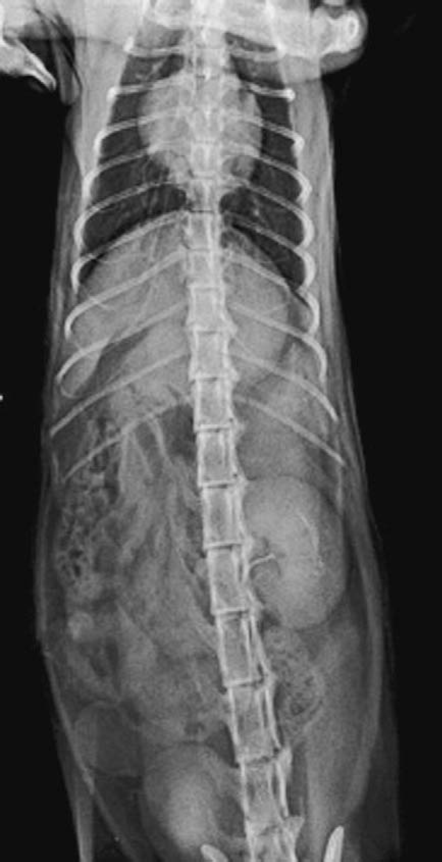
Right renal aplasia and absence of ipsilateral ureter demonstrated by intravenous urography in a Persian cat.
Gross findings of the submitted material revealed unilateral aplasia of the caudal segment of the right uterine horn. The distended segment was filled with turbid watery contents (Fig 2A). Based on the low specific gravity (1.010), low protein (0.8 mg/gl) and the absence of white blood cells on sediment analysis, a diagnosis of hydrometra was made. The left uterine horn and both ovaries were considered to be within normal limits on gross examination.

(A) Dilated right uterine horn with blind end, normal sized left uterine horn, and both normal ovaries are grossly observed after ovariohysterectomy. (B) Microscopically, the endometrium exhibits marked atrophy and loss of endometrial glands. Both layers of uterine muscle are present. Hematoxylin and eosin. Bar=200 μm, a=circular myometrium, b=longitudinal myometrium, c=endometrium.
Upon microscopic examination, the muscular wall of the right uterine horn was markedly thin, although all layers (endometrim, circular myometrium and longitudinal myometrium) were identified. The endometrium exhibited marked atrophy, with few remaining glands (Fig 2B). The left uterine horn was considered within normal limits except for a hemorrhagic region under the superficial glandular epithelium. Both ovaries showed follicular activity including plentiful primodial and Graffian follicles, and hemorrhagic corpus lutea.
Disorders in the development of Müllerian ducts result in various types of segmental aplasia of uterus such as uterus unicornis, segmental aplasia of the uterine body, and segmental aplasia of the uterine horn. 2–4,7,8 Combinations of renal and uterine abnormalities have been often reported in humans; 9 however, reports of this condition are extremely rare in animals. A developmental anomaly of the uterus and concurrent unilateral renal agenesis in a cat was reported by Pierson and Grollman 7 and Robinson. 10 Pearson and Gibbs 11 described concurrent urogenital abnormalities in two calves. The urinary and genital systems are derived from common embryonic mesoderm, which suggests a common source for urogenital abnormalities, although the precise cause of most abnormalities is not known. Two paired Müllerian ducts ultimately develop into the fallopian tubes, uterus, cervix, and upper two-thirds of the vagina. The ovaries and lower one-third of the vagina have separate embryologic origins not derived from the Müllerian system. 12 Unilateral defects of the paramesonephric duct and metanephros may result in ipsilateral defects in both the urinary and genital systems. 13 The etiology and pathogenesis of the syndrome described here and its embryologic origin remain unclear.
Footnotes
Acknowledgements
The authors wish to thank veterinarians and pet owners collecting the material. This research was supported by a grant (code: CBM 31-B3003–01–01–00) from the Center for Biological Modulators of the 21st Century Frontier R&D Program, the Ministry of Science and Technology, Korea.