
Abstract
The selective desorption/ionization of analytes using nanomaterials is investigated using metallic nanoparticles. By replacing the sodium citrate capping of gold nanoparticles with self-assembled monolayers, we are able to both enhance analyte ionization and selectively capture analytes. Capping gold nanoparticles with a monolayer of 4-mercaptobenzoic acid enhances analyte ionization while greatly decreasing chemical noise resulting from alkali adducted species. Selective capture and sequential desorption/ionization of the peptide bradykinin (1–7) from a two peptide mixture is achieved using β-cyclodextrin capped gold nanoparticles. Finally, by switching from gold to silver nanoparticles, we are able to ionize both folic acid and amphotericin B. These results demonstrate that through careful control of nanoparticle surface chemistry and composition one can achieve selective analyte ionization for MS applications.
Keywords
Introduction
Selective capture of low abundance analytes from complex samples is one of many important topics in the development of biosensors and array-based platforms. Nanoparticles offer an excellent candidate to develop such a platform because they possess a high surface area, are readily functionalized with self-assembled monolayers (SAMs), and can be separated out of solution or immobilized on substrates. The surface chemistry of nanoparticles can be modified with SAMs to perform a wide array of functions. In matrix-assisted laser desorption/ionization MS (MALDI-MS), the use of SAMs on gold substrates has proved to be effective as affinity-capture surfaces and for on-probe sample cleanup. 1 -5 Recently, methods have been developed for MS applications that use surface functionalized nanoparticles to pull down and selectively extract analytes either through centrifugation or by magnetic separation. 6 -8 However, these methods typically require the addition of an organic matrix to facilitate the desorption and ionization of analytes for MS analysis.
David H. Russell, Ph.D.

Nanomaterials also provide a means to carry out laser desorption/ionization (LDI) ofanalytes without the use of organic matrices. The early beginnings of MALDI-MS made use of nanometer-sized cobalt particles suspended in glycerol matrix as a means of ionizing and desorbing biological analytes. 9 More recent nanomaterial systems include porous silicon, 10 carbon nanotubes, 11 silicon nanowires, 12 nanostructured silicon, 13,14 size-selected gold nanoparticles (AuNPs), 15 silicon microcolumn arrays, 16 and nanostructure-initiator MS (NIMS). 17,18 We are currently designing novel nanomaterials for LDI-MS with focus on using tailored metallic nanoparticles to provide both analyte selectivity and enhanced ionization capabilities. Gold nanoparticles (AuNPs) offer several unique advantages over organic matrices for LDI-MS including simplified sample preparation, and relatively uncomplicated low mass spectra (lacking noise from matrix clusters). 15
In our research, the nanoparticles act as an absorption chromophore for the ionizing laser irradiation. Ionized analyte and metal clusters are then separated and detected in the mass spectrometer. Figure 1a is a schematic illustrating the use of nanoparticles in LDI-MS. Although the mechanism of analyte desorption and ion formation in this system is not well understood, we suspect the mechanism to be governed by thermal, electronic, and field effects arising from interactions between electrons in the particles and the incident laser irradiation. The nanoparticle chemistry that governs selectivity is controlled by one of two techniques: (1) modifying the nanoparticle composition (i.e., Au, Ag) or (2) decorating the exterior of the nanoparticles with SAMs. Both techniques offer viable routes to provide either selective analyte capture or selective analyte ionization. Furthermore, the properties of the SAM can be tailored to act as a proton source in the desorption/ionization event. 19 Figure 1b illustrates a decorated nanoparticle with mixed SAMs for selective analyte capture and enhanced ionization properties.
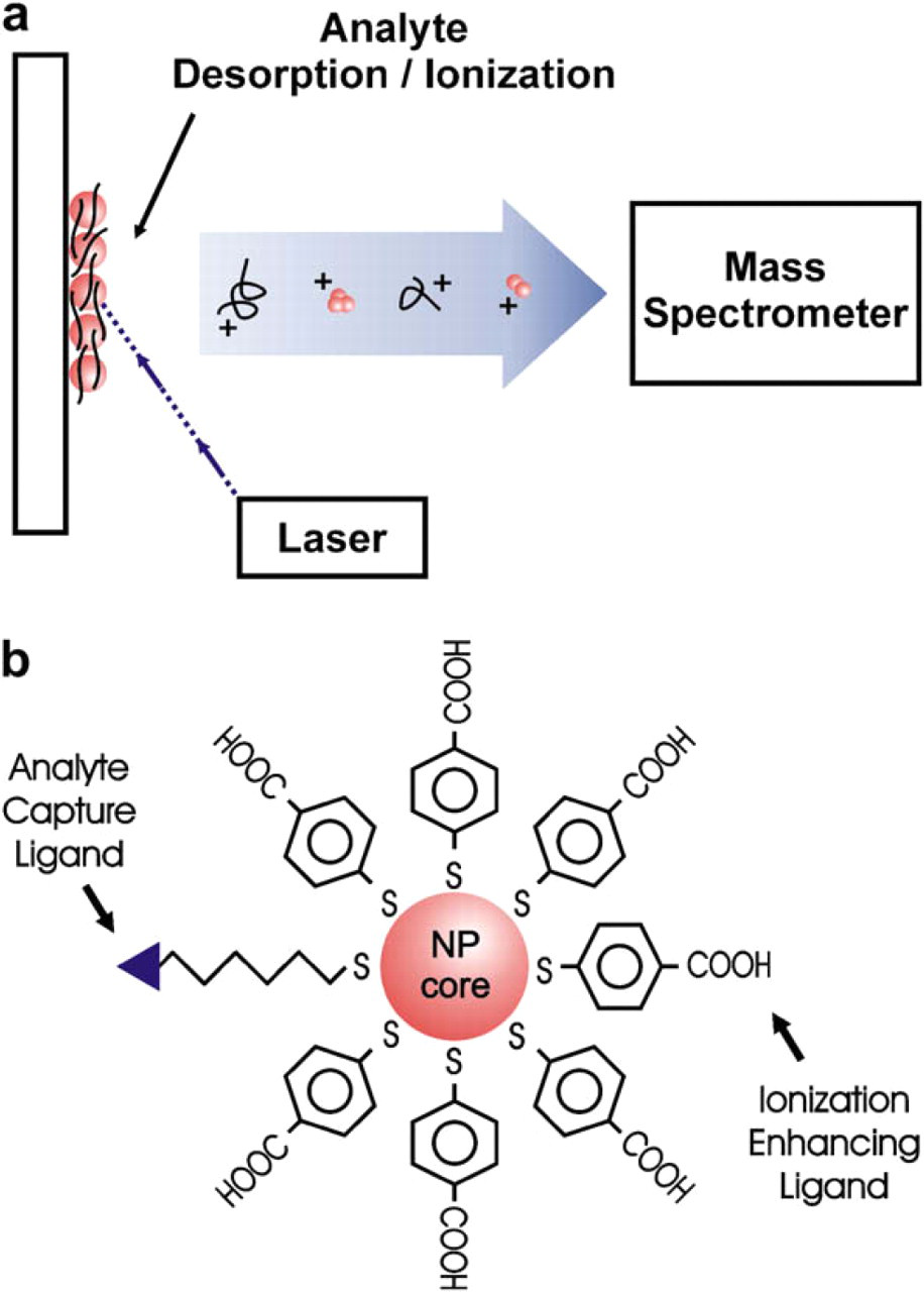
(a) Schematic representation of the nanoparticle-based LDI process. Ions are produced by 337 nm irradiation of a sample spot containing a mixture of nanoparticles and analyte. (b) Illustration of a hybrid nanoparticle capped with both ionization enhancing and analyte capture ligands.
Materials and Methods
Laser Desorption/Ionization Mass Spectrometry
All laser desorption/ionization time-of-flight mass spectrometry (LDI TOF-MS) experiments were carried out on a Voyager DE-STR (Applied Biosystems, Foster City, CA) under optimized conditions in reflected mode. LDI was performed by irradiating the sample with 1.0–2.5 mJ/cm 2 of 337 nm laser light. Care was taken to insure that all samples and control experiments were integrated under the same experimental conditions (e.g., laser fluence).
Citrate-Capped AuNPs Synthesis
All glassware was cleaned with aqua regia, rinsed with 18 MΩ water, and baked dry before use. AuNPs with an average diameter of 10 nm were prepared according to the citrate reduction method. 20 Briefly, 250 mL of 0.25 mM HAuCl4 (Sigma Aldrich, Milwaukee, WI) was brought to a boil and 25 mL of 38.8 trisodium citrate (Sigma Aldrich) was added. After a rapid color change to dark red, the solution was refluxed for an additional 10 min, removed from the heat and allowed to cool to room temperature. The cooled nanoparticle solution was then filtered through a 0.2 μm membrane. The nanoparticles were characterized by UV-vis and transmission electron microscopy.
Monolayer Capped AuNPs
4-MBA monolayer–capped AuNPs were prepared by incubating 1 mL of the citrate-capped AuNP solution with 0.5 mL of 5 mM 4-MBA solution in 6:1 methanol:acetic acid overnight. The particles were then purified by five cycles of centrifugation, removal of the supernatant, and resuspension of the particles in 0.1% trifluoroacetic acid (TFA). Mild sonication aided in the resuspension of the particles.
β-cyclodextrin capped nanoparticles were synthesized as follows. A 0.5 mM amino (6-monodeoxy-6mono-) β-cyclodextrin hydrochloride (Cyclodextrin Technologies Development, Inc., High Springs, FL) stock solution was made in 18 MΩ water and the pH was adjusted to 10 with sodium hydroxide. To a 1 mL of citrate-capped AuNP solution, 0.5 mL of 5 mM amino β-cyclodextrin solution was added and allowed to incubate overnight. The particles were then purified by five cycles of centrifugation, removal of the supernatant, and resuspension of the particles in 18 MΩ water. Mild sonication was used to aid in the resuspension of the particles.
Enhancing Laser Desorption
The analyte angiotensin II (American Peptide Company, Sunnyvale, CA) and either citrate-capped AuNPs or 4-MBA–capped AuNPs were mixed at a 500:1 molar ratio. A 2 μL aliquot of the analyte nanoparticle mixture was spotted directly onto a stainless steel sample plate, dried under vacuum, and loaded into the mass spectrometer. As a control experiment, 10 μL of 1 mM 4-MBA in 0.1% TFA was mixed with 2 μL of a 1 mg/mL angiotensin II solution and also spotted on the sample plate.
Selective Extraction With β-Cyclodextrin Capped AuNPs
Extraction experiments were carried out by first preparing a solution of 200 nM bradykinin (1–7) and 2 μM C-telopeptide (American Peptide Company). To this solution, a 20 μL aliquot of β-cyclodextrin–capped AuNPs was added. The particle–analyte mixture was allowed to incubate for 30 min followed by two cycles of centrifugation and resuspension in 18 MΩ water. The final resuspension brought the solution to ∼ 50 μL, from this, a 2 μL aliquot was spotted onto the sample plate for MS analysis. For comparison, a 250 μM bradykinin (1–7) and 250 μM C-telopeptide was mixed with β-cyclodextrin capped AuNP in a 500:1 analyte to nanoparticle molar ratio of which, a 2 μL aliquot was spotted onto the sample plate. An extraction was also attempted with 4-MBA–capped AuNPs.
Ionization of Olefins Using AgNPs
Folic acid and amphotericin B were purchased from Sigma Aldrich and used without further purification. The 20 nm silver colloids were purchased from Ted Pella, Inc. (Redding, CA). Stock solutions of vitamin or drug were dissolved in dimethyl sulfoxide at 100 pmol/μL. The AgNPs were spun down for 20 min at 7000 rpm followed by resuspension in methanol. The AgNP solution (in MeOH) was mixed 10:1 (v/v), NP solution: vitamin or drug. One microliter of each sample solution was spotted onto the sample plate and dried in vacuo.
Results
Enhancing Ionization With Self-Assembled Monolayers
One of the many reasons for capping the outer surface of nanoparticles with small molecules is to prevent flocculation. In the synthesis of AuNPs, sodium citrate serves a dual purpose: as a reducing agent during the AuNPs synthesis and as a capping agent to prevent nanoparticle flocculation. However, when citrate-capped AuNPs are used for LDI-MS, the resulting spectra sometimes lack protonated analyte ions and are convoluted with peaks arising from multiple alkali adducts. This is problematic because the presence of multiple alkali adducts in a mass spectra of a complex mixture (e.g., a protein digest) complicates peak assignment.
Here we demonstrate that capping AuNPs with a SAM of 4-MBA increases protonated analyte ion yields while drastically decreasing the presence of peaks arising from alkali adduction. These effects are observed as a result of the 4-MBA monolayer (1) acting as a source of protons and (2) allowing us to remove the source of alkali contamination. Figure 2a contains LDI-MS data of the peptide angiotensin II acquired with citrate-capped AuNPs. We observe peaks corresponding to the [M + H]+ ion, [M + Na]+ ion, [M + K]+ ion, and several other peaks arising from analyte molecules clustered with multiple alkali ions. Figure 2b contains LDI-MS data of the angiotensin II acquired using 4-MBA–capped AuNPs. We observe an increase in the abundance of the protonated analyte ion signal and a dramatic decrease in the signal corresponding to [M + alkali]+. Also, the peaks resulting from analyte with multiple alkali adductions are essentially eliminated. It is important to note that in this study, at the laser fluence levels used, mixtures of analyte and either 4-MBA or 0.1% TFA produced no discernible analyte ion signal above the background.
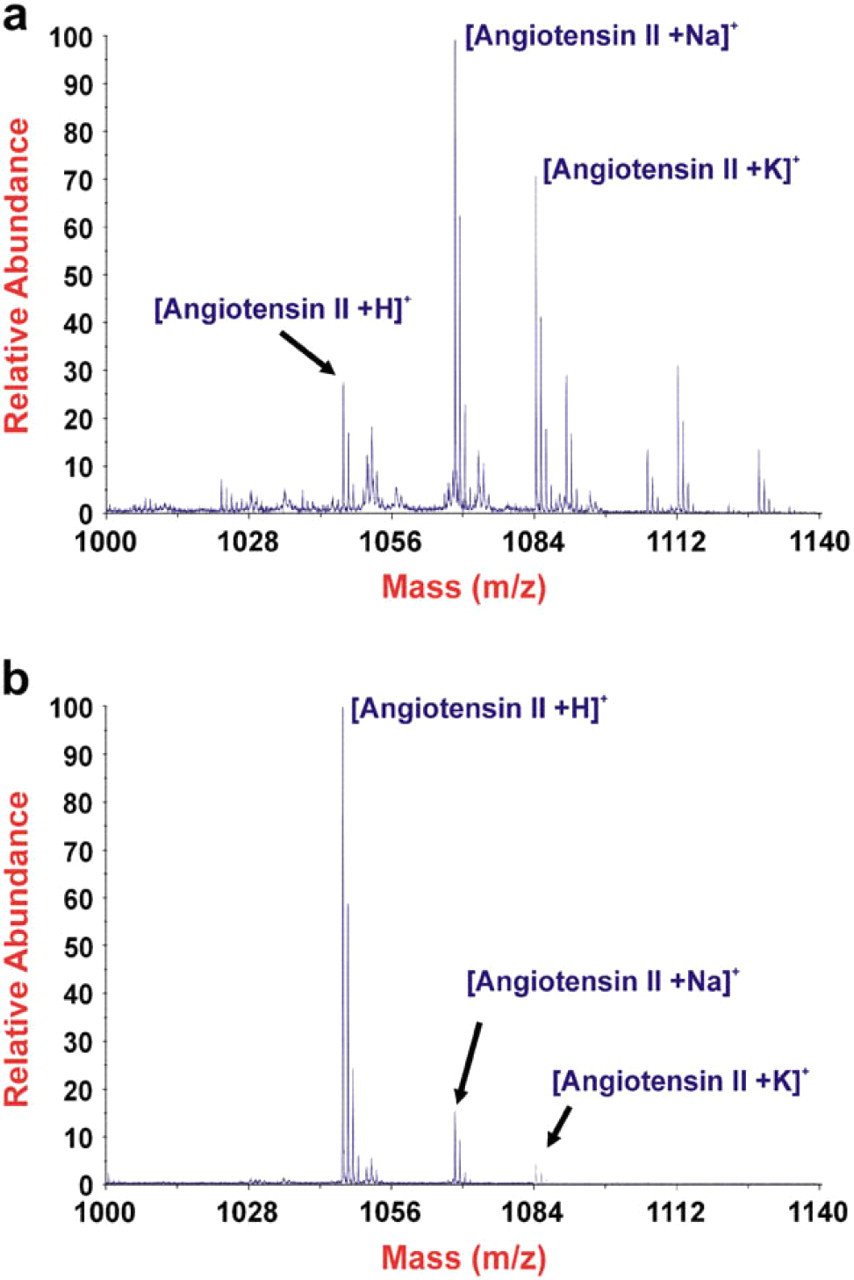
LDI time-of-flight mass spectra of angiotensin II using both citrate-capped AuNP (a) and 4-MBA–capped AuNPs (b).
Selective Analyte Capture and Ionization With Self-Assembled Monolayers
In addition to enhancing analyte ionization, capping nanoparticles with SAMs also allow us to impart selectivity into our system. Through the use of commercially available materials, the surface chemistries of SAMs are easily modified to present a myriad of functionalities. This allows us to tailor our system for analyte pull down experiments where it is desirable to selectively capture and ionize low abundance analytes from complex mixtures.
We chose to modify the surface of our nanoparticles with β-cyclodextrins owing to their ability to form inclusion complexes with side chains of the aromatic amino acids tryptophan and phenylalanine. 21 , 22 To demonstrate selective capture and ionization of a low abundance analyte, the peptides C-telopeptide (EKAHDGGR) and bradykinin (1–7) (RPPGFSP) were mixed in a 10:1 molar ratio. The sequence of bradykinin (1–7) contains a phenylalanine (F), whereas C-telopeptide does not. After incubation (with the two peptides) and extraction using the AuNP capped β-cyclodextrin, the captured analyte (bradykinin [1–7]) was ionized without the use of an organic matrix. Figure 3 (blue spectrum) contains the data from a selective analyte capture and ionization experiment using β-cyclodextrin capped AuNPs. LDI-MS data from the control experiment where a mixture of bradykinin (1–7) and C-telopeptide were desorbed and ionized using β-cyclodextrin capped AuNPs without extraction is also shown (Fig. 3, red spectrum). Extraction experiments using 4-MBA–capped AuNPs were negative for both peptides (data not shown). We have successfully demonstrated the selective capture and ionization of a lower abundance peptide from a two peptide mixture.
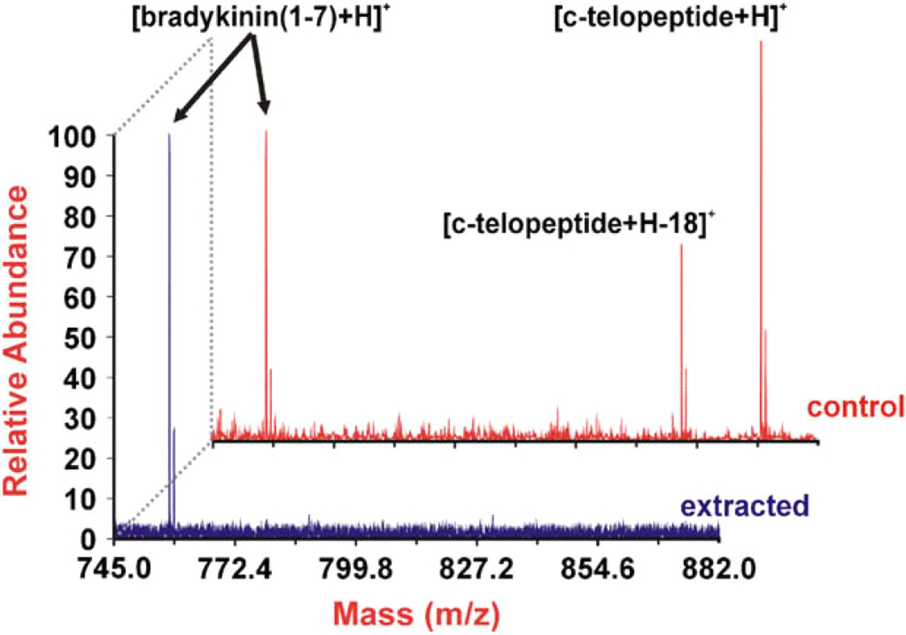
LDI time-of-flight mass spectra using β-cyclodextrin–capped AuNPs. The blue spectrum (extracted) is acquired after extracting bradykinin (1–7) from a two peptide mixture. The red spectrum (control) is acquired using a mixture of the peptides bradykinin (1–7) and C-telopeptide.
Ionization of Olefins Using AgNPs
Although we have modified the surface chemistry of nanoparticles to capture and ionize specific analytes, it is also possible to use the nanoparticle composition to selectively ionize specific functional groups. In this work, the silver–olefin interaction is exploited to ionize olefin containing species. The silver–olefin interaction has been used since the early 1960s to separate fatty acids and lipids in silver ion chromatography 23 -26 and the interaction between silver ion and olefin has been described by the Dewar model. In brief, the Dewar 24,27 model predicts that silver and olefin interact by both a σ and π bond. Figure 4 contains data illustrating the ionization of olefins using silver nanoparticles. Specifically, folic acid (Fig. 4a) and amphotericin B (Fig. 4b) are analyzed using 20 nm silver nanoparticles for LDI-MS. In both vitamin (folic acid) and drug (amphotericin B) samples, we observe only [M + Ag]+ ion species (i.e., no [M + H]+ ion is observed). We interpret these results as evidence that the addition of the AgNPs facilitates ionization as the [M + Ag]+ ion of both folic acid and amphotericin B. These results also suggest the ability to selectively ionize olefins from a complex mixture owing to silver's preference to interact with olefins.
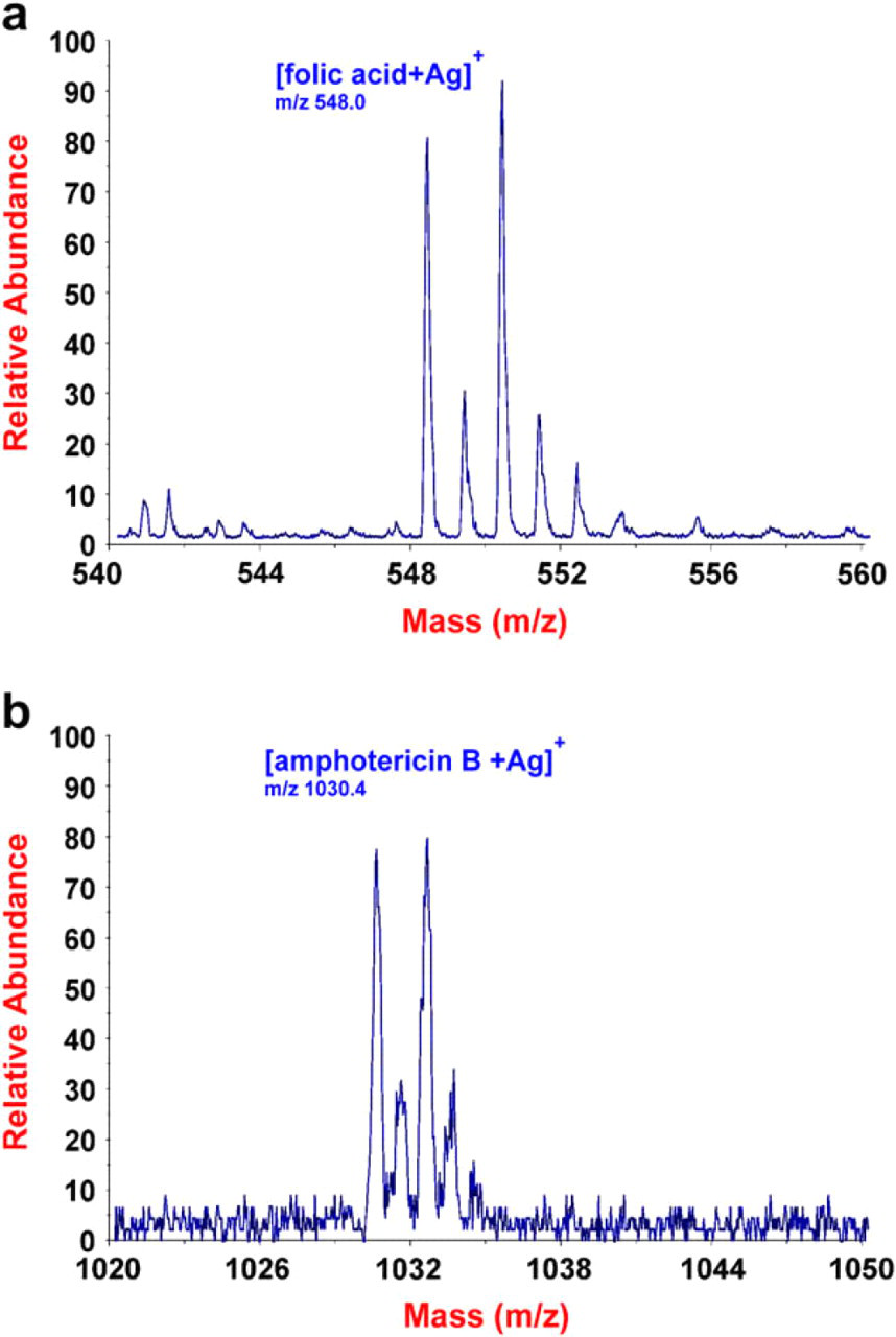
LDI time-of-flight mass spectrum of folic acid and amphotericin B obtained using 20 nm AgNPs. [M + Ag]+ ions of folic acid and amphotericin B are observed in the mass spectrum.
Conclusions
We have successfully demonstrated that controlling the surface chemistry is a key aspect in tailoring the properties of nanoparticles for MS applications. Through the use SAMs and by altering nanoparticle composition, we are able to (1) enhance the ionization of analytes, (2) reduce analyte alkali adduction, (3) selectively capture analytes, and (4) ionize analytes containing specific functional groups. This work highlights several benefits of nanomaterials in biological MS and illustrates the potential to provide selective analyte desorption/ionization of low abundance analytes from highly complex samples. That is, by modulating both nanoparticle material and surface chemistry it may be possible to simultaneously impart both analyte pull-down capabilities and enhance analyte ionization. In combination with high throughput imaging MS, these nanomaterial systems have the potential to aid in developing label-free screening assays for early identification of disease biomarkers.
Acknowledgment
This research was supported by a grant from the Robert A. Welch Foundation (A-1176) and the U.S. Department of Energy, Division of Chemical Sciences, BES (DE-FG02–04ER5520).