
Abstract
With biological discovery and technology advancing in parallel, AstraZeneca (AZ) announced that it would soon take delivery of what is thought to be the first fully automated high-throughput high-content screening system. This custom designed assay platform from RTS Life Science International (RTS) automates the IN Cell Analyzer 3000 sub cellular analysis system from Amersham Biosciences. RTS has integrated its advanced scheduling system with the imaging tool to enable AZ to evaluate the effect of drug compounds on cellular processes.
Introduction
Following AZ's embracing of ultra high-throughput screening (HTS) as detailed in JALA, 1 the company is also one of the first to adopt high-content screening (HCS). The company, along with many of its competitors in the drug discovery field, is focusing on the elimination of bottlenecks and production of high quality ‘hits’ in primary screening. This trend is only a few years old. Prior to that, efforts to streamline processes and select for quality leads were put into later screening. Many of the current cellular assay technologies employed by HTS do not allow characterization, analysis, and screening of target drugs in a truly biological context.
Potentially, the ability to deploy a robust HTS format to transfer a primary screen using live cells to a secondary screen without having to wait or reconfigure the assay was always going to become the next goal of the early drug discovery process. Conveniently, HCS combines high-context cellular assays with high-quantity input. For this reason, HCS has been the subject of much anticipation over the last couple of years. Its ability to use multiple fluorescent probes for parallel labeling and analysis of manifold parameters has become the next leap forward in the sphere of cell-based assays for drug discovery.
To succeed, the new technology draws on expertise in the fields of fluorescent microscopy, cell biology, and automation, creating a new set of screening tools. At the heart of this system lies a reader capable of converting multiple fluorescent signals from raw image data into kinetic analysis of multiple cellular activities within individual cells.
IN Cell Analyzer 3000
The IN Cell Analyzer 3000, the advanced HCS instrument from Amersham Biosciences, underwent beta testing at AZ in the summer of 2002 and the company has now placed an order for two systems. The team based at AZ's Advanced Science and Technology Laboratory found that the stand alone reader lived up to its claims, allowing scientists to probe deep inside a living cell and evaluate directly the effects of a drug compound on cellular processes in real time. The real-time element is deemed especially pertinent, as it theoretically allows the researcher to watch the cell undergo changes as they happen.
The IN Cell Analyzer 3000's detection technology permits sub-cellular imaging and the analysis of nearly 400 assays in as little as 15 minutes and of more than 12,000 cellular images in eight hours. At its heart is a proprietary, laser-based, confocal imaging system that includes three high-speed cameras. These view red, blue, and green emissions simultaneously, negating the need for consecutive reads.
The simultaneous use of three lasers makes this reader the fastest available. Confocal imaging means that the user can focus on cells within different layers of culture with ease and, in automatic mode, the reader is able to compensate for curvature or irregularities of well surfaces. Rapid feedback of the reader's view in space is provided by a fourth positioning laser.
The IN Cell Analyzer 3000 builds upon Amersham Bioscience's development of a live cell reporter gene system, which allows the differently colored fluorescent substrates to link to cellular reporters, including GFP. Sophisticated optics and software are then used to analyze these images in real time, allowing biologists to observe reactions at a sub-cellular level.
By maintaining cell culture conditions during the screening process, the IN Cell Analyzer 3000 facilitates the performance of time dependent kinetic assays on living cells and extended analytical assays.
Reducing secondary screening requirements, the IN Cell Analyzer 3000 provides better quality data while saving both time and reagent cost. AZ has found reagent costs spiraling in recent years, because although the formats have generally moved from 96 wells to 384 and even 1536, the sheer volume of assays a typical HTS department undertakes in a year means that demand has kept growing. As HCS looks at the stimulation and inhibition of receptors on an individual cell level, each cell can act as its own control, negating the need for control wells that further saves reagent cost.
The IN Cell Analyzer 3000's large field of view (0.75 mm × 0.75 mm) enables the user to view more cells in fewer images, thus helping the rapid acquisition of statistically significant data. This raw image data is converted automatically into signal distribution, area, morphology, and activity measurements, which can be stored for review and analysis at a later date.
Given the large number of different assay types the IN Cell Analyzer 3000 is capable of carrying out, flexibility was a key driver to its functionality. The system contains a large range of assay and image analysis algorithms, allowing it to handle:
Receptor internalization Cell signaling Cell cycle analysis Gene reporter assays Apoptosis
However AZ realized from the outset, and this was confirmed during the trial of the IN Cell Analyzer 3000, that the complexity of the biology is such that the processing could take longer than a working day, making automation essential if the reader technology is to be fully utilized.
The Creation of an Automated HCS System
As AZ already used the RTS Assay Platform for HTS, it approached RTS Life Science to integrate the IN Cell Analyzer 3000 with the other instruments needed to provide a fully automated HCS system (see Figures 1). The timing of compound addition and even dispensing speed is crucial if a good read is to be attained. Therefore AZ was aware scheduling and process control was going to be as important as the successful integration of the different instruments with the HCS reader. See Figures 1.
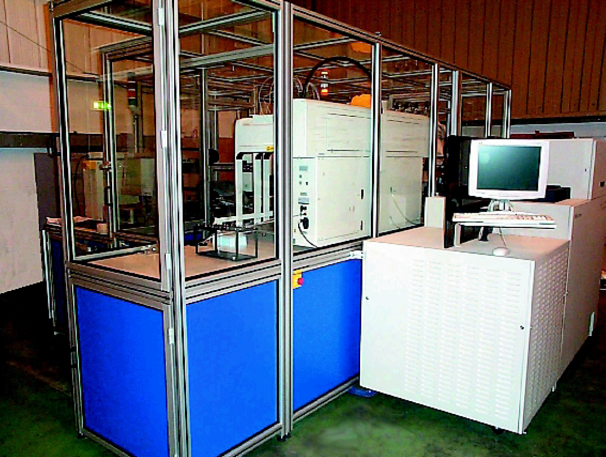
The RTS Assay Platform provides ready-to-read plates to the IN Cell Analyzer 3000 while the reader remains accessible for manual use.
Biological processes are dynamic and do not fit within the confines of a rigid automated system. RTS's mature SPRINT real-time scheduling software (see Figures 2) was a crucial factor in the choice of RTS to perform the system integrator role. SPRINT ensures that the use of the IN Cell Analyzer 3000 is optimized, so that the user maximizes its investment in the reader. For example, if the biology for a particular assay takes five minutes but the estimated read time is 10 minutes, then SPRINT schedules the throughput so that only half a plate is read at a time.
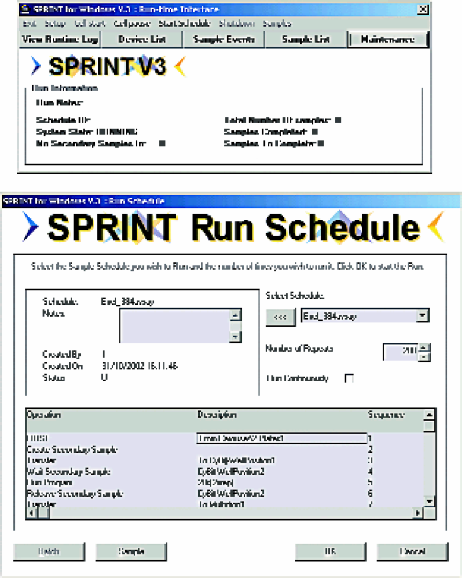
Windows based, SPRINT real-time scheduling software user interface examples.
Investment in each run and plate is high. Reliability is paramount and had to be built in from the inception of the design process. The completed system was rigorously tested in the RTS workshops. SPRINT monitors the status of all instruments and each plate. Any instrument error is reported, automatically handled, and any affected plate flagged. SPRINT will report errors to a remote operator via page or e-mail.
During the trials of the IN Cell Analyzer 3000 at AZ, it was realized that dispensing speed was crucial, as the reagents need to be dispensed as rapidly as possible for good mixing to be achieved, but not so fast that cells are dislodged off the bottom of the well. The Biomek FX was chosen as the dispenser within the integrated system.
The delivery of the system is scheduled for the second quarter of 2003. To further maximize the utilization of Amersham Biosciences' IN Cell Analyzer 3000, the system has been designed for both operator and robotic access. Operators and robots can each gain access from opposite instrument sides, so there is no need to remove the reader from the system.
A Staübli multi-axis robot sits at the center of the assay platform. All the instruments surrounding the robot - the Biomek FX which aliquots and dispenses, the Cytomat incubators and carousel which enable plate incubation and compound plate entry, and the two Multidrop dispensers - are mounted on slide tables with drag links and cable management systems. During maintenance, each instrument is easy to pull out and then slide back in.
Working on similar principles, the cabinet that encases the entire system except the computer, protecting both its operators and the cells involved in the assay, is formed of 10 separate sections. Different instruments can be added and maintenance can take place in a phased manner. The entire enclosure can be accessed easily via a manual walk-in area, facilitating easy maintenance and also making robot teaching more straightforward. This also allows the individual instruments to be used in bench top mode when the whole system is not in use.
The IN Cell Analyzer 3000 results are easily interpreted once complete because SPRINT produces a file similar to an Excel spreadsheet. Compound plates are barcode read and the codes imported to the IN Cell Analyzer 3000 via SPRINT. This allows full tracking of data making single and part plate reads possible. The reading of part plates is crucial, as it allows SPRINT to maintain the synchronous assay processing times vital for quality and noise free results, whether numeric or image based. The automated system also has the equipment needed to carry out kinetic assays and then fix them.
Conclusion
High-throughput high-content screening makes possible on an industrial scale what scientists have achieved for years using microscopes. This said, this automated system does not appear too remarkable, but it brings a subtlety into HTS that could, until recently, only be dreamed of.
The amount of data generated from such a system seems frighteningly large, but as the measurement of all the biological events take place on a cell-by-cell basis, it is possible to revisit it repeatedly, resetting the assay parameters if necessary. The fluorescent labels allow close analysis and even visualization of events taking place within the cells, such as a particular protein moving from its position in the cytoplasm to the nucleus. Simultaneously, it is also possible to observe the movement of the cells and any changes to their shape. Contrasting HCS with standard HTS, where only one biological event can be measured, is like moving from black and white into color. The laser positioning system is so accurate that when cells are reread later during the same assay, the reader reads 95% of the same cells it captured during the initial read.
The project to develop an integrated, automated, high-throughput HCS system has drawn on several emerging technologies. The sphere of assay development will be filled with complex choices in the years ahead. The development of the most appropriate assays for a flexible system, which potentially can yield such a wealth of information, could prove more challenging than initially envisaged. However, scientists using an HCS system will need to make fewer assumptions and so their work will have greater focus.