
Abstract
Objective
To evaluate the clinical and functional results of surgical treatment for fibrous long head of the triceps in children.
Materials and methods
Data were analyzed from 32 patients (38 shoulders) aged over 5 years of age from August 1995 to December 2004. The adduction contracture, elbow flexed angles when the scapula was held in the chest wall, and scapulo-humeral angles in radiographs were measured. Surgical release of the long head of the triceps was performed.
Results
There were 22 females and 10 males in this study. Bilateral shoulder involvement was found in six patients. Only the right shoulder was involved in 5 patients, and only the left in 21 patients. All 32 patients (38 shoulders) developed adduction contracture of the shoulder after repeated intramuscular injection of antibiotic(s) into the long head of the triceps. Thirty-four shoulders (29 patients) were classified as severe, and four shoulders (3 patients) were classified as moderate. In all, we attained excellent results in 36 shoulders (94.7%) and good results in two shoulders (5.3%). There have been no fair or poor results or complications so far.
Conclusion
Generally, surgical treatment of adduction contracture of the shoulder has achieved good results, with improved shoulder function. Releasing the long head of the triceps is a simple and safe surgical technique.
Keywords
Introduction
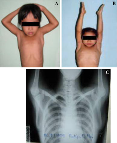
Fibrous change in muscles may be congenital or secondary to intramuscular injection or direct trauma [1–7]. In Asian countries, intramuscular injections are used commonly to treat children who have an infection or even only a fever. Shoulder adduction contracture is due to a fibrous long head of the triceps muscle, as identified intraoperatively and confirmed histopathologically. Cosmetic problems include adduction contracture of the shoulder and outward movement of the scapula when the elbow is flexed, dimpling of the skin, and a palpable long head of the triceps (Fig. 2a–d).
Materials and methods
A retrospective study was undertaken to evaluate the results of surgical techniques performed from August 1995 to December 2004 in 36 patients (42 shoulders) with fibrous long head of the triceps muscle. Four patients (four shoulders) were excluded from the study because of insufficient follow-up. The remaining 32 patients (38 shoulders) formed the basis of this study. In six of these patients, their condition was also associated with fibrous contractures of quadriceps, triceps (opposite shoulder), deltoid, and rectus femoris muscles, likely due to a similar process.
Preoperatively, information was obtained about the type and quantity of medication that had been injected, history of trauma, age at the onset of symptoms, duration of symptoms, associated contractures, localized skin changes, pain, cosmetic problems, and changes in functional activities.
In all patients, measurements were taken with a goniometer of the adduction contracture angle and range of motion (ROM) of the elbow with the scapula held at the chest wall (Fig. 2e), the radiographic scapulo-humeral angle, as well as the range of motion (Fig. 2f). Anteroposterior radiographs of both shoulders with both arms in a maximum abduction were done systematically for each patient. Rotation of the scapula was assessed on anteroposterior radiographs of the chest with both arms in a maximum abduction position. Upper extremity function was evaluated according to functional points of Quick DASH (Disabilities of the Arm, Shoulder and Hand) [8–11].
Ultrasound confirmed fibrous long head of the triceps. Axial radiographs, arthrograms, computerized tomography scans, or magnetic resonance images were made when a fibrous long head of the triceps muscle was associated with a fibrous deltoid and there was suspicion of rotator-cuff disease or subluxation or dislocation of the humeral head.
Severity was classified according to the criteria shown in Table 1.
Degree of severity of the disease
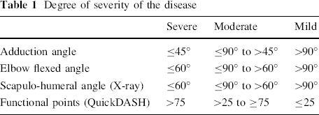
To be considered for operative intervention, patients were at least 5 years old and had at least 60° of adduction contracture. In addition, they showed evidence of progressive deformity during growth or change in bony anatomy, chronic discomfort, and concern about the appearance.
One surgeon (the author) performed all operations.
Operative procedure
To perform Henry's posterior exposure [12], place the patient prone on the operating table with the upper extremity on an arm board, the arm and shoulder free. The lateral decubitus position with the arm supported on bolsters also works well. Under general anesthesia, abduct the shoulder with maximum elbow flexion.
Identify the long head of the triceps. Using this as a landmark, make an incision 4 cm inferior to the acromion, extending distally along the medial edge of this muscle for 6 cm (Fig. 1a).
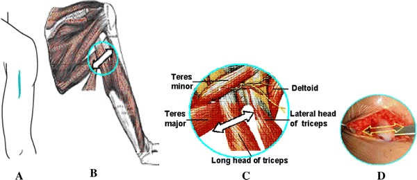
Develop the skin and subcutaneous tissue to expose the long and lateral heads of the triceps. Digitally develop the proximal interval between these two heads of the triceps. Lift the seam free from the underlying tissue and split it proximally. Identify the contractrure and fibrosis of the long head of the triceps by elbow flexion and extension.
Notice the relation of the radial nerve and its branches to the three heads of the triceps. Protect the median nerve, ulnar nerve, and brachial artery medially, and the radial nerve and profunda brachii artery laterally. Confirm that branches of the radial nerve are identified and protected before release of the long head of the triceps at the level of the deltoid tubercle of the humerus and inferior edge of the teres major (Fig. 1b–d).
Upon completion of the release, coagulate all sources of bleeding to achieve complete hemostasis. Close the skin without drainage; place the arm in adduction with the elbow at 90° and held by a silk band.
Postoperative rehabilitation
The shoulder was exercised starting on the 3rd postoperative day. During the first 2 weeks, the scapula was held at the chest wall with maximum elbow flexion, and a passive range of shoulder abduction up to 100° was performed with the help of a physiotherapist. After 2 weeks the range of motion was increased to 130°, and after 3 weeks the range of motion was increased to normal. A full range of shoulder abduction was allowed between the 4th and 6th weeks after the operation.
Postoperatively, we documented the following: dimpling of the skin, range of motion, adduction contracture, elbow extension angles, elbow flexion angles with the scapula held at the chest wall, functional use of the upper extremity for grooming, radiographic scapulo-humeral angles, results of manual muscle testing according to the Jones's classification, and complications (Table 2).
Criteria used to assess surgical results
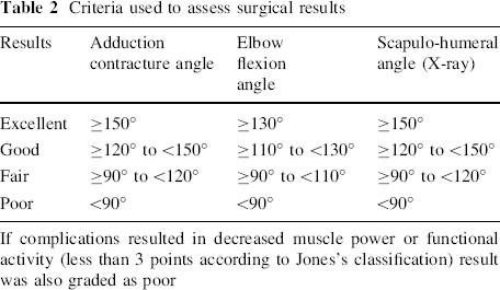
If complications resulted in decreased muscle power or functional activity (less than 3 points according to Jones's classification) result was also graded as poor
We evaluated the patients independently at 1 week, 3 weeks, 6 weeks, 3 months, and every 6 months thereafter. An anteroposterior radiograph and a scapular radiograph of the shoulder, as well as an anteroposterior radiograph of the chest that included both shoulder joints, were made 1 week and 6 weeks after the operation, and every 6 months thereafter if any changes in the shoulder joint occurred.
Results
From August 1995 to December 2004, 32 patients (38 shoulders) were operated. In all patients, the fibrous long head of the triceps muscle had developed after repeated intramuscular injections of antibiotic(s) into the posterior proximal arm. Most of these injections were done in the first 24 months of life. Injected pharmaceutical agents were all antibiotics: penicillin in 21 cases (65.6%), penicillin and gentamycin in eight cases (25.0%), and others (lincomycin, streptomycin, cloxacilin) in three cases (9.4%). In 19 patients (24 shoulders), the fibrous long head of the triceps developed between the ages of 4 and 9 years. In 11 patients (12 shoulders), the fibrous long head of the triceps developed between the ages of 10 and 14 years, and in 2 patients (2 shoulders), the fibrous long head of the triceps developed between the ages of 15 and 19 years. No patients had a history of birth trauma, direct trauma or other congenital anomalies.
The average age at the time of the operation was 6 years and 10 months (range 5 years 2 months to 16 years 7 months). There were 22 females and 10 males. Unilateral involvement was most common, with the left shoulder affected in 21 patients and the right in 5. Both shoulders were involved in six patients.
The average duration of follow-up was 7 years 2 months (range 2 years 6 months to 10 years 8 months).
Preoperatively, manual muscle strength tested with the shoulder in flexion and extension was normal in all patients. Three months after the procedure, there was no decrease in manual muscle strength. Postoperatively, all the patients recovered muscle power equal to or better than four points according to the Jones's classification.
Thirty-four shoulders (29 patients) were classified as severe and four shoulders (3 patients) were classified as moderate.
Long-term follow-up showed excellent results in 36 shoulders (94.7%, 30 patients) and good results in two shoulders (5.3%, 2 patients). There were no fair or poor results or complications in any of the shoulders.
Preoperatively, the scapula was held in the chest wall, and the shoulder could not abduct beyond 90° (Fig. 2a).
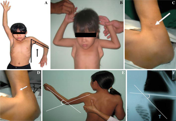
The preoperative adduction contracture (maximum abduction of the shoulder) average was 42°, range 35°–60° (Fig. 2a).
The postoperative adduction angle (maximum abduction of the shoulder, Fig. 4b) was greater than 150° in 37 shoulders (97%, 31 patients), with 140° in one shoulder (2.6%, 1 patient).
The preoperative elbow flexed angle when the scapula is held in the chest wall was on average 39°, range 25°–55° (Fig. 2e).
The postoperative elbow flexed angle when scapula is held in the chest wall (Fig. 4a) was greater than 130° in 36 shoulders (94.7%, 30 patients), with 120° and 125° in two elbows (5.3%, 2 patients).
Postoperative elbow extension angles were normal in all patients.
The preoperative scapulo-humeral angle in radiographs was on average 61° (range 40°–72°; Fig. 2f).
The postoperative scapulo-humeral angle in radiographs (Fig. 4c) was greater than 150° in 37 shoulders (97.4%, 31 patients) and 145° in one shoulder (2.6%, 1 patient).
Ultrasound showed the fibrous long head of the triceps in all patients (Fig. 3b).
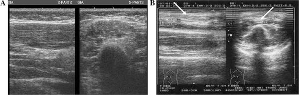
Ultrasound views of: a normal long head of the triceps; b fibrous long head of the triceps
Muscle biopsy performed intraoperatively showed marked fibrous tissue of the long head of the triceps, as is also seen in fibrosis of the quadriceps, deltoid, triceps, rectus femoris, and gluteal muscles after intramuscular antibiotic injections.
Preoperatively, dimpling of the skin on the medio-lateral proximal third of the arm was seen clearly when the elbow was flexed (Fig. 2c). A palpable long head of the triceps contracture (Fig. 2d) was observed in 38 shoulders (100%).
Postoperatively, dimpling of the skin and a palpable long head of the triceps contracture resolved completely in all patients, and movement of the scapula outward when the elbow was flexed also resolved completely (Fig. 4a) in 38 shoulders (100%, 32 patients).
Postoperative:
There were no wound infections or neuromuscular complications.
Discussion
Hněvkovský (1961) and Miki (1962) were the first authors to describe fibrous change after intramuscular injection of antibiotic(s) in children [6, 13]. Since that time, most published cases of muscle contracture seem to be related to injections in childhood, such as in the fibrous quadriceps, triceps, deltoid, rectus femoris, and fibrous gluteus maximus muscle [1–6, 8, 10, 13–35].
Lénárt [23], McCloskey [24], and Ogata [36] stated that the injection site also is correlated with disease development. Hence, the ratio of affected muscles depends on the customary injection sites in each country. Thus, injection into the posteriomedial proximal third of the arm may also result in a fibrous long head of the triceps.
Fibrotic muscular change has been most commonly related to a post-injection etiology. In our patients, antibiotics had been injected, including penicillin, gentamycin, lincomycin, streptomycin, and cloxacilin.
Intramuscular injections can result in local ischemic necrosis and chemical myositis, which can lead to muscle fibrosis. Repeated needle punctures and local trauma to the muscle can cause hemorrhage, necrosis, phagocytosis of muscle fibers, accumulation of mononuclear cells, and cell infiltrates even without the injection of a substance [37]. The contracture usually develops after repeated injections of medication into these muscles. Furthermore, the chemical composition of some medications or the non-physiological pH of the carrier medium can produce various degrees of histotoxic effects and can stimulate the formation of fibrous tissue. This property and the rapid injection of a large bolus of drugs can lead to muscle fibrosis. Electromyography has shown no electrical activity in the involved part of the muscle, while the findings on nerve-conduction-velocity studies have been normal [37]. Frasch (1976) [18] and McCloskey (1977) [24] proposed that if long-term antibiotic treatment is anticipated in children, intramuscular injection of antibiotic(s) should be avoided and the intravenous route be employed if possible, and we agree with their opinion.
Adduction contracture of the shoulder and outward movement of the scapula due to a long head of the triceps contracture must be differentiated from adduction contracture of the shoulder due to other causes. Neonatal brachial plexus palsy is the result of injury to one or more cervical or thoracic nerve roots (C5-T1) that occurs before, during, or after the birth process. Overall, about half completely resolve spontaneously during the first year. Most improvement occurs in the first 3 months. Brachial plexus palsy also results in an adduction contracture of the scapula; however, this usually involves the infraspinatus and teres minor muscles, and dyskinesia (abnormal involuntary movement) is a prominent feature [34]. Brachial plexus palsy does not result in dimpling of the skin or palpable long head of the triceps contracture. Paralysis of the long thoracic nerve results in loss of function of the serratus anterior muscle. This permits unopposed action of the trapezius, which tends to move the scapula outward when the arm is elevated, without dimpling of the skin or palpable long head of the triceps contracture. Osteochondroma of the scapula, when located on the anterior surface, causes apparent movement of the scapula outward that resolves after excision [7, 34].
According to Gray [20], the triceps arises anatomically from the dorsal arm and consists of three heads forming a tripennate structure: the lateral head from the upper half of the posterior surface of the shaft of the humerus above the spiral groove and the medial head from the posterior surface of the lower half of the shaft of the humerus below the spiral groove. The long head arises from the infraglenoid tuberosity of the scapula. The common tendon, a bilaminated aponeurotic structure beginning at the middle of the muscle, inserts into the upper surface of the olecranon with a small band of fibers continuing distally and laterally over the anconeus to join with the deep fascia [38, 39]. If the long head is fibrosed, shoulder abduction will not be possible beyond 90° when the scapula is held against the chest wall (Fig. 2a) and the scapula will move outward when the elbow is flexed (Fig. 2b), which happened in 38 shoulders (100%, 32 patients). The fibrous triceps results in triceps contracture leading to extension contracture of the elbow. The fibrous muscle commonly could be seen at the middle of the muscle, and the dimpling of the skin also could see at the mid third of the posterior arm. maximum abduction of the shoulder is still possible. However, the scapula does not move outward at shoulder abduction. The deformity could be corrected by operation for excision of fibrous tissue, and lengthening of the triceps was necessary to restore adequate conditions [1, 33].
Cosmetic problems include adduction contracture of the shoulder and outward movement of the scapula, especially on attempted elbow flexion. Long-standing fibrous long head of the triceps muscle produces dimpling of the skin (Fig. 2c) and the palpable long head of the triceps contracture (Fig. 2d) that we observed in all patients. The only functional disturbance observed was limitation of abduction of the shoulder due to adduction contracture (Fig. 2a). Operative intervention reduced the adduction contracture, movement of the scapula outward, and chronic discomfort. Postoperatively, dimpling of the skin and the palpable head of the triceps contracture resolved completely in all patients. Outward movement of the scapula with elbow flexion resolved completely in all as well (Fig. 4a).
The deformity can be corrected by surgical treatment, which was centered on the release of contracture of the long head of the triceps. Postoperative results with adduction angle were resolved completely in 37 shoulders (97.4%, 31 patients; Fig. 4b), elbow flexed angles were greater than 130° in 36 shoulders (94.7%, 30 patients; Fig. 4a), and scapulo-humeral angles in radiographs were greater than 150° in 37 shoulders (97.4%, 31 patients; Fig. 4c).
Normal muscle fibers were preserved to avoid decreasing the strength of abduction, flexion, and extension of the shoulder, and flexion and extension of the elbow. In this series, shoulder function was improved with good to excellent results in all, elbow extension was not affected, and there were no complications in any patients.
Conclusion
Adduction contracture of the shoulder is a rare deformity of the upper limb resulting from intramuscular injection of antibiotic(s). The principal signs are: shoulder abduction limited to 90° if the scapula is held in the chest wall; outward movement of the scapula when the elbow is flexed; dimpling of the skin on the medio-lateral proximal third of the arm, and palpable long head of the triceps contracture. Muscle biopsy shows marked fibrous tissue of the long head of the triceps, and ultrasound also shows a fibrous long head of the triceps. Operative release of the long head of the triceps is simple and safe, and affords marked improvement of shoulder function.