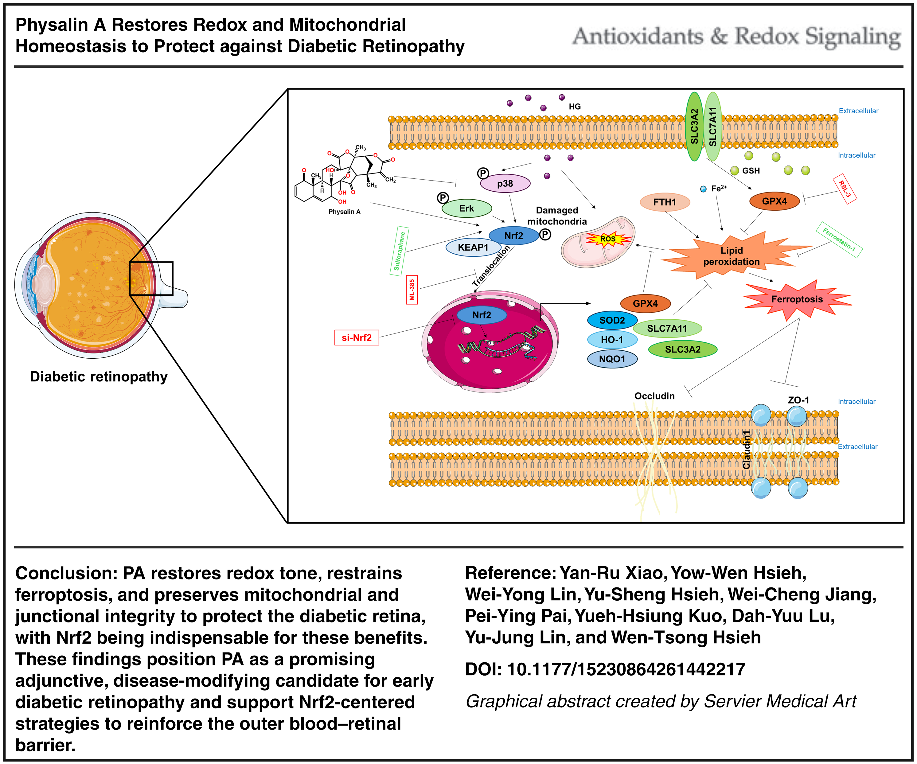
To determine whether physalin A (PA) safeguards the outer blood–retinal barrier under diabetic stress by engaging nuclear factor erythroid 2-related factor 2 (Nrf2) to restore redox balance and restrain ferroptosis in human retinal pigment epithelial (hRPE) cells and C57BLKS/J Iar −+Leprdb/+Leprdb mice.
Results:
In high-glucose challenged hRPE cells, PA dose-dependently preserved viability, maintained claudin-1/occludin/zonula occludens-1 abundance and membrane localization, and reversed ferroptosis hallmarks (restored solute carrier family 7 member 11 [SLC7A11], SLC3A2, and glutathione peroxidase 4; reduced ferrous iron [Fe2+] overload and lipid peroxidation). PA restored glutathione levels, reduced malondialdehyde (MDA), and enhanced the antioxidant defense pathway mediated by Nrf2, including upregulation of heme oxygenase 1, NAD(P)H quinone dehydrogenase 1, and superoxide dismutase 2. Silencing Nrf2 abolished the effects of PA on barrier integrity and ferroptosis suppression, with rebounds in reactive oxygen species, MDA, Fe2+, and tight junction loss. In db/db mice treated systemically for 20 weeks, PA reduced Evans Blue leakage, increased retinal thickness, restored RPE tight junction proteins, and normalized mitochondrial architecture by transmission electron microscopy. PA rebalanced mitochondrial dynamics (dynamin 1-like, optic atrophy 1, fission 1, mitofusin 1, FUN14 domain containing 1), increased retinal mitochondrial DNA copy number, and partially stabilized glycemia and weight.
Conclusion:
PA restores redox tone, restrains ferroptosis, and preserves junctional integrity to protect the diabetic retina, with Nrf2 being indispensable for these benefits. These findings position PA as a promising adjunctive candidate for early diabetic retinopathy and support Nrf2-centered strategies to reinforce the outer blood–retinal barrier. Antioxid. Redox Signal. 45, 5–27.
Research article
Restricted accessResearch articleFirst published July, 2026pp. 28-43
Sepsis-associated acute kidney injury (SA-AKI) is a frequent and severe complication in critically ill patients, yet effective targeted therapies are lacking. Ferroptosis has been implicated in various forms of organ injury, but its role in SA-AKI and underlying regulatory mechanisms remain unclear.
Methods:
A SA-AKI mouse model was established using cecal ligation and puncture (CLP). Renal histopathology, kidney function assays, and spatial proteomics were employed to assess ferroptosis activation. In vivo and in vitro models were subjected to lipopolysaccharide (LPS) stimulation to evaluate ferroptosis-related markers, including reactive oxygen species (ROS), lipid peroxidation, ferrous iron levels, and mitochondrial membrane potential. DNA pull-down coupled with mass spectrometry identified potential upstream regulators of HO-1. Chromatin immunoprecipitation-quantitative polymerase chain reaction (ChIP-qPCR) and dual-luciferase reporter assays were used to validate transcriptional regulation by SMAD4. Functional studies assessed the impact of SMAD4 on HO-1 expression, ferroptosis, and renal function.
Results:
Ferroptosis was markedly activated during SA-AKI progression. LPS stimulation induced significant ROS accumulation, lipid peroxidation, elevated ferrous iron levels, mitochondrial membrane potential disruption, and robust upregulation of heme oxygenase-1 (HO-1). SMAD4 was identified as a transcriptional repressor of HO-1. ChIP-qPCR and dual-luciferase assays confirmed SMAD4 binding to the HO-1 promoter and suppression of its transcription. SMAD4 overexpression reduced HO-1 expression, alleviated ferroptosis, and improved renal function in both in vivo and in vitro models.
Conclusions:
SMAD4 mitigates ferroptosis by transcriptionally repressing HO-1, exerting a protective effect in SA-AKI. This study identifies a novel SMAD4–HO-1 regulatory axis and suggests a potential therapeutic target for sepsis-induced kidney injury. Antioxid. Redox Signal. 45, 28–43.
Review article
Restricted accessReview articleFirst published July, 2026pp. 44-62
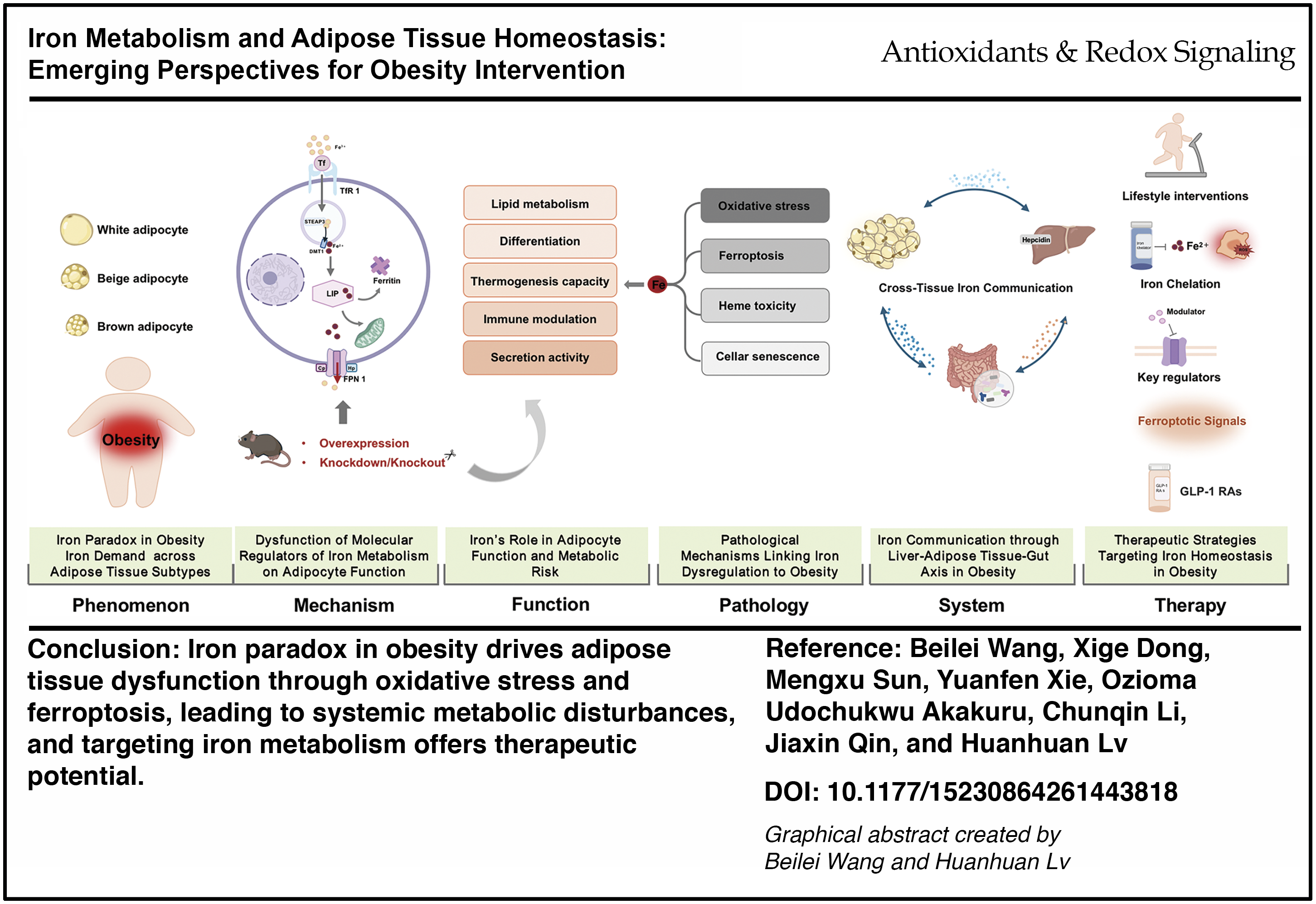
The global obesity epidemic is a significant risk factor for chronic metabolic diseases. Iron, an essential micronutrient involved in numerous biological processes, exhibits a dual role in health and disease. Understanding the mechanistic interplay between iron metabolism and adipocytes is crucial for elucidating the pathogenesis of obesity.
Recent Advances:
Emerging evidence indicates that iron plays a key role in adipose tissue physiology, and its dysregulation contributes substantially to obesity pathogenesis. Within adipocytes, iron exerts multifaceted effects, influencing lipid metabolism, differentiation, thermogenic capacity, secretory function, mitochondrial activity, and insulin sensitivity. Conversely, the metabolic status of adipose tissue reciprocally affects systemic iron homeostasis.
Goal of Review:
This review underscores the phenomenon of iron dysregulation in obesity. It examines the cellular mechanism describing iron demand and its dysregulation across adipose tissue subtypes. Following, it explores the functional pathology connecting iron imbalance to impaired energy metabolism, thermogenesis capacity, immune modulation, and secretion activity in adipose tissue, as well as iron-related oxidative stress, ferroptosis, heme toxicity, and cellular senescence within adipocytes in obesity. Then, it expands to the systemic level, highlighting the interorgan crosstalk in iron homeostasis, particularly among the liver, gut, and adipose tissue. Finally, it discusses the therapeutic strategies targeting iron homeostasis regulation to alleviate obesity and its related complications.
Future Direction:
Despite advancements, questions remain regarding depot-specific iron regulation and its relationship with metabolic dysfunction. Future studies should employ tissue-specific genetic models, well-designed human trials, and multiomics approaches to establish causality and translate the findings into targeted iron-modulating therapies for obesity. Antioxid. Redox Signal. 45, 44–62.
Research article
Restricted accessResearch articleFirst published July, 2026pp. 63-77
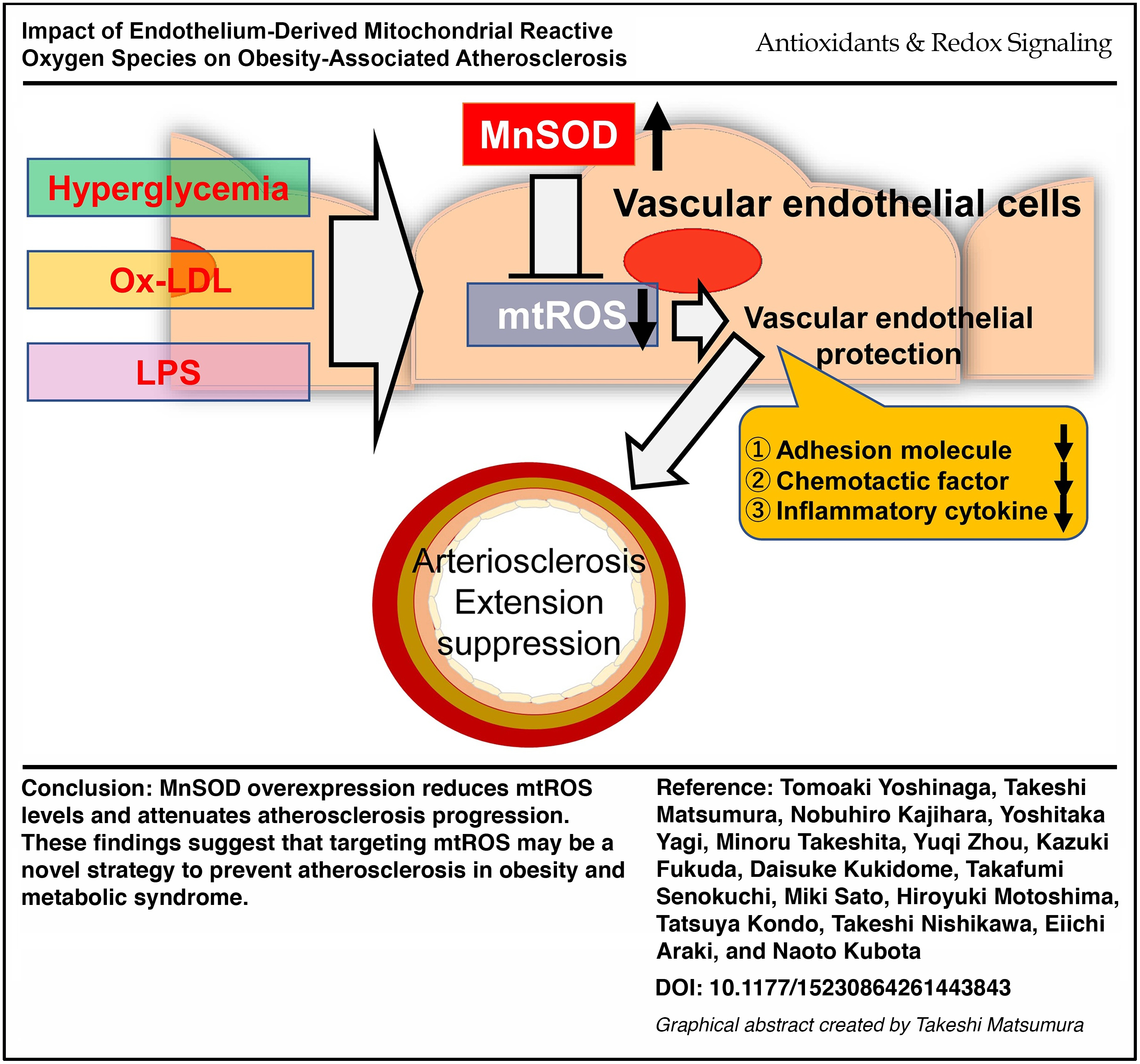
Obesity, a risk factor for atherosclerosis development and progression, is marked by excessive reactive oxygen species (ROS) production. We previously demonstrated that high-glucose (HG) conditions induce mitochondrial ROS (mtROS) production in aortic endothelial cells (ECs). However, the link between elevated mtROS levels in obesity and atherosclerosis progression remains unclear. This study aimed to investigate whether endothelial-specific mtROS suppression by overexpressing manganese superoxide dismutase (MnSOD) could attenuate atherosclerosis progression in high-fat diet (HFD)-induced obese apolipoprotein E-deficient (ApoE KO) mice.
Results:
Atherosclerotic lesion formation did not differ significantly between normal chow-fed control ApoE KO mice and endothelial cell-specific MnSOD-overexpressing ApoE KO (eMnSOD-Tg/ApoE KO) mice. However, in HFD-fed groups, eMnSOD-Tg/ApoE KO mice exhibited reduced atherosclerotic lesion size, decreased relative ROS levels, and lower Icam1 and Ccl2 expression compared to HFD-fed control ApoE KO mice. In obese ApoE KO mice, serum glucose, lipopolysaccharide (LPS), and low-density lipoprotein (LDL) levels were elevated. In human aortic endothelial cells, exposure to LPS, oxidized LDL, and high glucose increased relative mtROS levels, which was effectively attenuated by MnSOD overexpression. MnSOD also suppressed intercellular adhesion molecule-1 and monocyte chemoattractant protein-1 expression under these conditions.
Innovation:
Our findings demonstrate that endothelial-specific MnSOD overexpression suppresses obesity-related atherosclerosis in ApoE KO mice.
Conclusion:
mtROS plays a pivotal role in obesity-associated atherosclerosis, and targeting endothelial mtROS may offer a therapeutic strategy for preventing vascular complications in obesity. Antioxid. Redox Signal. 45, 63–77.
Review article
Restricted accessReview articleFirst published July, 2026pp. 78-112
Cellular proteostasis is essential for cellular proteome integrity, which is exquisitely sensitive to the redox environment. Heat shock proteins (HSPs) are the central chaperones that sense and adapt to these redox fluctuations. Emerging evidence demonstrates dysregulation of cellular HSPs-modulated redox-proteostasis in protein aggregation diseases, including cancers, senescence, neurodegenerative diseases, limb-girdle muscular dystrophy type D1, and β-thalassemia, making HSPs promising therapeutic targets in disease treatment.
Recent Advances:
Redox post-translational modifications (PTMs) serve as master switchboards to dynamically modulate the structure and chaperone function of HSPs. Redox PTMs allow HSPs to participate in protein synthesis and folding, conformational maintenance, and degradation, thereby maintaining cellular proteostasis. Beyond their chaperone functions, HSPs also play critical roles in organelle-specific stress responses, such as mitochondrial unfolded protein response, endoplasmic reticulum (ER) stress, and unfolded protein response.
Critical Issues:
Despite the well-known contributions of HSPs to redox-proteostasis, the double-edged functions of HSPs in protein aggregation diseases remain unclear. The main issues covered in this review include the regulation of HSPs by redox PTMs, the important role of HSPs in proteostasis and organelle-specific stress responses, dual modulation of HSPs in protein aggregation diseases, and pharmacological agents targeting HSPs.
Further Directions:
The functional diversity of HSPs in redox-proteostasis makes them promising therapeutic targets in disease treatment. Further studies should focus on exploiting agents that precisely target cysteine residues modifications on HSPs with good blood–brain barrier (BBB) penetration and low toxicity. Antioxid. Redox Signal. 45, 78–112.
Research article
Restricted accessResearch articleFirst published July, 2026pp. 113-132
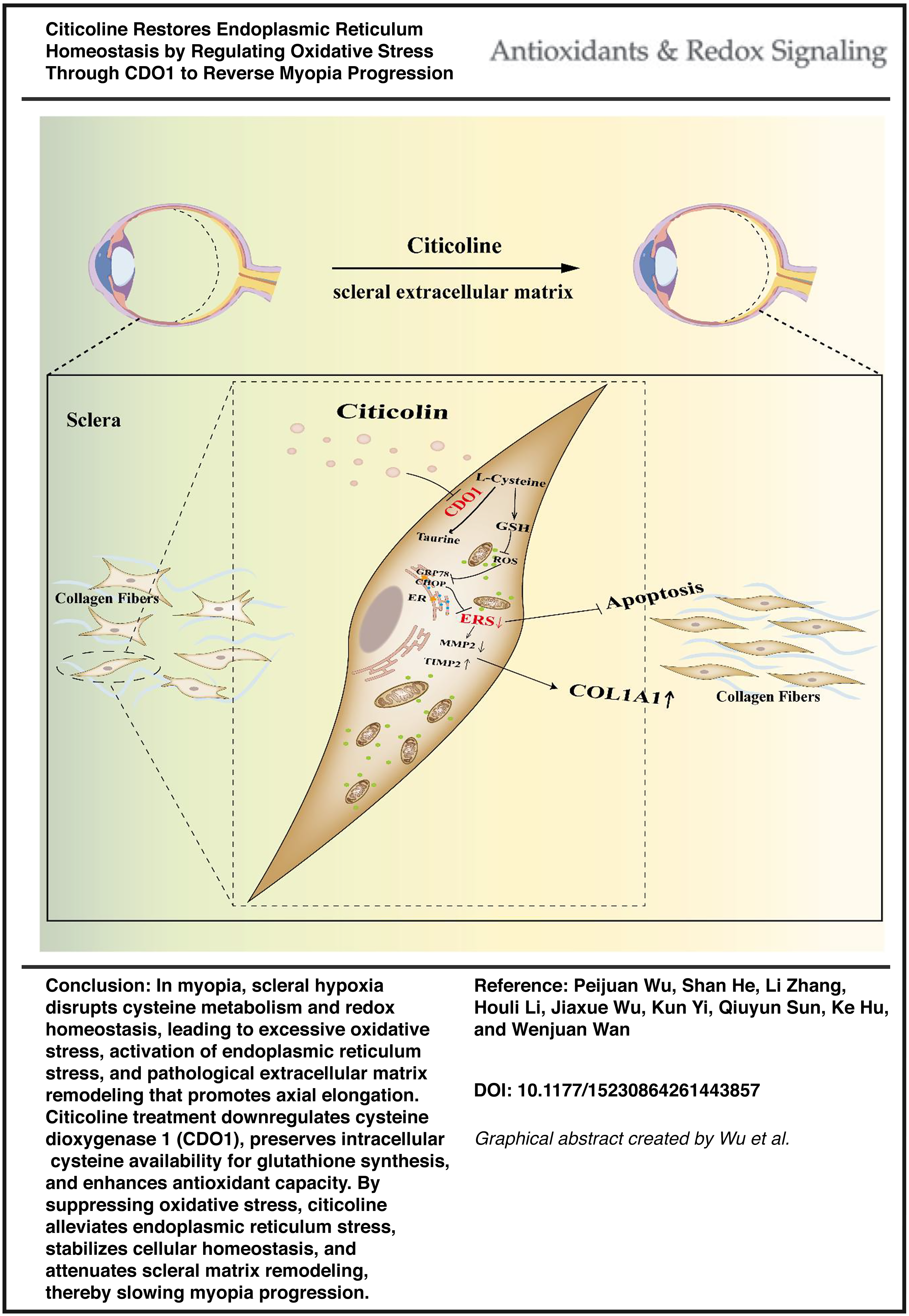
To investigate whether citicoline alleviates endoplasmic reticulum (ER) stress by modulating oxidative stress in myopia.
Results:
A form-deprivation myopia model in guinea pigs and a hypoxia-induced model in human scleral fibroblasts revealed that citicoline downregulated cysteine dioxygenase 1 (CDO1) expression, increased glutathione, and reduced reactive oxygen species. These changes mitigated oxidative stress, suppressed ER stress, maintained cellular homeostasis, and inhibited extracellular matrix remodeling, leading to reduced axial elongation.
Innovation:
Transcriptomic profiling identified CDO1 as a novel downstream effector of citicoline, linking cysteine metabolism to oxidative and ER stress regulation in myopia.
Conclusion:
Citicoline shows potential to delay myopia progression by alleviating ER stress through CDO1-mediated redox regulation, providing preclinical mechanistic insight into its possible role in myopia control. Antioxid. Redox Signal. 45, 113–132.
Research article
Restricted accessResearch articleFirst published July, 2026pp. 133-148
Liver ischemia-reperfusion injury (IRI) is a sterile inflammatory process that contributes significantly to graft rejection following liver transplantation. Although SET domain bifurcated histone lysine methyltransferase 1 (SETDB1) is known to preserve genomic stability and restrain inflammation under oxidative stress, its immunoregulatory function in myeloid cells during liver IRI has not been elucidated. This study aimed to investigate the role and mechanism of SETDB1 in regulating macrophage-driven inflammatory responses in liver IRI.
Results:
Myeloid-specific SETDB1 knockout (SETDB1 cKO) mice exhibited exacerbated liver injury, increased infiltration of pro-inflammatory macrophages and neutrophils, and amplified inflammatory responses compared with SETDB1fl/fl controls. Depletion of macrophages alleviated liver damage, reduced neutrophil infiltration and hepatocyte apoptosis, and eliminated the excessive injury observed in SETDB1 cKO mice. Mechanistically, SETDB1 suppressed the expression of purinergic receptor P2X7 (P2RX7). Pharmacological inhibition of P2RX7 with oxidized adenosine triphosphate significantly attenuated liver injury and macrophage infiltration in SETDB1 cKO mice. In vitro assays confirmed that SETDB1 inhibited the P2RX7/Caspase-1/Gasdermin D (GSDMD) pathway in macrophages, thereby limiting pyroptosis and inflammation.
Innovation:
This study identifies SETDB1 as a previously unrecognized regulator of macrophage pyroptosis during liver IRI. By linking epigenetic regulation to suppression of the P2RX7/Caspase-1/GSDMD pathway, our findings provide novel mechanistic insight into how SETDB1 protects against sterile liver inflammation.
Conclusion:
SETDB1 plays a pivotal role in protecting the liver from IRI by restraining macrophage-mediated pyroptosis and inflammation. These findings suggest that targeting the SETDB1/P2RX7/Caspase-1/GSDMD axis may represent a promising therapeutic strategy for mitigating liver IRI and improving transplant outcomes. Antioxid. Redox Signal. 45, 133–148.
Research article
Restricted accessResearch articleFirst published July, 2026pp. 149-168
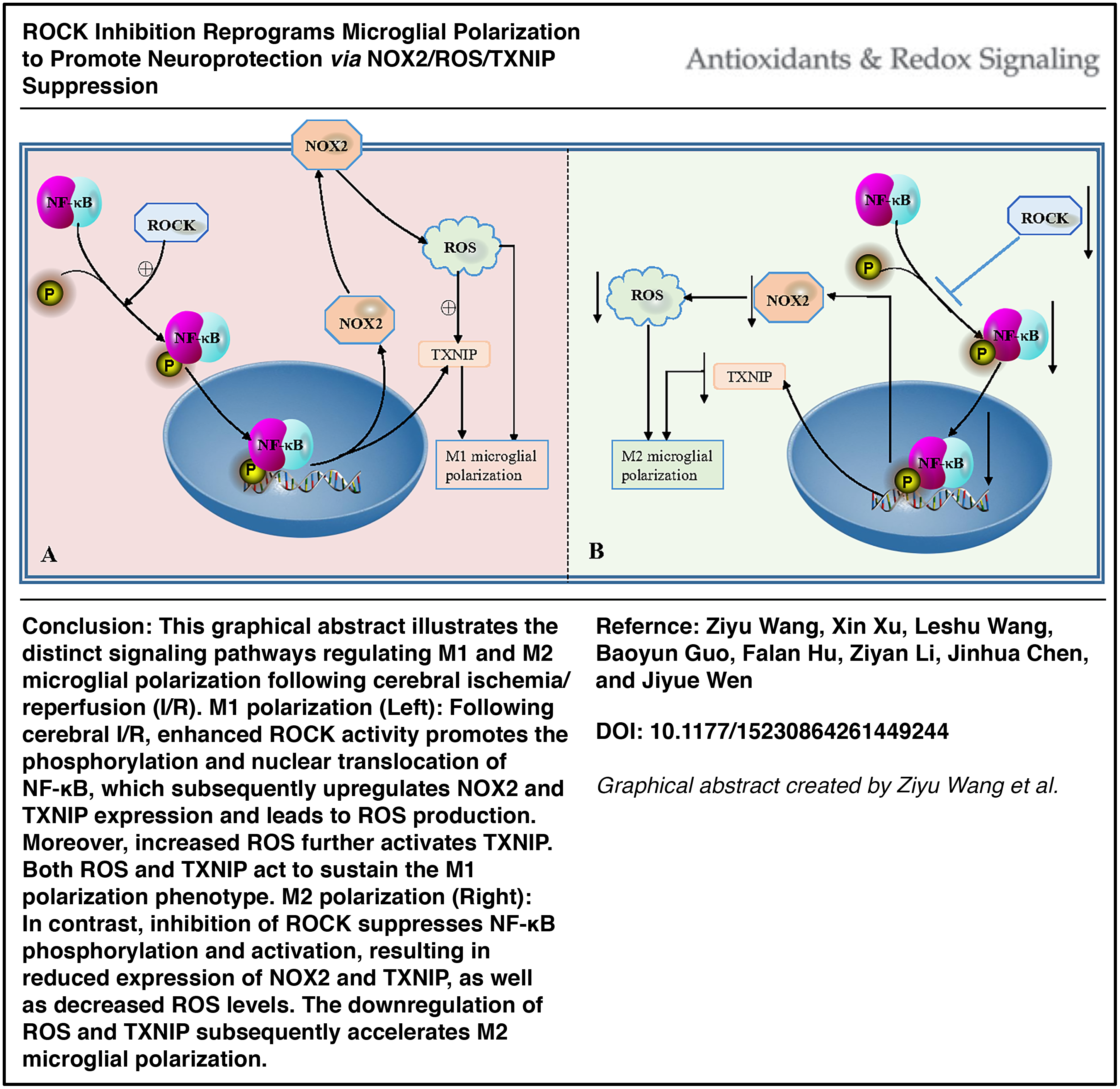
Cerebral ischemia/reperfusion (I/R) injury represents a significant challenge to recanalization therapy for ischemic stroke and is critically influenced by microglial polarization. Although inhibition of Rho-associated protein kinase (ROCK) has been shown to mitigate cerebral I/R injury and associated neuroinflammation, its specific effect on the balance between M1 and M2 (anti-inflammatory) microglial polarization remains incompletely understood. This study aimed to elucidate the role and underlying mechanism of ROCK inhibition in regulating M1 and M2 microglial polarization, using the classical antidepressant fluoxetine as a positive control.
Results:
ROCK inhibitor fasudil and positive control fluoxetine effectively alleviated cerebral I/R injury and facilitated a shift in microglial polarization from the M1 to the M2 phenotype, both in vivo and in vitro. In the hippocampal tissues of cerebral I/R mice exposed to lipopolysaccharide, we observed an upregulation of thioredoxin-interacting protein (TXNIP) and nicotinamide adenine dinucleotide phosphate (NADPH) oxidase 2 (NOX2). ROCK2 knockdown promoted the M2 microglial polarization, suppressed the expression of NOX2 and TXNIP, and inhibited the activation of NF-κB P65 in mouse hippocampal tissue. Notably, pharmacological inhibition of NF-κB reduced the expression of NOX2 and TXNIP, as well as the production of reactive oxygen species (ROS), in microglia subjected to oxygen-glucose deprivation/reoxygenation. Correspondingly, inhibition of NOX2 also decreased TXNIP expression.
Conclusion and Innovation:
ROCK inhibition promotes a shift in microglial polarization from the M1 to the M2 subtype by suppressing the NF-κB/NOX2/ROS/TXNIP signaling pathway. This study provides the first evidence demonstrating the mechanism by which ROCK inhibition drives microglial polarization toward the M2 phenotype. Antioxid. Redox Signal. 45, 149–168.