
Abstract
Introduction
Brain metastasis is one of the most common and feared cancer complications in patients with solid tumors. The first 5 ranking causes of brain metastases are: lung cancer (40%-50%), breast cancer (20%-30%), melanoma (5%-15%), renal cancer (6%-10%), and colorectal cancer (0.3%-9%) (1-2-3-4-5-6-7-8). Increased intracranial pressure and neurological and/or neurocognitive impairment represent the most common characteristics in these patients; these conditions lead to decreased quality of life, impose a considerable economic burden on both healthcare providers and society compared with non-metastatic disease, and have also been shown to influence prognosis, or even shorten survival. Although the application of brain-directed management, including whole brain radiotherapy, stereotactic radiosurgery, or prophylactic cranial irradiation, is able to induce prolonged remission or prevent brain metastases in many cases (9-10-11-12), cancer patients with brain metastases still have less favorable outcomes than those without brain metastases. Furthermore, brain metastasis incidence seems to be increasing probably due to improved imaging modalities and systemic treatments. As such, accurate approaches for the identification of cancer patients at ‘high risk' of developing brain metastases at initial diagnosis are required for early selection of those most likely to benefit from targeted therapy to prevent or delay brain metastasis, and/or intensive monitoring, as to optimize the likelihood of timely and successful treatment.
With the advent of tumor molecular profiling, established cancer-specific biomarkers help to elucidate the underlying tumor biology. Activating mutations in oncogenes, including the epidermal growth factor receptor (EGFR) gene and echinoderm microtubule-associated protein-like 4-anaplastic lymphoma kinase (EML4-ALK) in lung cancer, the Kirsten rat sarcoma viral oncogene homolog (KRAS) in colorectal cancer, and the human epidermal growth factor receptor-2 (HER2) in breast and gastric cancer, are used as predictors, since they are associated with prognosis, survival, and response to treatment. The sub-classification of patients with solid tumors with the use of molecular diagnostics has allowed us to re-examine the characteristics and outcomes of patients with solid tumors (13). Besides, it is hypothesized that the biology of the tumor may, at least in part, regulate the patterns of metastatic spread. That is, the cancer preference for giving rise to distant site metastasis may depend on specific genetic pathways (14-15-16). Partly based on this hypothesis, the patterns of metastatic spread comparing different dominant oncogenes recognized in solid tumors have been analyzed. Gene expression signatures derived from cell lines that repeatedly metastasize to the brain suggest that this process is programmed rather than being a stochastic result, and that it is possible to predict brain metastasis at initial diagnosis (17-18-19). However, the search for genetic difference has just begun, with no definitive conclusion yet as to which patients might benefit from such a strategy. The present review gives a broad overview on the rationales of oncogene status for the prediction of brain metastases based on the available evidence.
Search Methods
Eligible studies were identified by searching the PubMed and Cochrane databases for relevant reports, published before May 2014, using the search criteria “brain metastasis”. Only English-language literature and the most recent or the most complete studies were used. Meeting abstracts, unpublished reports, and review articles were not considered. Since lung cancer, breast cancer, and melanoma are the most common solid tumors that metastasize to the brain, and very few potentially genetic markers have been identified in brain metastases from other solid tumors, in this review we decided to focus on brain metastases from these 3 solid tumors.
Relationship between Brain Metastasis Formation and Oncogenes Expressed in Cancer Cells
The mechanism of brain metastasis formation remains unknown. However, what has been proven up to now is that “seeds” (i.e., cancer cells) exhibit specific preferences for the “soil” (i.e., brain) such that interactions between metastatic tumor cells and the brain micro-environment exist (20). Generally, when tumor cells reach the brain via the arterial blood supply, they might arrest in sites of slow flow within the capillary bed at vascular branch points (21). Then, the interactions between cancer cells and brain endothelial cells, or transendothelial migration, are mediated by the interaction of tumor cell-surface receptors with endothelial cell adhesion molecules, including integrins, selectins, and chemokines (22). After overcoming the blood-brain barrier (BBB), tumor cells are confronted with components of the local microenvironment including the extracellular matrix, resident brain parenchymal cells (astrocytes and microglia), and paracrine signaling molecules including cytokines and growth factors (22). The successful tumor cell survival requires the adaptation to and interaction with these factors, enabling them to thrive in the new milieu and develop clinically relevant secondary tumors. Also, to offset increasing metabolic demands associated with unrestrained cell division tumor cells synthesize proangiogenic proteins that instruct adjacent microvascular endothelial cells to form new vascular networks (i.e., angiogenesis). Kienast et al (23) used multiphoton laser scanning real-time microscopy to follow single steps of brain metastasis formation, and found that melanoma cells, as opposed to lung cancer cells, did not induce early angiogenesis. This particular finding further suggested that distinct signaling pathways, or the tumor biology, might affect brain metastasis formation, i.e., certain proteins are over or under-expressed in particular cancer cells for this phenomenon to occur.
Lung Cancer
Since a significant difference was found between the incidence of brain metastasis in non-small cell lung cancer (NSCLC) and small cell lung cancer (SCLC), we here present in different sections the possible oncogenes involved in these 2 lung cancer histotypes.
NSCLC
In NSCLC, differentially expressed oncogenes or signaling pathways have been found. As an example, in NSCLC cell lines harboring either wild-type or mutant EGFR, Breindel et al (24) reported that the inhibition of EGFR or MAPK reduced c-N-Methyl-N'-nitro-N-nitroso-guanidine HOS transforming gene (c-MET) activation and protein levels; also, c-MET signaling promoted EGFR-driven migration and invasion, and c-MET attenuation decreased the incidence of brain metastasis. A second study by Huang et al (25) demonstrated the migration of lung cancer cells via the C-X-C chemokine receptor type 4 (CXCR4)-mediated activation of extracellular signal-related kinase (ERK)-activated IKKa/b and NF-κB, resulting in the activation of integrins (ITGB1 and ITGB3) and in the production of MMPs in lung cells. This kind of directed migration was previously reported for breast cancer cells overexpressing CXCR4, which facilitated their transmigration through brain endothelial cells (26).
SCLC
The small GTP-binding protein Rho and its best-characterized downstream effector Rho-associated serine-threonine protein kinase (ROCK) participate in actin cytoskeleton organization, and are linked to the pathogenesis and progression of several human tumors. It has been shown that inhibition of ROCK decreases the migration of SCLC cells through the brain endothelium (27). This effect provides new information supporting a role of Rho/ROCK signaling in the decrease of transendothelial migration of tumor cells. Another possible oncogene involved is the placental growth factor (PLGF), a member of the VEGF family. It has been found to be mainly involved in cellular cytoskeleton rearrangements, which leads to cell migration in human breast cancer cell lines and human non-SCLC cell lines (28, 29). Li et al (30) reported that PLGF derived from SCLC cells triggered VEGF receptor-1-Rho-extracellular regulated protein kinase 1/2 signaling axis activation, resulting in disassembly of tight junctions in brain endothelial cells, and promoting SCLC cell transendothelial migration. Furthermore, down-regulation of PLGF suppresses SCLC cell metastasis to the brain in an experimental brain metastasis model (30). Current ongoing studies focusing on the potential roles of oncogenes in SCLC brain metastasis might provide a deeper understanding of the critical pathway that drive this condition.
Breast Cancer
The PI3K/Akt pathway
The PI3K/Akt signaling pathway regulates many normal cellular processes including cell proliferation, survival, growth, and motility; all these processes are critical for tumorigenesis. Aberrant activation of the PI3K/Akt pathway has been widely implicated in many cancers. In a recent study, a novel inhibitor of downstream PI3K was found to effectively control metastatic growth of HER2-positive breast cancer cells and resulted in a large number of mice free from brain metastases (31). Sandra et al (32) reported that the type I insulin-like growth factor receptor (IGF-IR), a mediator of the PI3K/Akt signaling pathway, can not only promote cell motility and pro-metastatic behavior in breast cancer cells, but can also mediate the onset of brain metastasis in breast cancer. The main effects observed by knockdown of IGF-IR were decreased migration and invasion of MDA-MB-231-BR brain-seeking cells (the brain metastatic derivative of the MDA-MB-231 cell line), as well as reduced potential to establish brain metastases in an in vivo experimental brain metastasis model. All these results further support the hypothesis that specific gene expression signatures may be crucial for brain metastasis growth.
HER2/neu
HER2-overexpressed breast tumors have been shown to have higher frequencies of brain metastasis than other breast cancers, with occurrences as high as 30%-35% among patients with advanced breast cancers (33, 34). Palmieri et al (35) also demonstrated that after transfecting human MDA-MB-231-BR cells with HER2, the HER2-overexpressing cells showed a three-fold increase in the number of brain metastases compared with the control group. Moreover, signaling through this receptor pathway is active and results in cell proliferation in the brain microenvironment (35).
The Notch Signaling
The Notch signaling pathway is a highly conserved cell signaling system present in most multicellular organisms. It has been shown that Notch signaling is activated in human breast cancer, which may be correlated with poor overall survival and chemoresistance (36). This pathway has also been shown to play a role in cancer stem cell (CSC) maintenance (37-38-39). McGowan et al (40) reported that cells sorted for a reduced “stem-like” phenotype (CD44hi/CD24lo) had a reduced ability to form brain metastases compared with unsorted or CD44hi/CD24lo cells (p<0.05). Adding to this, in vivo, Notch1 knockdown reduced the expression of CD44hi/CD24lo phenotype by about 20%. In vitro, Notch1 shRNA resulted in a reduction in cellular growth at 24, 48, and 72 hours time points (with respectively p=0.033, p=0.002, and p=0.009, calculated by ANOVA); additionally, a reduction in matrigel invasion of about 60% was observed (p<0.001, ANOVA). Given the relationship of the “stem-like” phenotype with both brain metastasis and Notch signaling inhibition, it is suggested that the CSC phenotype contributes to the development of brain metastases from breast cancer and this may arise in part from increased Notch activity.
Other Single Oncogenes
There are some other single oncogenes implicated in the proliferation, invasion, or migration of breast cancer cells that might contribute to our current understanding of brain metastasis occurrence. Kim et al (41) reported that the inflammatory lipid sphingosine-1-phosphate (S1P) activated the transcription of C-reactive protein (CRP), one of the main integrins in breast cancer brain metastasis development, in MDA-MB-231 cells and nude mice bearing MDA-MB-231-derived tumors, through signaling pathways involving reactive oxygen species (ROS)/ERKs and matrix metallopeptidase-9 (MMP-9) upregulation. Chiu et al (42) found that strategies to increase or decrease Caveolin-1 (Cav-1) expression were sufficient to attenuate or promote breast cancer cell invasion. Increased expression of Cav-1 phenocopied the effects of STAT3 activation in blocking primary tumor growth and abrogating the formation of brain metastases (42). Rodriguez et al (43) reported that the neuropeptide substance P (SP) secreted by human MDA-MB-231 and MDA-MB-231BRM2 cells induced transmigration of breast cancer cells across the BBB, leading to the activation of brain microvascular endothelial cells and secretion of TNF-α and Ang-2, which are involved in BBB impairment and colonization of tumor cells in brain. Another study by Nie et al (44) showed that EGFR inhibition or activation strongly affected MDA-MB-231-BR cell migration/invasion activities as assessed by an adhesion assay, a wound-healing assay, a Boyden chamber invasion assay, and cytoskeleton staining. Also, EGFR inhibition significantly decreased brain metastases of MDA-MB-231-BR cells in vivo. Recently, Malin et al (45) found that αB-crystallin, a molecular chaperone linked to many biologic characteristics of breast cancer, may be a novel regulator of brain metastasis in breast cancer. In his study, stable overexpression of αB-crystallin in MDA-MB-231 breast cancer cells enhanced adhesion to human brain microvascular endothelial cells, transendothelial migration, and BBB transmigration in vitro and in vivo, whereas silencing αB-crystallin inhibited these events. Although the molecular mechanisms accounting for brain metastasis specifically from breast cancer are still largely unknown, the above findings support the hypothesis that some alterations may occur early at the primary tumor site that promote spread of cancer cells to the brain.
Melanoma
In malignant melanoma, which is another common tumor generating brain metastasis, specific pathways activated in tumor cells have been found.
The JAK-STAT Pathway
The JAK-STAT pathway, which promotes survival, growth, and angiogenesis, was reported to increase melanoma brain metastasis mainly via STAT3 activation by phosphorylation or downregulation of its inhibitor SOCS-1 (46). The main effects observed in the above study were the increased expression of MMP-2, bFGF, and VEGF. However, a recent study showed that melanoma brain metastases exhibited the highest level of p-STAT3 expression and that p-STAT3 expression was not associated with either an increased risk of developing brain metastasis or time to brain metastasis (47).
Rho/ROCK Signaling
As for Rho/ROCK signaling, an important signaling pathways involved in tumor-endothelial interactions in the brain, Wilhelm et al (48) investigated the effect of the inhibition of ROCK on the attachment of melanoma cells to monolayers of cerebral endothelial cells. The results indicated that ROCK inhibition in melanoma cells increased the number of melanoma cells attached to the brain endothelium, strengthened the adhesion force between melanoma and endothelial cells, raised the number of melanoma cells migrating through the brain endothelial monolayer and promoted the formation of parenchymal brain metastases in vivo.
Other Solid Tumors
In colorectal cancer, it has been shown that S100A4, a member of the S100 family of calcium-binding proteins, not only promotes motility and invasion of tumor cells, resulting in aggressive metastasis (49), but also reduces the expression of occludin, stimulates p53 expression in brain microvascular endothelial cells, and disturbs the normal construction of the BBB (50). More work is needed to determine whether S100A4 physically and functionally interact with brain metastasis specificity.
Based on preclinical studies, we can conclude that the functional significance of oncogene signaling expression in primary tumors contribute to our current understanding of brain metastasis occurrence. Therefore, identifying oncogene status that could be potentially involved in carcinoma metastasis to the brain may offer insights into the prediction of brain metastases.
Clinical Evidence of Biomarkers in the Prediction of Brain Metastases
Recently, much of the work on promising oncogenes for brain metastases at initial diagnosis has been conducted in patients. These studies have identified multiple possible markers of brain metastases, especially in lung cancer, breast cancer, and melanoma.
Lung Cancer Oncogene Status
In lung cancer, approximately 10%-25% of patients have brain metastases at initial diagnosis and about 40%-50% of patients develop brain metastases during the course of disease (51). Although the incidence of brain metastasis is higher in SCLC than NSCLC, very few potential genetic markers have been identified in SCLC. Therefore, in this section, we will mainly focus on NSCLC brain metastasis.
NSCLC Molecular Subtypes
NSCLC is a heterogeneous disease with respect to the adenocarcinoma subgroup, and is perhaps best exemplified by the molecular features identified as “driver mutations”, including EGFR, KRAS, EML4-ALK fusions, HER2, PIK3CA, AKT, BRAF, MAP2K1, and c-MET (52). Recently it was found that driver mutations in the adenocarcinoma subgroup may be, at least in part, associated with brain metastases from NSCLC. As an example, a few reports have been published on the relationship between brain metastases and EGFR mutations, which are of several types and include the exon 19 deletion and the exon 21 point mutation. Among them, Lee et al (53) reported that the brain was a common organ for recurrence in patients with resected NSCLC harboring EGFR mutations, and some other researchers showed that the EGFR-mutated NSCLC patients were prone to brain metastases or developed brain metastases earlier than those without the mutation (54-55-56-57), see Table I. Sekine et al (58) found that NSCLC patients with the exon 19 deletion may have a peculiar pattern of brain metastases, with multiple small metastases and small brain edema, compared to patients with wild-type EGFR (see Tab. I). NSCLC with exon 19 deletions showed a higher incidence of CNS involvement than tumors bearing a L858R mutation (21% vs 3%) (59), see Table I. Moreover, the EGFR inhibitors gefitinib and erlotinib have not only shown evidence for an activity against established brain metastases, but also for prophylaxis of CNS relapse (21). In such a situation, it might be expected that EGFR-mutated NSCLC would metastasize more often to brain than non-mutated NSCLC. However, after retrospectively evaluating 79 patients with 469 brain metastases between 2005 and 2012, Johung et al (59) reported that the rates of distant brain recurrence did not significantly differ across EGFR mutations. Hendriks et al (60) reached a similar conclusion through a retrospective case-control study of 189 NSCLC patients. Therefore, data from more clinical studies are needed. Interestingly, ALK translocations, which represent other “druggable” alterations, besides activating EGFR mutations to date in NSCLC, appear to be correlated with brain metastasis formation (61, 62), see Table I. On the other hand, Doebele et al (63) retrospectively analyzed a total of 209 consecutive patients with stage IV nonsquamous NSCLC with EGFR mutations (n=39), KRAS mutations (n=49), ALK gene rearrangements (n=41), or wild-type for all 3 (triple negative, n=80). The authors found that patients with ALK gene rearrangements were predisposed to liver metastases, compared to the triple negative cohort (odds ratio, OR=5.50; 95% confidence interval, 95% CI, 1.76, 17.18; p=0.003), but not to brain metastases. A similar conclusion was also reached by Johung et al (59). Despite this negative result on brain metastases, the association of distinct patterns of metastatic spread with certain targetable oncogenes is promising.
POSSIBLE ONCOGENES THAT MAY PREDICT BRAIN METASTASIS IN CANCER PATIENTS
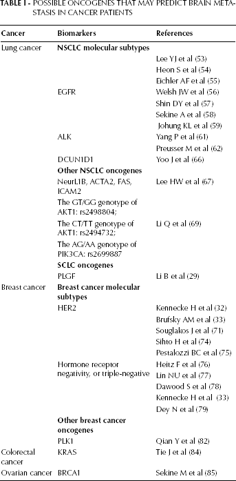
The successful identification of druggable targets in lung adenocarcinoma has stimulated screening of squamous cell carcinoma specimens and has led to the identification of possible “driver mutations”, including FGFR1, PIK3CA, DDR2, MDM2, and PTEN (64, 65). So far, to the best of our knowledge, most of these candidate driver genes have not been investigated in brain metastasis formation except for DCUN1D1. In a recent study Yoo et al (66) found that DCUN1D1-positive primary NSCLCs carry a biological predisposition for brain metastasis in 14/16 NSCLC patients (87.5%) with DCUN1D1-positive resulting in brain metastases (OR=3.112, p=0.009) (see Tab. I). As such, further studies are needed to clarify the predictive implications of the above genes in brain metastasis from squamous cell carcinoma.
Other Oncogene Statuses of NSCLC
Several recent studies focusing on the functional significance of other oncogenes have also contributed to the prediction of brain metastasis development. Using molecular inversion probe technology, Lee et al (67) reported that amplified regions of primary lung tissues from 12 lung adenocarcinoma patients (5q35, 10q23 and 17q23-24) were detected to be associated with the development of brain metastases within 3 months after initial diagnosis of NSCLC. Those regions harbor several candidate genes, including NeurL1B, ACTA2, FAS, and ICAM2. It has recently been found that studying multiple single-nucleotide polymorphisms (SNPs) in signaling pathways that are regulating cell proliferation and migration may be a more powerful way of pinpointing the genes and polymorphisms involved in conferring risk of brain metastasis (68, 69), see Table I. The PI3K-AKT-mTOR signaling pathway will be here taken as an example. Li et al (70) tested 16 SNPs in 5 core genes (PIK3CA, PTEN, AKT1, AKT2, and FRAP1) by using DNA from blood samples of 317 NSCLC patients without brain metastasis. The results indicated that the GT/GG genotype of AKT1 (rs2498804), the CT/TT genotype of AKT1 (rs2494732), and the AG/AA genotype of PIK3CA (rs2699887) were associated with a higher risk of brain metastasis at 24 months of follow-up (with respective HRs 1.860, 95% CI 1.199-2.885, p=0.006; HR 1.902, 95% CI 1.259-2.875, p=0.002; and HR 1.933, 95% CI 1.168-3.200, p=0.010). Moreover, these SNPs had a cumulative effect on brain metastasis risk, with that risk being the highest for patients carrying both these unfavorable genotypes (p=0.003). With a clearer understanding of how these gene regions influence brain metastasis development, we believe that these oncogene biomarkers may provide another method to identify patients at high risk of brain metastasis at initial diagnosis. Further prospective trials are needed for confirmation.
Oncogene Statuses in SCLC
Besides investigating the PLGF-mediated SCLC cell migration capacity across the BBB in vitro, Li et al (29) screened the levels of PLGF in the serum of 79 SCLC patients who attended the Liaoning Cancer Hospital in China from 2006 to 2009. The authors found that SCLC patients with high levels of PLGF are prone to brain metastasis, with 77.78% of sensitivity, 66.67% of specificity, and 59.3% of accuracy. This finding may be useful in future efforts to identify SCLC patients at high risk of developing brain metastases.
Breast Cancer Oncogene Status
Breast Cancer Molecular Subtypes
Breast cancer is another clinically heterogeneous tumor, and the most common type of cancer affecting women worldwide. With respect to the molecular features, breast cancer is perhaps best exemplified by the molecular subgroups including basal-like, luminal A (hormone receptor positive), luminal B, HER2 amplified/over-expressed, and normal breast-like subtypes (70). As discussed above, patients with HER2-positive tumors are at a high risk of developing brain metastases, with an incidence ranging from 30% to 35% across studies (32, 33). Compared with HER2-positive/amplified primary breast tumors, HER2 mRNA levels were increased five-fold in breast cancer brain metastases (71). However, when considering trastuzumab as standard treatment in patients with breast cancer, several studies reported that trastuzumab is a risk factor for the development of brain metastases in HER2-positive metastatic breast cancer patients (72, 73). Accordingly, large retrospective series contribute to the issue. At present, the question of whether or not trastuzumab is associated with increased brain metastasis still needs further confirmation. Besides, in breast cancer, HER2 positivity, hormone receptor negativity, and even triple-negativity are important risk factors for the development of brain metastasis (see Tab. I). Sihto et al (74) conducted a retrospective study with 2,032 cases identified, and found that breast tumors with first metastases in the brain expressed nestin, prominin-1, and CK5, while only infrequently expressed estrogen receptor (ER) and progesterone receptor (PR). In a cohort of more than 9,000 women with early stage breast cancer who were enrolled into the International Breast Cancer Study Group clinical trials between 1978 and 1999, Pestalozzi et al (75) reported that the incidence of CNS metastases as the first site of recurrence in women who had ER-negative disease was higher than that of other subtypes (2-year incidence of 1.1%). In a single institution study of over 3,000 women, Hietz et al (76) reported higher odds of developing brain metastases among women with triple-negative breast cancer (OR, 4.16; 95%CI, 2.26-7.64; p<0.001) than among women with other breast tumor subtypes. The similar metastatic patterns were further validated by large cohorts analyzed by Lin et al (77), Dawood et al (78), and Kennecke et al (33). Dey et al (79) demonstrated that triple-negative breast cancer patients identified by the Wnt/beta-catenin classifier had a greater risk of lung and brain metastasis, but not bone metastases. Next, given the heterogeneity of HER2 amplified/overexpressed or triple receptor-negative breast cancer, it would be important to identify a specific subgroup among these patients at highest risk of developing brain metastases.
Other Oncogene Status
Polo-like kinase-1 (PLK1) is a crucial driver of cell cycle progression and its downregulation plays an important checkpoint role in the response to DNA damage. Numerous studies have now established that PLK1 is upregulated in various tumors and is a prime target for drug development in proliferative diseases such as breast cancer (80, 81). Qian et al (82) found that the transcriptional expression of Plk1 is higher in breast cancer patients with brain metastases than with extracranial metastases (p=0.0018). The Plk1 inhibitor GSK461364A prevented the growth of large brain metastasis by 62% and resulted in an improved survival by 17% in a mouse model of brain metastases derived from breast cancer, further indicating that Plk1 might be a promising biomarker for predicting and detecting this disease (82), see Table I. However, published data are far too limited to draw any firm conclusion on its predictive value. Therefore, more data is needed for validation.
Melanoma Oncogene Status
Schoenewolf et al (83) retrospectively studied 310 stage-IV melanoma patients with regard to the potential correlations between frequency and occurrence of metastasis and the genetic background and pathological/clinical melanoma subtypes. They found superficially spreading (SSM) and nodular melanomas (NMM) spread to the brain more frequently than acrolentiginous (ALM) and mucosal (MM) melanomas (p=0.0012), while the skeletal metastasis was significantly higher for ALM and MM than for SSM and NMM (p=0.0049). However, BRAF-mutant versus wildtype tumors showed no significant differences concerning localization of the metastasis. More work is needed to determine which oncogenes interact with brain metastasis.
Other Oncogenes for the Prediction of Brain Metastasis
Although brain metastases are less common in other solid tumors, specific and sensitive biomarkers for brain metastases are of great importance, as brain imaging is not routinely undertaken at initial staging (see Tab. I). Tie et al (84) searched for differences in oncogene mutation profiles between colorectal cancer metastases from different sites and evaluated these as markers for sites of relapse. The results indicated that the KRAS mutation prevalence was significantly higher in lung (62.0%) and brain metastasis (56.5%) than in liver metastases (32.3%; p=0.003). Mutation status was highly concordant between primary cancer and metastasis from the same individual. In addition, after finding 7 cases with brain metastases in 340 ovarian cancer cases (7/340, 2.1%), Sekine et al (85) reported 4 loss of heterozygosity-positive cases with a germline mutation of BRCA1 in 2 of the 4 cases, and absent staining of the BRCA1 protein in the remaining 2 cases. That is, the loss of BRCA1 function may be involved in the phenomenon of brain metastasis from ovarian cancer. Studies on larger patient cohorts will be required to formally confirm this association.
Future Directions
The brain microenvironment can protect tumor cells from chemotherapeutics and antitumoral immune response, and it also contains soluble factors favoring tumor cells' survival. Therefore, identifying oncogenes predictive of brain metastases may allow for the selective use of effective interventions in patients who would benefit the most, without affecting quality of life and/or radiological surveillance, as to optimize the likelihood of timely and successful treatment options. The oncogenes discussed above hold prospect in the prediction of brain metastasis for they can be measured easily and sensitively at initial diagnosis, and they may provide information on the status of brain metastasis in preclinical and/or clinical studies. However, currently the possibility for the above oncogenes to provide predictive information still requires further investigation in prospective trials. Moreover, whether these markers related to brain metastases more than other types of metastasis, among which bone and liver metastases, still needs confirmation. The gene expression levels can sometimes be affected by other factors and may not be so precise. Despite all this, a better comprehension of the mechanisms of brain metastasis together with prospective studies are expected for further stratifying the rationales of oncogene status in the prediction of brain metastases. Identifying which patients are at the highest risk for brain recurrence through oncogene analysis holds great promise in cancer management.