
Abstract
Background
This in vitro study evaluated the influence of adhesive systems on the color match of a resin composite with different translucencies.
Methods
Sixty disk-shaped specimens were made with A2 and opaque A2 (OA2) shades of nanohybrid resin composite Z250 XT. Specimens of each shade (n = 30) were randomly reassigned to 3 subgroups according to adhesive system: a 2-step etch-and-rinse adhesive (Adper Single Bond Plus), a 2-step self-etch system (Clearfil SE Bond) and a universal adhesive (Scotchbond Universal Adhesive). The bonding agents were applied to resin composite specimens following the manufacturers’ recommendations. Additionally, 5 disk samples of each adhesive system were prepared. Colorimetric evaluation (CIE L*a*b* system) was performed immediately after polishing the sample and application of the adhesive systems. Color changes (ΔE and ΔE00) were calculated between 2 measurements. Color coordinates L*, a* and b* of the adhesive disks were also assessed. The data obtained were analyzed using ANOVA and Tukey's post hoc test (α = 0.05).
Results
The application of Scotchbond Universal Adhesive to the resin composite A2 shade resulted in the highest color change (p<0.01; ΔE = 3.1 ± 0.7 and ΔE00 = 1.8 ± 0.4). However, no significant difference was observed among adhesive systems when applied to the resin composite OA2 shade (p>0.05). Scotchbond Universal Adhesive revealed augmented yellowing and greening in comparison with other experimental groups.
Conclusions
The universal adhesive tested resulted in higher visually perceptible color changes when using a more translucent resin composite shade, but this was clinically acceptable.
Introduction
Many factors can influence the esthetic appearance of resin composite restorations, such as inherent optical properties of the material, including color and translucency, the thickness employed and the color and opacity of underlying dental substrate (1, 2). Therefore, the fulfillment and stability of color match with enamel and dentin is the main goal when resin restorations are performed (3).
Although there is evidence to suggest that shade options for resin cements may affect the final appearance of the ceramic indirect restorations (4-5-6), it is not well established in the scientific literature if the adhesive systems could have a similar effect on resin composite restorations.
While some studies have shown that the adhesive system plays an important role in changing the color of direct composite restorations (7, 8), the results of a recent investigation suggested the opposite (3). Furthermore, considering the wide variation in color and composition of the commercially available adhesives, studies evaluating different adhesive systems are necessary. In this sense, this investigation attempted to elucidate whether a new generation of adhesive systems, referred to as universal adhesives, could influence on the color of esthetic restorations.
Likewise, the effect of the adhesive system could be dependent on the translucency of the restorative material. Therefore, this in vitro study aimed to investigate the influence of adhesive systems on the color match of a resin composite with different translucencies.
Methods
For this study, 3 adhesive systems were used: a 2 etch-and-rinse adhesive (Adper Single Bond Plus; 3M ESPE, St. Paul, MN, USA), a 2 self-etch system (Clearfil SE Bond; Kuraray, Noritake, Tokyo, Japan) and a universal adhesive system (Scotchbond Universal Adhesive; 3M ESPE, St. Paul, MN, USA). The nanohybrid resin composite (Filtek Z250 XT; 3M ESPE, St. Paul, MN, USA) was tested in the A2 and opaque A2 (OA2) shades. A detailed description of the materials is presented in Table I.
Detailed description of the materials tested
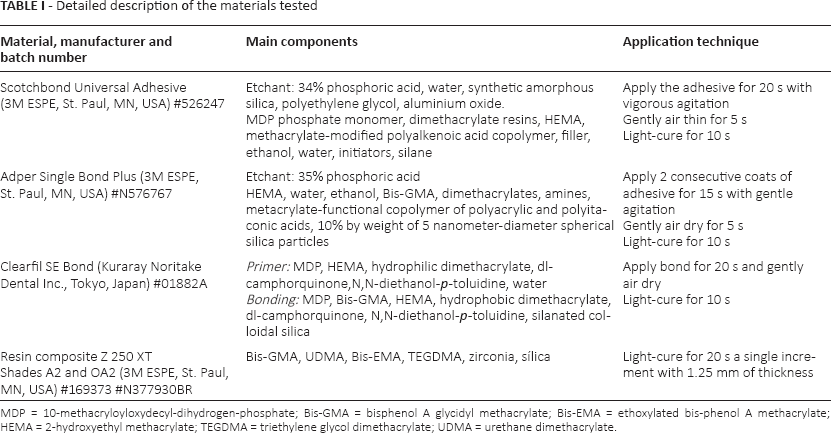
MDP = 10-methacryloyloxydecyl-dihydrogen-phosphate; Bis-GMA = bisphenol A glycidyl methacrylate; Bis-EMA = ethoxylated bis-phenol A methacrylate; HEMA = 2-hydroxyethyl methacrylate; TEGDMA = triethylene glycol dimethacrylate; UDMA = urethane dimethacrylate.
Specimen preparation
Sixty disk-shaped samples (8 mm in diameter and 1.25 mm in thickness) were made from the 2 shades of resin composite (A2 and OA2) with the aim of simulating direct laminate veneers.
A single increment of resin composite was placed in a metallic mold, pressed between 2 glass plates and light-cured for 20 seconds from the upper and the bottom surface, with a light-emitting diode curing unit (Emitter B; Schuster, Santa Maria, RS, Brazil) with a light output of at least 1,250 mW/cm2. Light intensity output was monitored with a Demetron Curing Radiometer (Kerr, Orange, CA, USA). The specimens were stored in distilled water at 37°C for 24 hours to ensure stabilization of the polymer network. After storage, specimens were polished using fine and extra-fine abrasive discs (SofLex Pop On; 3M ESPE, St. Paul, MN, USA) for 20 seconds each. The thickness of each specimen was confirmed with a digital caliper (Absolute Digimatic; Mitutoyo, Tokyo, Japan).
Composite disks of each shade (A2 and OA2) were randomly reassigned to 3 subgroups according to the adhesive system. This resulted in a 2 × 3 factorial experimental design, with 10 specimens for each of the 6 subgroups formed from the crossing of the 2 factors.
After polishing, the bonding agents of the different adhesive systems were applied to resin composite disks according to the manufacturers’ instructions (Tab. I). A single trained operator performed all procedures.
Additionally, 15 disk-shaped samples (8 mm in diameter and 1.25 mm in thickness) were made solely from the 3 adhesive agents tested (n = 5). Each material was dispensed in a silicone mold and light-cured for 20 seconds on each side of the disk. The thickness of each specimen was also measured with a digital caliper.
Color analysis
Colors were measured according to the CIE L*a*b* color scale relative to the standard illuminant D65 over a white background using a spectrophotometer (SP60; X-Rite, Grand Rapids, MI, USA). The color measurements were made immediately after polishing and after the application of the adhesive systems. The surface of the specimens with adhesive was turned down in the opposite side to the aperture of the spectrophotometer reader.
Three color readings were performed in each assessment with the spectrophotometer to establish the mean values. Color changes were calculated using the following formula: ΔE = (ΔL2 + Δa2 + Δb2)1/2.
The isolated coordinates were also separately analyzed to compare the variation of the color axes following the formulae: ΔL = L*f − L*i; Δa = a*f − a*I; and Δb = b*f − b*i, in which i represents the initial analysis (after polishing) and f the final analysis (after adhesive application) of each specimen.
CIELab values were also converted into CIEDE2000 color notation formula, which was additionally used to calculate color difference by using the equation (9): ΔE00 = [(ΔL’/kLSL)2 + (ΔC’/kCSC)2 + (ΔH’/kHSH)2 + RT (ΔC’/kCSC)(ΔH’/kHSH)]1/2, where ΔL’, ΔC’ and ΔH’ are differences in lightness, chroma and hue between 2 color readings, and RT is the rotation function that accounts for the interaction between chroma and hue differences in the blue region. SL, SC, and SH are weighting functions used to adjust the total color difference for variation in perceived magnitude with variation in the location of the color coordinate difference between 2 color readings. kL, kC, and kH are correction terms for the experimental conditions. Color coordinates L*, a* and b* of the adhesive disks were also calculated considering an average value of 3 measurements.
Statistical Analysis
The normal distribution of the data was confirmed using the Kolmogorov-Smirnov test. The ΔE00, ΔE, ΔL*, Δa* and Δb* values were subjected to 2-way analysis of variance (ANOVA), using “adhesive system” and “resin composite” as variables. Color coordinates L*, a* and b* of the adhesive agents disks were analyzed by 1-way ANOVA. Tukey's multiple comparisons statistical test at a 0.05 significance level was used. Statistical analysis were performed using the Minitab software (Minitab Inc., State College, PA, USA).
Results
Means and standard deviations of ΔE00, ΔE, ΔL*, Δa* and Δb* values considering the variables “adhesive system” and “resin composite” are presented in Table II. The color change (ΔE and ΔE00) was influenced by the factors “adhesive system” (p<0.01) and “resin composite” (p<0.01), as well as the cross-product interaction “adhesive system” vs. “resin composite” (p<0.01).
ΔE00, ΔE, ΔL*, Δa* and Δb* values for all experimental groups
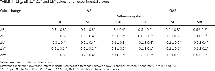
Values are means ± standard deviation.
Different superscript lowercase letters indicate significant differences between rows, considering each Δ separately (n = 10; p<0.05).
SB = Adper Single Bond Plus; SE = Clearfil SE Bond; SBU = Scotchbond Universal Adhesive.
The application of Scotchbond Universal Adhesive to the resin composite A2 shade resulted in the highest color change. The Δa* and Δb* mean values revealed a greater variation in green-red and blue-yellow coordinates, indicating increased greening and yellowing in comparison with other experimental groups. However, no significant difference was observed among adhesive systems when applied to the resin composite OA2 shade.
Means and standard deviations of L*, a* and b* values of the adhesive systems are summarized in Table III. The color of adhesives was only influenced by a* (p<0.01) and b* (p<0.01) parameters. Scotchbond Universal Adhesive showed a higher trend to green and yellow.
L*, a* and b* values for each adhesive system
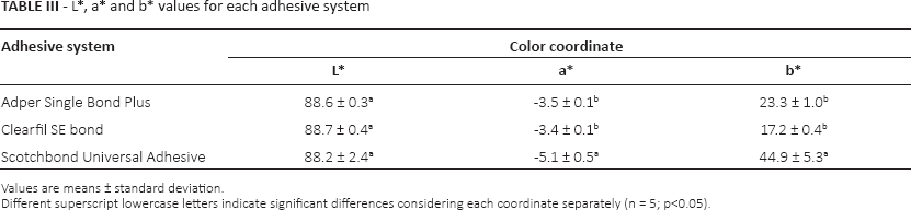
Values are means ± standard deviation.
Different superscript lowercase letters indicate significant differences considering each coordinate separately (n = 5; p<0.05).
Discussion
The color of direct esthetic restorations is affected by the optical properties and thickness of the material selected and the background color. Considering that the commonly used clinical background of resin composite restorations is composed of a hybrid layer – demineralized collagen and infiltrated adhesive resin – it could be assumed that the adhesive systems could interfere with the color of these restorations. To eliminate the interference of other factors on color data, such as subjacent substrate (10, 11), resin composite disks were not bonded to tooth structure – i.e., the bonding agents were only applied under restorative material. In our study, the effect of the adhesive systems on the color of the nanohybrid resin composite was dependent on the material tested. Scotchbond Universal Adhesive showed the highest color change along the a* and b* coordinates – i.e., it induced color alteration toward green and yellow when applied to the resin composite A2 shade. Previous studies have reported that an adhesive can compromise the color of direct composite restorations, showing changes detected on the a* and b* values (7), or still no influence on in vitro color stability (after 300 hours of accelerated artificial aging) has been found (3).
Methodological differences relating to variations in sample thickness (range of 0.7-1.0 mm) may explain these controversial findings. However, the thickness value used in this study (1.25 mm) had a significant effect on the color changes. Thus, it was expected that the color changes in the resin composites were mainly related to the differences in the composition of the adhesive systems. Oxidative reactions of camphorquinone and amine, occurring in Scotchbond Universal Adhesive, could result in darkness and a greater yellowing effect (12). The greater yellowing effect could also be attributed to a lower conversion degree (13). To avoid this yellowing effect and consequently reduce the influence of the adhesive layer on resin composite color, the use of substitutes for camphorquinone, such as acylphosphine oxides, has been suggested (13). Despite this, camphorquinone is still associated with higher color stability of resin composites (14).
No significant color changes were observed for any of the adhesives tested when applied to the resin composite OA2. The translucency of resins depends on the material thickness, absorption coefficient and scattering filler particles, pigments and opacifiers (15, 16). In our study, a single resin composite with varying translucency was used. The resin composite OA2 (opaque resin) presents higher opacity compared with the A2 shade. Thereby, universal adhesive may compromise only the initial color performance of esthetic restorations restricted to dental enamel.
Considering the association between color change and clinical significance, there is no consensus in the scientific literature regarding the magnitude of the color difference that must be regarded as visually detectable or visually unacceptable. However, it has been shown that the perceptibility and acceptability thresholds range from ΔE = 1-1.2 and ΔE = 2.7-3.7, respectively (17, 18). Although in the majority of dental studies, color differences are represented by ΔE, the CIEDE2000 color difference formula provides a better fit than the CIELAB formula to evaluate the color difference thresholds of dental materials (19, 20). In this sense, ΔE00 = 0.8 is considered clinically perceptible, but to be esthetically unacceptable, ΔE00 must be higher than 1.8 (19). Since 3.1 ΔE and 1.8 ΔE00 units were found when using universal adhesive associated with resin composite A2, this material can have a greater impact on the perceptibility of the initial color of esthetic restorations in comparison with other adhesive systems investigated; however, without jeopardizing the clinical appearance.
Color changes capable to be perceptible in a clinical situation (1) could be a consequence of the surface degradation of resin composite or the adhesive layer (10, 21). This effect seems to be proportional to the concentrations of hydrophilic monomers on the adhesive system, because the hydrophilicity is increased, and a considerably higher amount of water is expected to be absorbed (22). Further studies evaluating the influence of different adhesives, mainly the new 1-bottle systems, on the long-term color stability of resin composites, are required (23).
In conclusion, the effect of adhesive systems on the immediate color of resin composites is material-dependent. The tested universal adhesive resulted in higher visually perceptible color changes when a more translucent resin composite was used, but this was clinically acceptable.
Footnotes
Financial support: No grants or funding have been received for this study.
Conflict of interest: None of the authors has any financial interest related to this study to disclose.